Oral
(Artificial) Intelligence in the Body
ISMRM & SMRT Annual Meeting • 15-20 May 2021

| Concurrent 5 | 14:00 - 16:00 | Moderators: Teodora Chitiboi & Graham Kemp |
0529.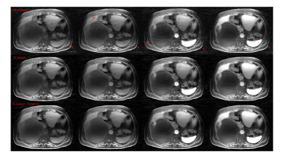 |
Deep learning based radial de-streaking for free breathing time resolved volumetric DCE MRI
Sagar Mandava1, Xinzeng Wang2, Ty Cashen3, Tetsuya Wakayama4, and Ersin Bayram2
1GE Healthcare, Atlanta, GA, United States, 2GE Healthcare, Houston, TX, United States, 3GE Healthcare, Madison, WI, United States, 4GE Healthcare, Hino, Japan
Radial imaging is becoming increasingly popular due to its ability to support highly accelerated imaging. However, it is plagued by streak artifacts that often arise from undersampling which can lead to poor image quality. The problem is particularly acute in time resolved imaging where the need for high spatio-temporal sampling usually leads to large amount of streaks. In this work, we propose a method for separate spatial and temporal deep learning for streak artifact reduction. The utility of the method is demonstrated on free breathing time resolved volumetric DCE MRI acquired using the stack-of-stars trajectory.
|
||
 |
0530.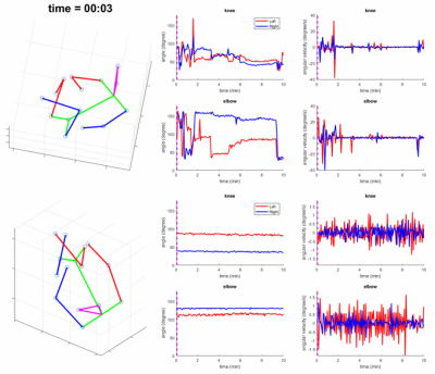 |
Motion Analysis in Fetal MRI using Deep Pose Estimator
Junshen Xu1, Esra Abaci Turk2, Borjan Gagoski2, Polina Golland3, P. Ellen Grant2,4, and Elfar Adalsteinsson5
1Department of Electrical Engineering and Computer Science, Massachusetts Institute of Technology, Cambridge, MA, United States, 2Fetal-Neonatal Neuroimaging and Developmental Science Center, Boston Children’s Hospital, Boston, MA, United States, 3Computer Science and Artificial Intelligence Laboratory, Massachusetts Institute of Technology, Cambridge, MA, United States, 4Harvard Medical School, Boston, MA, United States, 5Massachusetts Institute of Technology, Cambridge, MA, United States
Fetal motion is an important measure for monitoring fetal health and neurological function. However, current clinical MRI and ultrasound assessments of fetal motion are qualitative and cannot reflect detailed 3D motion of each body part. In this work, we propose a method for fetal motion analysis in MRI using a deep pose estimator. We train a neural network to estimate fetal pose from MR volumes, and extract quantitative metrics of motion from the time series of fetal pose. In the experiments, we study how different conditions affect fetal motion, such as gestational age and maternal position during scan.
|
|
0531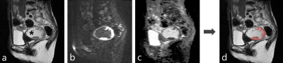 |
Automatic segmentation of uterine endometrial cancer on MRI with convolutional neural network Video Permission Withheld
Yasuhisa Kurata1, Mizuho Nishio1, Yusaku Moribata2, Aki Kido1, Yuki Himoto1, Koji Fujimoto3, Masahiro Yakami2, Sachiko Minamiguchi4, Masaki Mandai5, and Yuji Nakamoto1
1Diagnostic Imaging and Nuclear Medicine, Kyoto university hospital, Kyoto, Japan, 2Preemptive Medicine and Lifestyle-Related Disease Research Center, Kyoto university hospital, Kyoto, Japan, 3Real World Data Research and Development, Graduate School of Medicine Kyoto University, Kyoto, Japan, 4Diagnostic Pathology, Kyoto university hospital, Kyoto, Japan, 5Gynecology and Obstetrics, Kyoto university hospital, Kyoto, Japan
Endometrial cancer is the most common gynecological malignant tumor in developed countries, and accurate preoperative risk stratification is essential for personalized medicine. For realizing tumor feature extraction by radiomics approach, the segmentation of the tumor is usually required. The model developed in this study has achieved high-accuracy automatic segmentation of endometrial cancer on MRI using a convolutional neural network for the first time. Using multi-sequence MR images were important for high accuracy segmentation. Our model will lead to efficient medical image analysis of a large number of cases using the radiomics approach and/or deep learning methods.
|
||
 |
0532.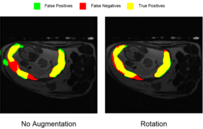 |
Evaluation of Data Augmentation Methods for Autonomous Segmentation of Placental Volume for Detecting Viral Complications
Thomas Lilieholm1, Ruiming Chen1, Ruvini Navaratna1, Daniel Seiter1, Walter F Block1,2,3, and Oliver Wieben1,2,3
1Medical Physics, University of Wisconsin at Madison, Madison, WI, United States, 2Biomedical Engineering, University of Wisconsin at Madison, Madison, WI, United States, 3Radiology, University of Wisconsin at Madison, Madison, WI, United States
Quantitative investigation of placental volumes can be used for characterization of Zika virus (ZIKV) infection, which causes several complications for developing fetuses. To provide more rapidly available image segmentation for analysis, efforts are being made to produce Convolutional Neural Networks (CNN) for autonomous segmentation of placental volume images. We investigated a number of data augmentation techniques for training machine learning models to determine which methods may be most suited for further development of ZIKV-quantifying placental segmentation models. We found rotational and reflective data augmentation to produce the greatest improvement in machine-segmentated Dice Coefficient comparisons.
|
|
0533.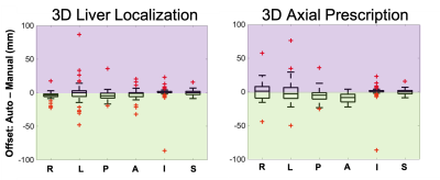 |
Automated Image Prescription for Liver MRI using Deep Learning
Ruiqi Geng1,2, Mahalakshmi Sundaresan3, Jitka Starekova1, Collin J Buelo1,2, Nikolaos Panagiotopoulos1, Marcin Ignaciuk1, Thekla Helene Oechtering1, Scott B Reeder1,2,4,5,6, and Diego Hernando1,2
1Radiology, University of Wisconsin-Madison, Madison, WI, United States, 2Medical Physics, University of Wisconsin-Madison, Madison, WI, United States, 3Electrical and Computer Engineering, University of Wisconsin-Madison, Madison, WI, United States, 4Medicine, University of Wisconsin-Madison, Madison, WI, United States, 5Emergency Medicine, University of Wisconsin-Madison, Madison, WI, United States, 6Biomedical Engineering, University of Wisconsin-Madison, Madison, WI, United States
To enable fully free-breathing, single button-push liver exams, an automated AI-based method for image prescription of liver MRI was developed and evaluated. A total of seven classes of rectangular bounding boxes covering the liver, torso, and arms for each localizer orientation were manually and automatically labeled to enable 3D prescription in any orientation. The intersection over union (IoU) between manual and automated 2D liver detection had a median > 0.88 and interquartile range < 0.11 for all classes. The shift in the resultant 3D axial prescription was less than 9 mm in S/I dimension for 91% of the test dataset.
|
||
|
0534.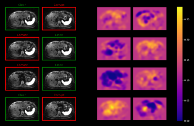 |
Deep Learning-based Adaptive Image Combination for Signal-Dropout Suppression in Liver DWI
Fasil Gadjimuradov1,2, Thomas Benkert2, Marcel Dominik Nickel2, and Andreas Maier1
1Pattern Recognition Lab, Friedrich-Alexander-Universität Erlangen-Nürnberg, Erlangen, Germany, 2Magnetic Resonance, Siemens Healthcare GmbH, Erlangen, Germany
Signal-dropouts due to pulsation are one of the most prominent artifacts in diffusion-weighted imaging (DWI) of the liver. It can affect a significant portion of the repetitions acquired for a given slice. Instead of performing uniform averaging which might result in locally attenuated liver signal, this work proposes to train a convolutional neural network (CNN) to estimate smooth weight maps for individual repetitions. This allows to locally suppress signal-dropouts, resulting in more homogeneous liver signal while maintaining signal-to-noise ratio (SNR) in artifact-free image regions.
|
|
0535.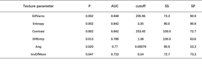 |
Utility of Texture Analysis on Quantitative Susceptibility Maps to Stage Hepatic Fibrosis
FengLing Gan1, Shuohui Yang2, Feng Xing3, Zheng Qu1, Gaiying Li1, Chenyao Yang2, Rongfang Guo2, Jiling Huang2, Fang Lu2, Caixia Fu4, Xu Yan4, Kelly Gillen5, Yi Wang5, Chenghai Liu3,
Songhua Zhan2, and Jianqi Li1
1Shanghai Key Laboratory of Magnetic Resonance, School of Physics and Electronic Science, East China Normal University, shanghai, China, 2Department of Radiology, Shuguang Hospital Affiliated to Shanghai University of Traditional Chinese Medicine, shanghai, China, 3Department of Liver Diseases, Shuguang Hospital Affiliated to Shanghai University of Traditional Chinese Medicine, shanghai, China, 4MR Collaboration NE Asia, Siemens Healthcare, shanghai, China, 5Department of Radiology, Weill Medical College of Cornell University, New York, NY, United States
Hepatic fibrosis is characterized by excessive accumulation of extracellular matrix (ECM) proteins including collagen, which would contribute strong diamagnetic susceptibility in the liver tissue due to enhanced density of orbiting electrons. This study measured the texture features on susceptibility maps in patients with chronic liver diseases. The results showed that some of the second-order texture parameters were significantly different between cohorts of significant hepatic fibrosis (Ishak-F ≥ 3) and non-significant hepatic fibrosis (Ishak-F < 3). The texture analysis on susceptibility maps may have the potential to stage hepatic fibrosis.
|
||
0536.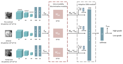 |
Discriminative feature learning and adaptive fusion for the grading of hepatocelluar carcinoma with Contrast-enhanced MR
Wu Zhou1, Shangxuan Li1, Wanwei Jian1, Guangyi Wang2, Lijuan Zhang3, and Honglai Zhang1
1School of Medical Information Engineering, Guangzhou University of Chinese Medicine, Guangzhou, China, 2Department of Radiology, Guangdong General Hospital, Guangzhou, China, 3Shenzhen Institutes of Advanced Technology, Chinese Academy of Sciences, Shenzhen, China
The combination of context information from multi-modalities is remarkably significant for lesion characterization. However, there are still two remaining challenges for multi-modalities based lesion characterization including features overlapping between different tumor grades and large differences in modal contributions. In this work, we proposed a discriminative feature learning and adaptive fusion method in the framework of deep learning architecture for improving the performance of multimodal fusion based lesion characterization. Experimental results of grading of clinical hepatocellular carcinoma (HCC) demonstrate that the proposed method outperforms the previously reported fusion methods, including concatenation, correlated and individual feature learning, and deeply supervised net.
|
||
0537.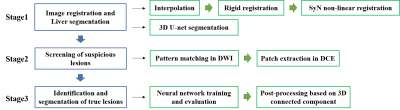 |
Automatic Detection of Small Hepatocellular Carcinoma (≤2 cm) in Cirrhotic Liver based on Pattern Matching and Deep Learning
Rencheng Zheng1, Luna Wang2, Chengyan Wang3, Xuchen Yu1, Weibo Chen4, Yan Li5, Weixia Li5, Fuhua Yan5, He Wang1,3, and Ruokun Li5
1Institute of Science and Technology for Brain-Inspired Intelligence, Fudan University, Shanghai, China, 2Department of Radiology, Shanghai Chest Hospital, Shanghai, China, 3Human Phenome Institute, Fudan University, Shanghai, China, 4Market Solutions Center, Philips Healthcare, Shanghai, China, 5Department of Radiology, Ruijin Hospital, Shanghai, China
This study presented an algorithm for small hepatocellular carcinoma (sHCC) detection and segmentation in cirrhotic liver based on diffusion-weighted imaging (DWI) and dynamic contrast-enhanced (DCE) images. The model included two-steps: screening of suspicious lesions in DWI using pattern matching algorithm; identification and segmentation of true lesions in DCE based on deep learning. The proposed model exhibited superior performance in sHCC (≤2 cm) detection and segmentation, which significantly outperformed the Liver Imaging Reporting and Data System (LI-RADS) based diagnosis.
|
||
0538.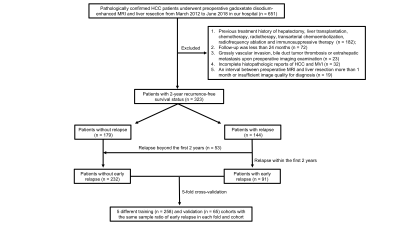 |
Peritumoral Dilation Radiomics of Gd-EOB-DTPA MRI Predicts Early Relapse in Hepatocellular Carcinoma Without Macrovascular Invasion
Huan-Huan Chong1,2, Yu-Da Gong3, Lei Chen4, Xian-Pan Pan4, Ai-E Liu4, Chun Yang2, and Meng-Su Zeng1,2,5
1Shanghai Institute of Medical Imaging, Shanghai, China, 2Department of Radiology, Zhongshan Hospital, Fudan University, Shanghai, China, 3Department of General Surgery, Zhongshan Hospital, Fudan University, Shanghai, China, 4Shanghai United Imaging Intelligence Co., Ltd., Shanghai, China, 5Department of Medical Imaging, Shanghai Medical College, Fudan University, Shanghai, China
Preoperatively identifying predisposing predictors of early relapse is crucial for stratifying patient risk, performing prompt intervention and improving long-term outcome. Whether peritumoral dilation radiomics can predict early recurrence in hepatocellular carcinoma(HCC) patients without macrovascular invasion remains unclear. Hence, we developed a bi-regional (the entire tumor and peritumoral zone within 1 cm) radiomics of gadoxetate disodium-enhanced (Gd-EOB-DTPA) MRI, which derived a satisfying discrimination in 2-year recurrence by the 5-fold cross-validation method.
|
The International Society for Magnetic Resonance in Medicine is accredited by the Accreditation Council for Continuing Medical Education to provide continuing medical education for physicians.