Oral
Liver
ISMRM & SMRT Annual Meeting • 15-20 May 2021

| Concurrent 5 | 14:00 - 16:00 | Moderators: Johannes Heverhagen & Jin Wang |
0311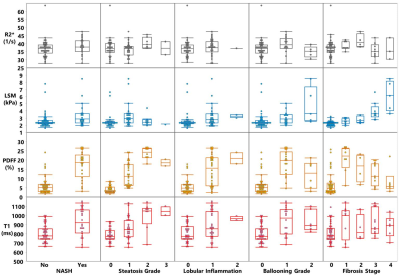 |
Diagnostic Performance of Multiparametric Models Using Fat Fraction, Liver Stiffness, and T1 for Detection of Nonalcoholic Steatohepatitis Video Permission Withheld
Xin Lu1, Jiahui Li1, Zheng Zhu1, Alina Allen2, Taofic Mounajjed3, Kevin J Glaser1, Jinhang Gao 4, Jingbiao Chen1, Jie Chen1, Safa Hoodeshenas1, Armando Manduca1, Richard L Ehman1, and Meng Yin1
1Department of Radiology, Mayo Clinic, Rochester, MN, United States, 2Gastroenterology and Hepatology, Mayo Clinic, Rochester, MN, United States, 3Anatomic Pathology, Mayo Clinic, Rochester, MN, United States, 4Division of Gastroenterology and Hepatology, Mayo Clinic, Rochester, MN, United States
NASH is traditionally diagnosed by liver biopsy, limited by subjective scoring and sampling error. This motivates identifying imaging-based biomarkers that might quantitatively characterize the pathophysiologic features of NASH. This prospective study established a streamlined imaging protocol for acquiring three candidate biomarkers ( fat fraction, liver stiffness, and T1 ) in a cohort of 66 patients with suspected NASH who underwent biopsy. The results indicate that a two-parameter model using fat fraction and liver stiffness has superior accuracy in diagnosing NASH, including ones that include the T1 relaxation time, which was found to have high collinearity with the fat fraction.
|
||
 |
0312.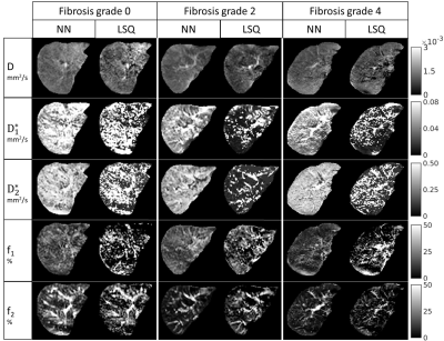 |
Physics-informed deep neural network for tri-exponential intravoxel incoherent motion fitting in non-alcoholic fatty liver disease.
Marian A. Troelstra1, Julia J. Witjes2, Anne-Marieke van Dijk2, Anne Linde Mak2, Jurgen H. Runge1, Joanne Verheij3, Max Nieuwdorp2, Adriaan G. Holleboom2, Aart J. Nederveen1, and Oliver J. Gurney-Champion1
1Department of Radiology and Nuclear Medicine, Amsterdam UMC, location AMC, Amsterdam, Netherlands, 2Department of Internal and Vascular Medicine, Amsterdam UMC, location AMC, Amsterdam, Netherlands, 3Department of Pathology, Amsterdam UMC, location AMC, Amsterdam, Netherlands
In this study we have developed an unsupervised physics-informed deep neural network (IVIM3-NET) to fit a tri-exponential model to intravoxel incoherent motion (IVIM) data from 35 non-alcoholic fatty liver disease (NAFLD) patients. Diagnostic performance was compared to a tri-exponential least squares (LSQ) fit. Visually, IVIM3-NET showed high-quality parameter maps with less noise than the LSQ-fit. IVIM3-NET showed slightly higher correlations between fit parameters and histology and more significant differences between levels of fibrosis and inflammation than the LSQ-fit. Correlations between f2 and fibrosis and inflammation grade, potentially highlighting NAFLD-induced vascular changes, warrant further investigation of the IVIM3-NET in NAFLD patients.
|
|
0313.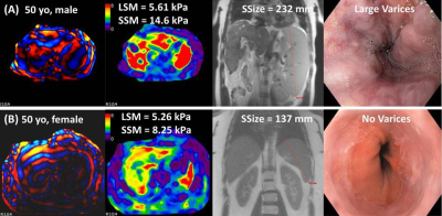 |
High-risk Esophageal Varices Screening with MR Elastography in Patients with Cirrhosis
Safa Hoodeshenas1, Mahmoud Adam Tahboub Amawi2, Jingbiao Chen1,3, Nimish Thakral2, Kevin J. Glaser1, Bogdan Dzyubak1, Jiahui Li1, Xin Lu1, Jie Chen1, Zheng Zhu1, Patrick S. Kamath2, Vijay Shah2, Richard L. Ehman1, Sudhakar K.
Venkatesh1, Douglas A. Simonetto2, and Meng Yin1
1Department of Radiology, Mayo Clinic, Rochester, MN, United States, 2Division of Gastroenterology and Hepatology, Mayo Clinic, Rochester, MN, United States, 3The Third Affiliated Hospital of Sun Yat-sen University, Guangzhou, China
In patients with end-stage chronic liver diseases, esophageal varices can be a life-threatening complication of portal hypertension because of bleeding risk. In this study, we explored the role of MR elastography (MRE) in stratifying portal hypertension severity and predicting high-risk varices by using upper gastrointestinal endoscopy as the reference standard. We concluded that a composite model including spleen size and stiffness plus liver stiffness and platelet count more accurately predicts the presence of high-risk varices, and thus could reduce unnecessary endoscopies more precisely than using liver stiffness and platelet count alone.
|
||
|
0314.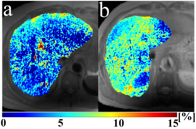 |
Macromolecular proton fraction mapping based on spin-lock for the non-invasive diagnosis of early stage liver fibrosis
Jian Hou1, Vincent Wai-Sun Wong2, Grace Lai-Hung Wong2, Baiyan Jiang1, Yi-Xiang Wang1, Anthony Wing-Hung Chan3, Winnie Chiu-Wing Chu1, and Weitian Chen1
1Department of Imaging and Interventional Radiology, The Chinese University of Hong Kong, Hong Kong, Hong Kong, 2Department of Medicine & Therapeutics, The Chinese University of Hong Kong, Hong Kong, Hong Kong, 3Department of Anatomical and Cellular Pathology, The Chinese University of Hong Kong, Hong Kong, Hong Kong
Liver fibrosis is characterized by excessive accumulation of extracellular matrix proteins, such as collagen. Macromolecular Proton Fraction (MPF) is an indicator of the relative amount of macromolecular content. It was reported recently that MPF map can be obtained based on spin-lock (MPF-SL). In this work, we investigated the diagnostic value of MPF-SL for detecting early stage liver fibrosis on a clinical study.
|
|
|
0315.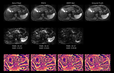 |
Partial Fourier Reconstruction in Liver DWI using a Recurrent Convolutional Network
Fasil Gadjimuradov1,2, Thomas Benkert2, Marcel Dominik Nickel2, and Andreas Maier1
1Pattern Recognition Lab, Friedrich-Alexander-Universität Erlangen-Nürnberg, Erlangen, Germany, 2Magnetic Resonance, Siemens Healthcare GmbH, Erlangen, Germany
Partial Fourier (PF) acquisition allows to reduce TE in single-shot echo-planar imaging in order to increase signal-to-noise ratio (SNR) in diffusion-weighted imaging (DWI). However, when applying it to motion-prone liver DWI, conventional PF reconstruction methods fail since they rely on smoothness priors of the phase. This work proposes to use an unrolled network architecture which aims to estimate a more appropriate regularization by learned recurrent convolutions. It can be shown that reconstructions produced by the network are superior in terms of quantitative measures as well as qualitative impression compared to conventional methods which tend to introduce artifacts.
|
|
|
0316.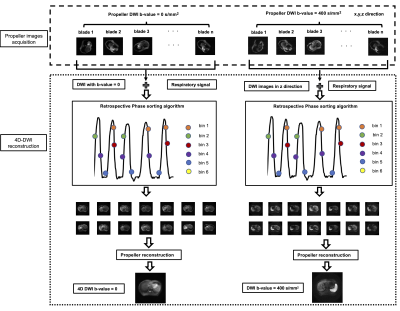 |
Motion-Resolved Four-Dimensional Abdominal Diffusion-Weighted Imaging using Propeller Echo-Planar Imaging (4D-DW-Propeller-EPI)
lu wang1, Tian Li2, Jing Cai2, and Hing-Chiu Chang1
1Department of Diagnostic Radiology, The University of Hong Kong, HongKong, China, 2Department of Health Technology and Informatics, The Hong Kong Polytechnic University, HongKong, China
Motion-resolved four dimensional diffusion-weighted echo-planar imaging (4D-DW-EPI) may better characterize the respiratory-induced motion of tumor than 4D-MRI with conventional T1 and T2 image contrasts. However, the existing 4D-DW-EPI technique can only provide limited geometric accuracy and low sampling rate over respiratory cycles. Thus, we proposed an advanced 4D-DW-Propeller-EPI technique with golden angle acquisition for improving the geometric accuracy and data sampling rate. Simulation study and in-vivo study were conducted to evaluate the performance of 4D-DW-Propeller-EPI. Our results demonstrate that 4D-DW-Propeller-EPI showed superior image quality to 4D-DW-EPI and had a potential for the application of MRI-guided abdominal radiotherapy.
|
|
0317.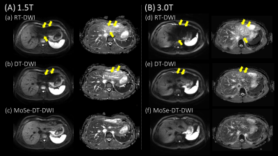 |
Improvement of Left Hepatic Lobe Diffusion Weighted Imaging using Double Triggering with Motion Sensitive CINE Imaging
Hiroshi Hamano1, Masami Yoneyama1, Akihiro Nishie2, Keisuke Ishimatsu2, Chiaki Tokunaga3, Hiroaki Watanuki3, Tatsuhiro Wada3, Isao Shiina4, Michinobu Nagao5, Yasuhiro Goto4, Kazuo Kodaira4, Yutaka Hamatani4, Takumi Ogawa4,
Takashi Namiki1, and Kenji Iinuma1
1Philips Japan, Tokyo, Japan, 2Department of Clinical Radiology, Graduate School of Medical Sciences, Kyushu University, Fukuoka, Japan, 3Division of Radiology, Department of Medical Technology, Kyushu University Hospital, Fukuoka, Japan, 4Department of Radiological Services, Tokyo Women’s Medical University, Tokyo, Japan, 5Department of Diagnostic Imaging and Nuclear Medicine, Tokyo Women’s Medical University, Tokyo, Japan
In the Liver DWI, the respiratory and cardiac motion induce signal loss and artificially increase ADC of the left hepatic lobe. On the other hand, Motion-Sensitive (MoSe) CINE imaging, based on T2FFE (also known as PSIF) sequence, could directly visualize the motion-insensitive cardiac timing thanks to that of motion sensitivity. We assumed that it is useful for determining optimal cardiac trigger delay (TD) in the liver DWI. We demonstrated that the respiratory and cardiac trigged DWI with optimal cardiac TD using MoSe CINE imaging leads to the robustness of image quality in DWI and ADC of the left hepatic lobe.
|
||
0318.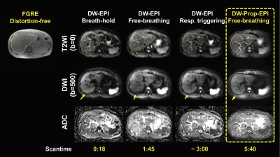 |
Repeatability of Liver Apparent Diffusion Coefficient Measurement Using Free-Breathing Diffusion-Weighted Propeller Echo-Planar Imaging
Hing-Chiu Chang1, Lu Wang1, Guangtao Chen1, Liyuan Liang1, Keith Wan-Hang Chiu1, Yi-Jui Liu2, Chun-Jung Juan3,4,5, and Hsiao-Wen Chung6,7
1Department of Diagnostic Radiology, The University of Hong Kong, Hong Kong, Hong Kong, 2Department of Automatic Control Engineering, Feng Chia University, Taichung, Taiwan, 3Department of Medical Imaging, China Medical University Hsinchu Hospital, Hsinchu, Taiwan, 4Department of Radiology, School of Medicine, College of Medicine, China Medical University, Taichung, Taiwan, 5Department of Medical Imaging, China Medical University Hospital, Taichung, Taiwan, 6Department of Electrical Engineering, National Taiwan University, Taipei, Taiwan, 7Graduate Institute of Biomedical Electronics and Bioinformatics, National Taiwan University, Taipei, Taiwan
The current limitations of liver DWI mainly relate to the image quality and the repeatability/reproducibility of ADC measurement using single-shot DW echo-planar imaging (DW-EPI). The discrepancy of ADC measurement can substantially cause difficulty in cross-sectional or longitudinal liver DW-EPI. A preliminary study reported that a free-breathing liver DW-Propeller-EPI technique can provide superior image quality to conventional liver DW-EPI methods. In this study, we further improved the robustness of free-breathing liver DW-Propeller-EPI by incorporating velocity-compensation (VC) diffusion gradient into data acquisition, and then evaluated the repeatability of liver ADC measurement for free-breathing DW-Propeller-EPI by comparing to three routine liver DW-EPI methods.
|
||
|
0319.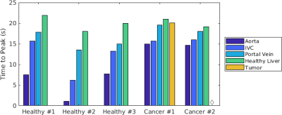 |
Characterization of Arterial and Portal Venous Contributions to Metabolic Imaging of the Human Liver Using Hyperpolarized 13C-pyruvate MRI
Philip Meng-en Lee1, Jeremy W Gordon1, Zhen J Wang1, Zihan Zhu1, Hsin-Yu Chen1, Pamela N Munster2, Rahul Aggarwal2, Daniel B Vigneron1, and Michael A Ohliger1
1Department of Radiology & Biomedical Imaging, University of California, San Francisco, San Francisco, CA, United States, 2Department of Medicine, University of California, San Francisco, San Francisco, CA, United States
Analysis of the delivery and metabolism of hyperpolarized 13C pyruvate within the liver is complicated by the organ’s unique dual blood supply. Distinguishing the hepatic arterial and portal venous contributions of pyruvate delivery would improve real-time acquisition triggering and kinetic modeling. Three healthy subjects and two metastatic cancer subjects underwent hyperpolarized 13C MRI on a clinical 3 T MR scanner. We observed differential arrival of [1-13C]pyruvate signal in the aorta, inferior vena cava, portal vein, healthy liver, and metastases matching physiologic expectations. Consistencies were observed within subject groups, which is crucial for accurate timing and modelling of metabolic hyperpolarized signals.
|
|
0320.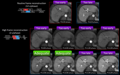 |
Intraindividual comparison of stack-of-stars acquisition for arterial phase imaging with and without breath-holding on dynamic MRI of the liver
Shintaro Ichikawa1, Utaroh Motosugi2, Tetsuya Wakayama3, Satoshi Funayama1, Daiki Tamada1, Sagar Mandava4, Ty A Cashen5, and Hiroshi Onishi1
1Department of Radiology, University of Yamanashi, Chuo, Japan, 2Kofu Kyoritsu Hospital, Kofu, Japan, 3GE Healthcare, Hino, Japan, 4GE Healthcare, Atlanta, GA, United States, 5GE Healthcare, Madison, WI, United States
We performed intra-individual comparison of arterial phase (AP) images between differential subsampling with Cartesian ordering (DISCO) and stack-of-stars acquisition without breath-holding (DISCO-Star) on dynamic magnetic resonance imaging of the liver. Patients showing inadequate scan timing of AP in DISCO and DISCO-Star (12 s/phase) dataset were 4.9% and 21.2%, respectively. One advantage of DISCO-Star was that the adequate scan timing of AP can be obtained by using additional high frame rate reconstruction (3 s/phase) in the patient with inadequate scan timing in routine reconstruction. DISCO-Star was useful to obtain adequate AP imaging in all patients.
|
The International Society for Magnetic Resonance in Medicine is accredited by the Accreditation Council for Continuing Medical Education to provide continuing medical education for physicians.