Oral
Cancer in the Body
ISMRM & SMRT Annual Meeting • 15-20 May 2021

| Concurrent 5 | 16:00 - 18:00 | Moderators: Hina Arif-Tiwari & Scott Reeder |
 |
0367.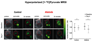 |
Hyperpolarized 13C-Pyruvate MRSI in Prediction of Response to Tyrosine Kinase Inhibition Therapy in Gastric Cancer
Shadi A Esfahani 1, Cody Callahan2, Nicholas M Rotile1, Peter D Caravan 1, Aaron K Grant 2, and Yi-Fen Yen1
1Athinoula A. Martinos Center for Biomedical Imaging, Department of Radiology, Massachusetts General Hospital, Boston, MA, United States, 2Department of Radiology, Beth Israel Deaconess Medical Center, Boston, MA, United States
Personalized treatment of gastric cancer remains a major challenge mainly due to lack of an optimal imaging method for detection of early response to treatment. We evaluated the role of Hyperpolarized [1-13C] Pyruvate Magnetic Resonance Spectroscopic Imaging (HP-13C MRSI) for quantitative measurement of early changes in glycolytic metabolism of gastric cancer models upon initiation of afatinib, a pan-receptor tyrosine kinase inhibitor. We showed that HP-13C MRSI is more sensitive for detection of early metabolic changes in gastric cancer after starting treatment with afatinib compared to 18F-FDG PET/MRI, and therefore can be used for early prediction of response to targeted therapies.
|
|
0368.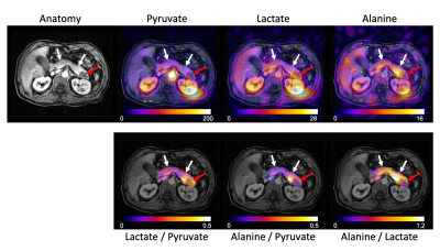 |
Hyperpolarized 13C Metabolic Imaging of Patients with Pancreatic Ductal Adenocarcinoma
Jeremy W Gordon1, Hsin-Yu Chen1, Philip Lee1, Robert Bok1, Michael Ohliger1, Andrew Ko2, Eric Collisson2, Margaret Tempero2, Pelin Cinar2, Peder Larson1,3, and Zhen Jane Wang1,3
1Department of Radiology & Biomedical Imaging, University of California, San Francisco, San Francisco, CA, United States, 2Department of Medicine, University of California, San Francisco, San Francisco, CA, United States, 3UC Berkeley-UCSF Graduate Program in Bioengineering, San Francisco, CA, United States
Metabolic imaging of hyperpolarized [1-13C]pyruvate was performed in four patients with pancreatic ductal adenocarcinoma prior to chemotherapy. Compared to the normal appearing pancreas, higher lactate/pyruvate and lower alanine/pyruvate ratios were seen in the primary tumors. The alanine/lactate ratio, which reflects the relative enzymatic activity and metabolite pool sizes, was reduced 2- to 20-fold in the tumors and provided improved contrast between tumor and normal appearing pancreas over either the lactate/pyruvate or alanine/pyruvate ratio. These results indicate the potential for HP 13C MRI to be a novel tool to stage disease and assess treatment response in pancreatic cancer patients
|
||
0369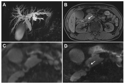 |
Zoomed Diffusion-Weighted Echo-Planar Imaging for the Evaluation of Periampullary Carcinomas Video Permission Withheld
Jingjing Liu1, Jingliang Cheng1, and Jinxia Zhu2
1Department of MR Imaging, the First Affiliated Hospital of Zhengzhou University, Zhengzhou, China, 2MR Collaboration, Siemens Healthcare Ltd, Beijing, China
We evaluated the efficacy of a zoomed diffusion-weighted echo-planar imaging (z-EPI) sequence to diagnose periampullary carcinoma compared with a conventional single-shot EPI (c-EPI) sequence. Better delineation of lesion conspicuities and margins were observed, as well as enhanced diagnostic confidence with z-EPI. The diagnostic accuracy increased by combining magnetic resonance cholangiopancreatography (MRCP) and z-EPI together. These findings showed remarkable image quality improvements for periampullary carcinomas using z-EPI. The ability to detect and delineate lesions using z-EPI was superior to that of c-EPI. Diagnostic accuracy was also attained, especially for small lesions.
|
||
0370.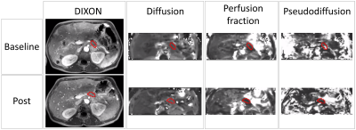 |
Assessment of pancreatic tumour response on LDE225, gemcitabine and nab-paclitaxel using Intravoxel Incoherent Motion MRI
Nienke P.M. Wassenaar1, Esther N. Pijnappel2, Remy Klaassen2, Femke Struik1, Jaap Stoker1, Jurgen H. Runge1, Hanneke W.M. van Laarhoven2, Johanna W. Wilmink2, Aart J. Nederveen1, and Oliver J. Gurney-Champion1
1Department of Radiology and Nuclear Medicine, Cancer Center Amsterdam, Amsterdam University Medical Centers, University of Amsterdam, Amsterdam, Netherlands, 2Department of Medical Oncology, Cancer Center Amsterdam, Amsterdam University Medical Centers, University of Amsterdam, Amsterdam, Netherlands
Stromal deposition can become a physical and biological barrier that prevents chemotherapy from reaching pancreatic ductal adenocarcinoma (PDAC). In this study, 34 patients were treated with LDE225, which specifically targets tumour stroma, gemcitabine and nab-paclitaxel. Pancreatic tumour response was assessed using intravoxel incoherent motion (IVIM) MRI. We found that the diffusion in PDAC increased after chemotherapy, which may be explained by reduction of stroma or tumour necrosis. Furthermore, a positive correlation was found between overall survival and the change in tumour perfusion, underlining the fact that reperfusion of PDAC by LDE225 improves prognosis.
|
||
0371.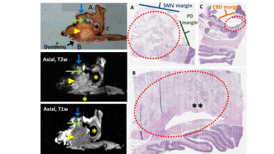 |
Ex Vivo Radiologic-Histologic Correlation of Pancreas Adenocarcinoma: A Feasibility Study
Alexandra W. Acher1, Joseph Krenzer1, Krisztian Kovacs1, Soudabeh Kargar1, Ali Pirasteh1, Jitka Starekova1, TJ Colgan1, Victoria Rendell1, Daniel E. Abbott1, Erin Brooks1, Rashmi Agni1, Emily Winslow2, and Scott B. Reeder1
1University of Wisconsin School of Medicine and Public Health, Madison, WI, United States, 2Georgetown University, Washington DC, MD, United States
The purpose of this study was to assess the feasibility of a novel radiologic-histologic correlation device RHCD and ex vivo MRI to facilitate direct correlation of radiologic and histologic features of pancreas cancer. Our approach is based on previous radiologic-histologic correlation of pancreatic anatomy using cadaveric pancreas specimens. The final protocol was applied to co-localize pancreas cancer margins radiologically and histologically, as well as nodal burden in pancreaticoduodenectomy specimens.
|
||
0372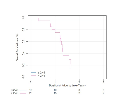 |
Prediction of Overall Survival in Patients with Pancreatic Cancer: Texture Analysis of ADC Value and Correlation with Intratumoral Necrosis Video Permission Withheld
Yoshifumi Noda1, Nobuyuki Kawai1, Hiroyuki Tomita2, Takuma Ishihara3, Yoshiki Tsuboi3, Masaya Kawaguchi1, Tetsuro Kaga1, Fuminori Hyodo4, Akira Hara2, and Masayuki Matsuo1
1Department of Radiology, Gifu University, Gifu, Japan, 2Department of Tumor Pathology, Gifu University, Gifu, Japan, 3Innovative and Clinical Research Promotion Center, Gifu University Hospital, Gifu, Japan, 4Department of Frontier Science for imaging, Gifu University, Gifu, Japan
The use of texture analysis in clinical images can provide surrogate information on tumor microenvironment and predict the prognosis of patients. In this study, we evaluate the utility of texture analysis of tumor ADC values to predict the overall survival in patients with pancreatic cancer and to correlate with pathological evaluated massive intratumoral necrosis. Our results showed that the kurtosis of tumor ADC values obtained from texture analysis is correlated with massive intratumoral necrosis and is associated with poor prognosis in patients with pancreatic cancer.
|
||
0373.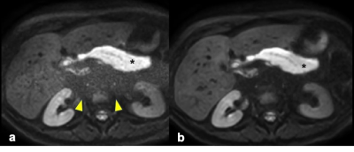 |
Noise reduction in diffusion weighted MRI of the pancreas using an L1-regularized iterative SENSE reconstruction
Omar Kamal1,2, Sean McTavish1, Felix Harder1, Anh T. Van1, Johannes M. Peeters3, Kilian Weiss4, Marcus R. Makowski1, Dimitrios C. Karampinos1, and Rickmer F. Braren1
1Department of Diagnostic and Interventional Radiology, Technical University of Munich, Munich, Germany, 2Department of Radiology, South Egypt Cancer Institute, Assiut, Egypt, 3Philips Healthcare, Best, Netherlands, 4Philips GmbH, Hamburg, Germany
DWI plays a growing role in the imaging of the pancreas. Current abdominal DWI scans have a large field of view with limited resolution. Approaches for a higher resolution single-shot DW-EPI with SENSE often suffer from g-factor-induced noise band-like artifacts from which the pancreas is significantly affected due to its central location. We use an L1-regularized iterative SENSE reconstruction to obtain high resolution single shot DW-EPI with reduced noise in patients with pancreatic cancer. The employed method reduces the noise band-like artifacts over the pancreas and eliminates the noise bias from the ADC measurements without lengthening scan times.
|
||
0374.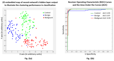 |
High-precision neural-network discrimination of human plasma samples to detect pancreatic cancer using specialized data-augmentation method
Meiyappan Solaiyappan1, Santosh Kumar Bharti1, Paul T Winnard Jr1, Mohamad Dbouk2, Michael G Goggins2,3,4, and Zaver M Bhujwalla1,3,5
1Department of Radiology, The Johns Hopkins University School of Medicine, Baltimore, MD, United States, 2Department of Pathology, The Johns Hopkins University School of Medicine, Baltimore, MD, United States, 3Department of Oncology, The Johns Hopkins University School of Medicine, Baltimore, MD, United States, 4Department of Medicine, The Johns Hopkins University School of Medicine, Baltimore, MD, United States, 5Department of Radiation Oncology and Molecular Radiation Sciences, The Johns Hopkins University School of Medicine, Baltimore, MD, United States
The insidious growth of pancreatic cancer is a major factor contributing to its lethality. Only ~20% of pancreatic cancers are resectable by the time they are detected. Early detection of pancreatic cancer through routine screening is clearly an unmet clinical need. Here we have applied neural-network analysis to 1H magnetic resonance spectra of human plasma samples to differentiate between healthy subjects (control), subjects with benign lesions, and subjects with pancreatic ductal adenocarcinoma (PDAC). Our data support developing a neural-network approach to identify PDAC from 1H MRS of plasma samples.
|
||
0375.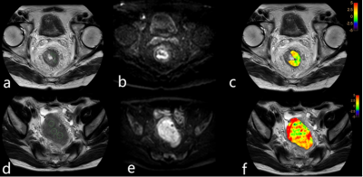 |
Application of APTw imaging in prediction of lymph node metastasis in rectal cancer
Mingxiao Wang1, AIlian Liu1, Yuhui Liu1, Anliang Chen1, wan Dong1, Xinao Wang1, Qingwei Song1, Xinru Zhang1, Liangjie Lin2, and Jiazheng Wang2
1The First Affiliated Hospital of Dalian Medical University, Dalian, China, Dalian, China, 2Philips Healthcare,Beijing,China, Beijing, China The purpose of this study was to evaluate the value of amide proton transfer weighted (APTw) imaging in the identification of lymph node metastasis in rectal cancer. Results showed that, for patients with rectal cancer, the APTw value in group with lymph node metastasis was significantly lower than those in group without lymph node metastasis. APTw imaging can be used as a promising non-invasive method to predict the lymph node metastasis in rectal cancer.
|
||
0376.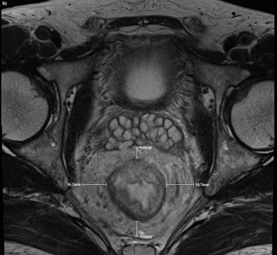 |
Prognostic significance of MRI-detected mesorectal fat thickness in rectal cancer; a risk factor for distance metastasis
Pratik Tripathi1, Zhen Li1, Yaqi Shen1, Xuemei Hu1, and Daoyu Hu1
1Department of Radiology, Tongji Hospital, Tongji Medical College of Huazhong University of Science and Technology, Wuhan, China
Our study got a step closer to probable cause of the poor prognosis in rectal cancer in obese patients. The correlation of the area within mesorectum with BMI and also the correlation between the mesorectal fat thickness and distant metastasis signifies the need of selection of optimum treatment modality for obese patients in rectal cancer. This preliminary study may shed some light on the current scenario and show probable direction for the solution. Selection of different treatment modalities and new surgical techniques may be required to improve the prognosis.
|
The International Society for Magnetic Resonance in Medicine is accredited by the Accreditation Council for Continuing Medical Education to provide continuing medical education for physicians.