Oral
Gynecologic & Prostate Cancers
ISMRM & SMRT Annual Meeting • 15-20 May 2021

| Concurrent 5 | 14:00 - 16:00 | Moderators: Masako Kataoka & Daniel Margolis |
 |
0757.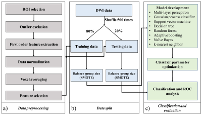 |
MOdel-free Diffusion-wEighted MRI (MODEM) with Machine Learning for Cervical Cancer Detection
Guangyu Dan1,2, Cui Feng1,3, Zheng Zhong1,2, Kaibao Sun1, Muge Karaman1,2, Daoyu Hu3, and Xiaohong Joe Zhou1,2,4
1Center for MR Research, University of Illinois at Chicago, Chicago, IL, United States, 2Department of Bioengineering, University of Illinois at Chicago, Chicago, IL, United States, 3Department of Radiology, Tongji Hospital, Wuhan, China, 4Departments of Radiology and Neurosurgery, University of Illinois at Chicago, Chicago, IL, United States
Characterization of diffusion-weighted imaging signal is typically performed by modeling the data based on biophysical, mathematical, and/or statistical models to estimate quantitative biomarkers. However, conventional nonlinear fitting, which is required for the estimation of model parameters, often suffers from instability and degeneracy. In this study, we propose a Model-free Diffusion-wEighted MRI technique (MODEM) with machine learning to detect cervical carcinomas by using diffusion signal intensities and the first-order statistical features extracted from the signal attenuation as the input. By using MODEM, superior diagnostic performance and stability can be achieved even with limited number of b-values in cervical cancer detection.
|
|
0758.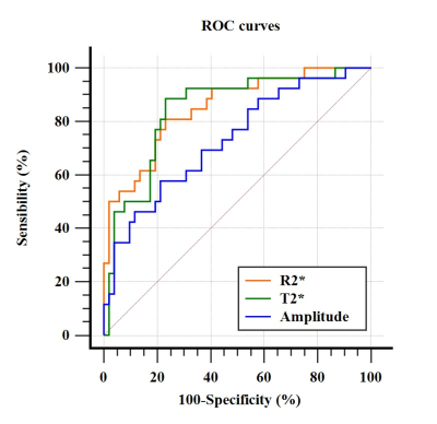 |
Differential diagnosis of cervical adenocarcinoma and squamous carcinoma by using multiple parameters of Enhanced T2* -Weighted Angiography
Dahua Cui1, Ailian Liu1, Shifeng Tian1, and Qingwei Song1
1The First Affiliated Hospital of Dalian Medical University, Dalian, China
The aim of this study was to explore the value of multiple quantitative parameters of enhanced T2 star-weighted angiography (ESWAN) in differentiating cervical adenocarcinoma (CA) from Squamous cell carcinoma (SCC). The areas under the ROC curve of amplitude, R2* and T2* in US group were 0.729, 0.851 and 0.843, respectively, promising to be a valuable diagnostic method for differentiation CA from SCC.
|
||
0759.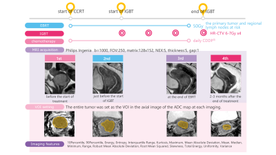 |
Prediction of prognosis after definitive radiotherapy for uterine cervical cancer using changes of ADC histogram during the clinical course
Akiyo Takada1, Hajime Yokota2, Miho Watanabe Nemoto2, Takuro Horikoshi1, Koji Matsumoto1, Yuji Habu3, Hirokazu Usui3, Katsuhiro Nasu1, Makio Shozu3, and Takashi Uno2
1Radiology, Department of Radiology, Chiba University Hospital, Chiba, Japan, 2Radiology, Department of Diagnostic Radiology and Radiation Oncology, Graduate School of Medicine, Chiba University, Chiba, Japan, 3Obstetrics and gynecology, Department of Reproductive Medicine, Graduate School of Medicine, Chiba University, Chiba, Japan
To realize precision medicine for cervical cancer, it is essential to predict the prognosis. We focused on changes in ADC histogram during the clinical course. Sixty-eight patients were included and they underwent MRI evaluation 4 times during the clinical course. Imaging features were extracted from the ADC histogram in the whole tumor VOI and their change rates were calculated. The change rates of kurtosis between the first and third, and between the first and second imaging could predict recurrence with high AUCs (0.90 and 0.83). By predicting the prognosis early during clinical course, a more personalized treatment can be provided.
|
||
0760.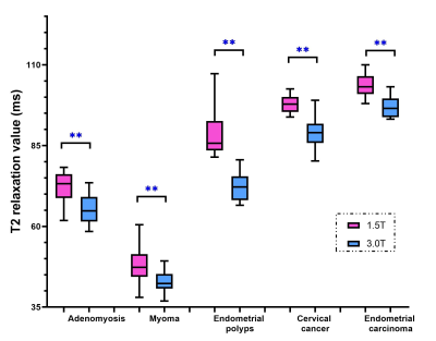 |
Preliminary study about T2 mapping technology in quantification of uterine benign and malignant tumors under 1.5T and 3.0T MRI
Liuhong Zhu1, Pu-Yeh Wu2, Hao Liu1, and Jianjun Zhou1
1Radiology, Xiamen Branch, Zhongshan Hospital, Fudan University, Xiamen, China, 2GE Healthcare, Beijing, China
MRI is the best imaging tool for evaluation of uterine tumors, but conventional MRI diagnosis results are subjective. T2 mapping is an objective quantification technique under certain magnetic field. Here we compared T2 values of common benign and malignant tumors under different field strengths. We found that T2 value of benign lesions was significantly lower than those of malignant lesions under both 1.5T and 3.0T, while diagnostic performance of T2 value under 3.0T were higher than 1.5T. We concluded that T2 mapping can be an effective quantitative tool in distinguishing between benign and malignant tumors, especially under 3.0T MR.
|
||
0761.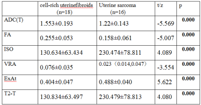 |
DTI quantitative parameters were used to differentiate uterine sarcoma from cell - rich uterine fibroids
Changjun Ma1, Ailian Liu1, Shifeng Tian1, and Jiazheng Wang2
1Radiology Department, The First Affiliated Hospital of Dalian Medical University, Dalian, China, Dalian,China, China, 2Philips Healthcare,Beijing,China, Beijing,China, China
DTI is a novel MRI sequence that enables quantitative assessment of various diseases. However, its potential for diagnosis of uterus has not been explored,and it may also help differential diagnosis of myoma of uterus and uterine sarcoma.In this study, the DTI was used for quantitative analyses of myoma of uterus and uterine sarcoma.
|
||
0762. |
Deep Learning for the Ovarian Lesion Localization and Discrimination Between Borderline Tumors and Cancers in MR Imaging
Yida Wang1, YinQiao Yi1, Haijie Wang1, Changan Chen2, Yingfang Wang2, Guofu Zhang2, He Zhang2, and Guang Yang1
1East China Normal University, Shanghai Key Laboratory of Magnetic Resonance, Shanghai, China, 2Department of Radiology, Obstetrics and Gynecology Hospital, Fudan University, Shanghai, China
We proposed a deep learning (DL) approach to segment ovarian lesion and differentiate ovarian malignant from borderline tumors in MR Imaging. Firstly, we used U-net++ with deep supervision to automatically define lesion region on conventional MRI; secondly, the segmented ovarian masses regions were classified with an SE-ResNet model. We compared the performance of classification model with those of radiologist’. The results showed the trained DL network model could help to identify and categorize ovarian masses with a high accuracy from MR images.
|
||
|
0763.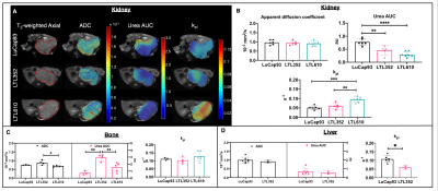 |
Hyperpolarized 13C MR imaging of prostate cancer patient derived xenograft models and their response to therapy
Shubhangi Agarwal1, Jinny Sun1, Emilie Decavel-Bueff1, Robert A Bok1, Romelyn Delos Santos1, Mark Van Criekinge1, Rahul Aggarwal2, Daniel B Vigneron1, Donna Peehl1, John Kurhanewicz1, and Renuka Sriram1
1Department of Radiology and Biomedical Imaging, University of California, San Francisco, San Francisco, CA, United States, 22Division of Hematology/Oncology, Department of Medicine, University of California, San Francisco, San Francisco, CA, United States
This study demonstrates for the first time, a successful study of three small cell neuroendocrine prostate cancer patient derived xenografts (PDX) models in liver and bone in pre-clinical setting. Kidney served as an optimum site for propagation with its rich blood supply and high take rate. Hyperpolarized 13C MRI was able to identify metabolic differences between kidney, bone and liver tumors. The same PDXs had different metabolism when implanted in bone and liver. Hyperpolarized [1-13C] pyruvate conversion to lactate may be used to indicate early response to carboplatin in small cell neuroendocrine prostate cancer models in the bone.
|
|
 |
0764.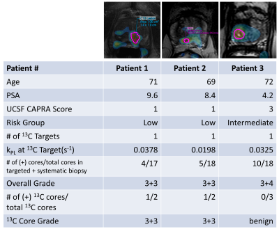 |
Improving Multiparametric MR - TRUS Guided Fusion Prostate Biopsies with Hyperpolarized 13C Pyruvate Metabolic Imaging : Technical Development
Hsin-Yu Chen1, Robert A. Bok1, Hao G. Nguyen2, Katsuto Shinohara2, Antonio C. Westphalen1, Zhen J. Wang1, Michael A. Ohliger1, Lucas Carvajal1, Jeremy W. Gordon1, Peder E.Z. Larson1, Rahul Aggarwal2, John Kurhanewicz1, and Daniel B. Vigneron1
1Radiology and Biomedical Imaging, University of California, San Francisco, San Francisco, CA, United States, 2Helen Diller Family Comprehensive Cancer Center, University of California, San Francisco, San Francisco, CA, United States
This project developed a new approach of integrating research biopsy targets, defined by metabolic abnormalities on hyperpolarized (HP) 13C pyruvate MRI, into clinical multiparametric MRI workflow to guide software-fusion transrectal ultrasound (TRUS) biopsy for improved prostate cancer risk stratification. Feasibility was investigated in 3 prostate cancer patient studies, for which research biopsy targets corresponding to high pyruvate-to-lactate conversion rates (kPL) were identified and outlined by experienced abdominal radiologists. Fusion biopsies identified histologically-confirmed cancer at two out of three “C13” targets, consistent with clinically low- to intermediate-risk disease, supporting continued active surveillance management for these patients.
|
|
|
0765.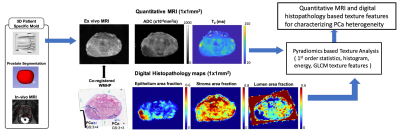 |
A Framework for Characterizing Prostate Cancer Heterogeneity Using Voxel-Wise Co-Registered Ex Vivo MRI and Whole-Mount Histopathology
Zhaohuan Zhang1, Jiayun Li1, Wenyuan Li1, Haoxin Zheng1, Sohrab Afshari Mirak1, Sepideh Shakeri1, Alan Priester1, Clara Magyar2, Anthony Sisk2, Robert Reiter3, Kyunghyun Sung1, Steven Raman1, Dieter R. Enzmann1, Corey Arnold1,
and Holden Wu1
1Department of Radiological Sciences, UCLA, Los Angeles, CA, United States, 2Department of Pathology, UCLA, Los Angeles, CA, United States, 3Department of Urology, UCLA, Los Angeles, CA, United States
Tumor heterogeneity is considered a key factor in determining the aggressiveness of prostate cancer (PCa). Quantitative MRI and digital whole-mount histopathology (WMHP) have potential to characterize heterogeneity in PCa in terms of MRI properties and tissue composition, respectively. Therefore, the purpose of this study is to develop a data acquisition and processing framework that produces high-resolution (1x1mm2) co-registered quantitative MRI and digital histopathology maps to enable texture features based quantification of PCa heterogeneity
|
|
0766.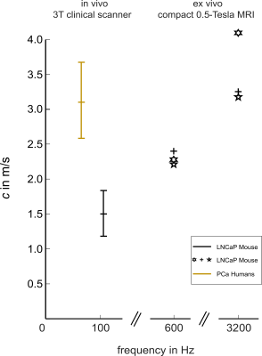 |
In-vivo Magnetic Resonance Elastography of implanted human prostate tumors in a murine model.
Joachim Snellings1, Kader Avan1, Marcus Markowski2, Bernd Hamm1, Patrick Asbach1, Carsten Warmuth1, Mehrgan Shahryari1, Heiko Tzschätzsch1, Ingolf Sack1, and Jürgen Braun3
1Institute of Radiology, Charité Üniversitätsmedizin, Berlin, Germany, 2School of Medicine & Klinikum Rechts der Isar, Technical University of Munich, Munich (TUM), München, Germany, 3Institut für Medizinische Informatik, Charité Üniversitätsmedizin, Berlin, Germany Prostate cancer (PCa) is the second leading cause of cancer-death in men in the western world. Advanced techniques of clinical MR-elastography (MRE), allow the characterization of PCa, based on the viscoelastic tissue-properties, which provide rich biophysical signatures of tumor progression. Using multifrequency MRE, we investigated PCa introduced LNCaP cell-lines, in a immunodeficient murine model, in-vivo, in a 3-Tesla MRI-scanner and, ex-vivo, by a 0.5-Tesla compact MRE-device. In-vivo and ex-vivo MRE values of LNCaP were in good agreement given the viscoelastic frequency-dispersion typical for soft-tissues. Compared with patient data in literature, LNCaP in mice are softer than PCa in humans. |
The International Society for Magnetic Resonance in Medicine is accredited by the Accreditation Council for Continuing Medical Education to provide continuing medical education for physicians.