Oral
MR Angiography & Vessel Wall
ISMRM & SMRT Annual Meeting • 15-20 May 2021

| Concurrent 3 | 18:00 - 20:00 | Moderators: Nicholas Burris & Michael Schar |
0185.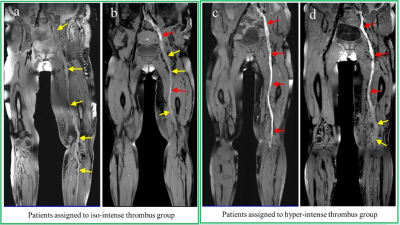 |
Assessing the Prognosis of Acute Deep Vein Thrombosis Using Magnetic Resonance Black-blood Thrombus Imaging
Guoxi Xie1, Hanwei Chen2, Chen Huang3, Xueping He2, Yueyuan Xie4, Xiaoyong Zhang5, Tianjing Zhang6, Yi Sun5, Debiao Li7, and Zhaoyang Fan8
1Department of Biomedical Engineering, Guangzhou Medical University, Guangzhou, China, 2Department of Radiology, Guangzhou Panyu Central Hospital, Guangzhou, China, 3Department of Minimally Invasive Interventional Radiology, Guangzhou Panyu Central Hospital, Guangzhou, China, 4Department of Anesthesiology, Mindong Hospital, Ningde, China, 5MR Collaborations, Siemens Healthcare Ltd, Shenzhen, China, 6Philips Healthcare, Guangzhou, China, 7Biomedical Imaging Research Institute, Cedars-Sinai Medical Center, Los Angeles, CA, United States, 8Department of Radiology, Keck School of Medicine, University of Southern California, Los Angeles, CA, United States
Patients with acute deep vein thrombosis (DVT) can be characterized as iso- or hyper-intense thrombus signals using a T1-weighted black-blood magnetic resonance imaging (BTI) technique. Patients with hyper-intense thrombus signals demonstrated a significant higher incidence of post-thrombotic syndrome (PTS) than those with iso-intense thrombus signals, regardless of the patient’s age, gender, the severity of DVT, and the treatment strategy of catheter-directed thrombolysis or conventional anticoagulant therapy. The results suggest that the thrombus signal characteristics obtained on BTI imaging are valuable for assessing the prognosis of acute DVT and may aid in guiding the clinical treatment plan.
|
||
0186.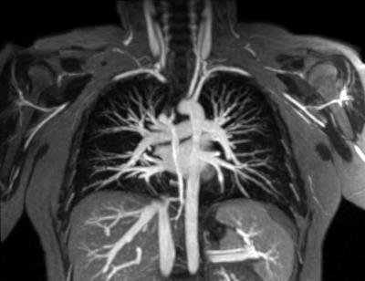 |
Ferumoxytol-enhanced Pulmonary MRA in Pregnancy: Evaluation of Initial Safety and Image Quality
Jitka Starekova1, Scott K Nagle1, Mark L Schiebler1, Scott B Reeder1,2,3,4,5, and Venkata N Meduri1
1Radiology, University of Wisconsin-Madison, Madison, WI, United States, 2Medicine, University of Wisconsin-Madison, Madison, WI, United States, 3Medical Physics, University of Wisconsin-Madison, Madison, WI, United States, 4Biomedical Engineering, University of Wisconsin-Madison, Madison, WI, United States, 5Emergency Medicine, University of Wisconsin-Madison, Madison, WI, United States
Ferumoxytol (Feraheme®, AMAG Pharmaceuticals, Waltham) is an injectable iron micro particle-solution approved by the FDA for treatment of iron-deficiency anemia. This medication also has favorable properties as intravascular blood pool agent (off-label) for contrast-enhanced MRA (Fe-MRA). Fe-MRA is an alternative modality for the assessment of pulmonary embolism without the need for ionizing radiation or gadolinium in pregnant patients. The purpose of this study was to summarize our clinical experience with 70 Fe-MRA exams in 66 pregnant patients. We aimed to demonstrate the safety and efficacy of Fe-MRA and provide guidance in establishing a clinical service.
|
||
0187.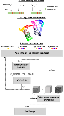 |
SIMBA 2.0: An enhanced SImilarity-driven Multi-dimensional Binning Algorithm for free-running ferumoxytol-enhanced whole-heart MRI
John Heerfordt1,2, Aurélien Bustin2,3,4, Ludovica Romanin1,2, Estelle Tenisch2, Milan Prsa5, Tobias Rutz6, Christopher W. Roy2, Jérôme Yerly2,7, Juerg Schwitter6,8, Matthias Stuber2,7, and Davide Piccini1,2
1Advanced Clinical Imaging Technology, Siemens Healthcare AG, Lausanne, Switzerland, 2Department of Radiology, Lausanne University Hospital and University of Lausanne, Lausanne, Switzerland, 3IHU LIRYC, Electrophysiology and Heart Modeling Institute, Fondation Bordeaux Université, Pessac-Bordeaux, France, 4Department of Cardiovascular Imaging, Hôpital Cardiologique du Haut-Lévêque, CHU de Bordeaux, Pessac, France, 5Division of Pediatric Cardiology, Department Woman-Mother-Child, Lausanne University Hospital and University of Lausanne, Lausanne, Switzerland, 6Division of Cardiology, Cardiovascular Department, Lausanne University Hospital and University of Lausanne, Lausanne, Switzerland, 7CIBM Center for Biomedical Imaging, Lausanne, Switzerland, 8Cardiac MR Center, Lausanne University Hospital, Lausanne, Switzerland
A SImilarity-driven Multi-dimensional Binning Algorithm (SIMBA) was recently proposed for fast reconstruction of motion-consistent clusters for free-running whole-heart MRA acquisitions. Originally, only the most populated cluster was used for the reconstruction of a motion-suppressed image. In this work we investigated whether the redundancy of information among the clusters can be exploited to improve image quality. Specifically, an adapted XD-GRASP reconstruction and a multidimensional patch-based low-rank denoising algorithm were compared. Four different reconstructions were quantitatively evaluated and compared using ferumoxytol-enhanced free-running datasets from 10 pediatric and adult CHD patients. Information sharing resulted in significantly sharper anatomical features and increased image quality.
|
||
 |
0188.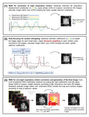 |
Focused navigation (fNAV) for cardiac and respiratory motion-compensated free-running 3D whole-heart coronary MRA
Giulia MC Rossi1, Nemanja Masala1, Jessica AM Bastiaansen1, Aurelien Bustin1,2, Jérôme Yerly1,3, John Heerfordt1,4, Davide Piccini1,4, Matthias Stuber1,3, and Christopher W Roy1
1Department of Radiology, Lausanne University Hospital (CHUV) and University of Lausanne (UNIL), Lausanne, Switzerland, 2LIRYC (Electrophysiology and Heart Modeling Institute), Bordeaux, France, 3CIBM Center for Biomedical Imaging, Lausanne, Switzerland, 4Advanced Clinical Imaging Technology (ACIT), Siemens Healthcare AG, Lausanne, Switzerland
A novel method for reconstructing motion-compensated 3D CMRA images from free-running data is proposed. We extend the use of a recent method for non-rigid respiratory motion correction called focused navigation (fNAV) to also encompass cardiac motion compensation that accounts for beat-to-beat heart-rate variability. Our combined fNAV approach is compared to the previously established cardiac and respiratory motion-resolved 5D imaging in vivo and is shown to provide overall similar image quality and comparable right coronary artery visualizations to 5D imaging in significantly shorter reconstruction times.
|
|
0189.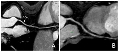 |
Preliminary study on free respiratory navigation whole-heart coronary magnetic resonance angiography based on Dixon at 3.0 T
Xin Li1, Di Tian1, Qingwei Song1, Ailian Liu1, and Zhiyong Li1
1The First Affiliated Hospital of Dalian Medical University, Dalian, China, Dalian, China
In comparison with the traditional whole-heart coronary magnetic resonance angiography (WH-CMRA) based on the spectral pre-saturation with inversion recovery (SPIR), the advanced WH-CMRA based on Dixon with non-contrast-enhanced free respiratory navigation is associated with advantages of improved image quality, increased blood signal-to-noise ratio, and the extra fat image. This study proved that it is feasible to visualize the coronary artery (image quality scores met the requirement of clinical diagnosis) using the Dixon based WH-CMRA without increasing scan time.
|
||
0190.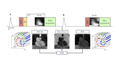 |
Gadolinium-free Multi-contrast 3D whole-heart MRI for improved anatomical assessment in patients with Congenital Heart Disease
Anastasia Fotaki1,2, Karl Kunze1, Harith Alam2, Yasodhara Emmanuel2, Alessandra Frigiola2, Kuberan Pushparajah1,2, René Botnar1, and Claudia Prieto1
1School of Biomedical Engineering and Imaging Sciences, King's College London, London, United Kingdom, 2Adult Congenital Heart Disease Department, Guy’s and St Thomas’s Hospital, London, United Kingdom
Bright- and black-blood MRI sequences provide complementary information on anatomical assessment for patients with Congenital Heart Disease(CHD).A free-breathing 3D whole-heart sequence (MTC-BOOST) has been recently proposed for simultaneous bright and black-blood high quality depiction of cardiac and vascular structures waiving the need for contrast agent injection. In this study, we clinically validated this sequence in a cohort of 35 patients with CHD against the conventional T2-prep bSSFP sequence. Quantitative and qualitative image quality assessment demonstrates that MTC-BOOST enhances pulmonary venous depiction, mitigates flow-related artefacts and has comparable image quality for intracardiac and vascular structures, promising potential integration into clinical workflow.
|
||
 |
0191.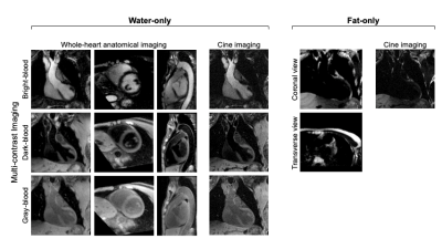 |
MR Multitasking based Multidimensional Assessment of Cardiovascular System (MT-MACS) with Extended Spatial Coverage and Water-Fat Separation
Zhehao Hu1,2, Jiayu Xiao1, Xianglun Mao1, Yibin Xie1, Alan Kwan1,3, Xiaoming Bi4, Shlee Song5, Alison Wilcox6, Debiao Li1,2, Anthony Christodoulou1,2, and Zhaoyang Fan1,6,7
1Biomedical Imaging Research Institute, Cedars-Sinai Medical Center, Los Angeles, CA, United States, 2Department of Bioengineering, University of California, Los Angeles, Los Angeles, CA, United States, 3Smidt Heart Institute, Cedars-Sinai Medical Center, Los Angeles, CA, United States, 4Siemens Medical Solutions USA, Inc., Los Angeles, CA, United States, 5Department of Neurology, Cedars-Sinai Medical Center, Los Angeles, CA, United States, 6Department of Radiology, Keck School of Medicine, University of Southern California, Los Angeles, CA, United States, 7Department of Radiation Oncology, Keck School of Medicine, University of Southern California, Los Angeles, CA, United States
Non-invasive imaging of cardiac anatomy plays an important role in diagnosis, risk stratification, and planning of procedures in patients with cardiovascular disease. MR imaging has the potential to provide a comprehensive evaluation of cardiac chambers and thoracic vessels. However, the clinical workflow for the acquisition of conventional cardiac MR imaging is complex and time-consuming. MR MultiTasking based 3D Multi-dimensional Assessment of Cardiovascular System (MT-MACS) technique has recently been demonstrated in thoracic aortic diseases without need for ECG- and navigator-gating. In this work, we further extend the application of MT-MACS to the assessment of the whole heart and great thoracic vessels.
|
|
0192.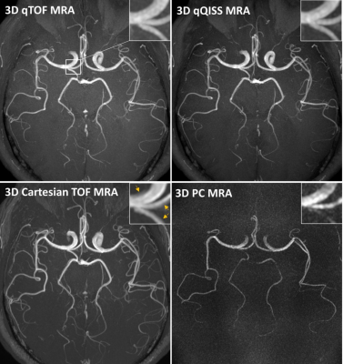 |
Quantitative Time-of-Flight (qTOF) and Quiescent Interval Slice-Selective (qQISS) Intracranial MR Angiography
Ioannis Koktzoglou1,2, Rong Huang1, Nondas Leloudas1, and Robert R Edelman1,3
1Radiology, NorthShore University HealthSystem, Evanston, IL, United States, 2University of Chicago Pritzker School of Medicine, Chicago, IL, United States, 3Northwestern University Feinberg School of Medicine, Chicago, IL, United States
Time of flight (TOF) and quiescent interval slice selective (QISS) magnetic resonance angiography (MRA) provide accurate anatomic evaluation blood vessels but do not readily quantify blood velocity or flow. Addressing this deficiency, we report two multi-echo stack-of-stars variants of TOF and QISS MRA (which we refer to as “quantitative TOF” (qTOF) and “quantitative QISS” (qQISS) MRA) that provide for simultaneous high-resolution anatomic and hemodynamic evaluation of the intracranial arteries.
|
||
|
0193.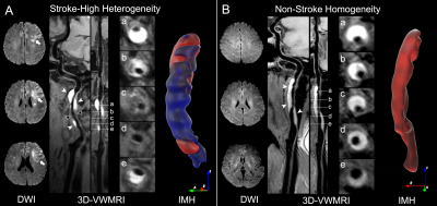 |
The Heterogeneity of Intramural Hematoma is Associated with Acute Ischemic Stroke in Patients with Proven Cervical Artery Dissection
Yuehong Liu1, Fang Wu2, Xiuqin Jia3, Haibin Li4, Xunming Ji5, and Qi Yang1
1Radiology, Beijing Chaoyang Hospital, Capital Medical University, Beijing, China, 2Radiology, Xuanwu Hospital, Capital Medical University, Beijing, China, 3Department of Radiology, Beijing Chaoyang Hospital, Capital Medical University, Beijing, China, 4Epidemiology, Beijing Chaoyang Hospital, Capital Medical University, Beijing, China, 5Neurosurgery, Xuanwu Hospital, Capital Medical University, Beijing, China
This study investigates the relation between the heterogeneity of intramural hematoma (IMH) on three-dimensional vessel wall MRI (3D-VWMRI) images and acute ischemic stroke in patients with proven cervical artery dissection (CAD). We found the high heterogenous IMH on 3D-VWMRI was associated with acute ischemic stroke in proven and non-occluded CAD. The heterogeneous signal of an IMH on 3D-VWMRI may indicate the asynchronous progress of CAD, possibly leading to thrombosis or hemodynamic instability, therefore results in ischemic stroke. The current finding may hold promise to better elucidate the pathophysiological mechanisms and occurrence risk of ischemic stroke for patients with CAD.
|
|
0194.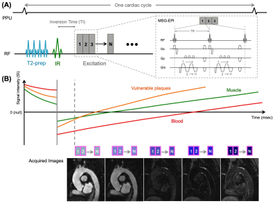 |
3D Multi-Contrast Blood Imaging with a Single Acquisition: Simultaneous Non-Contrast-Enhanced MRA and Vessel Wall imaging
Yoshihiko Tachikawa1, Hiroshi Hamano2, Hikaru Yoshikai1, Kento Ikeda1, Yasunori Maki1, Yukihiko Takahashi1, and Kunishige Matake1
1Karatsu Red Cross Hospital, Saga, Japan, 2Philips Japan, Tokyo, Japan
Assessment of vessel lumen and wall and plaques lesions is important in management of patients with atherosclerosis. Conventional MR imaging typically requires long scan times to acquire MRA and vessel wall imaging (VWI), respectively. In this work, we present a new multi-contrast blood imaging method named BRIDGE (bright and dark blood images with multi-shot gradient-echo-planar imaging), which simultaneously acquires MRA and VWI in a single acquisition. Initial results with volunteers and patients showed that comparable image quality to conventional methods could be acquired in a short time, allowed simultaneous assessment of luminal changes and vulnerable plaques in a single acquisition.
|
The International Society for Magnetic Resonance in Medicine is accredited by the Accreditation Council for Continuing Medical Education to provide continuing medical education for physicians.