Oral
Latest Advances in Hyperpolarized MRI
ISMRM & SMRT Annual Meeting • 15-20 May 2021

| Concurrent 2 | 12:00 - 14:00 | Moderators: James Grist & John Kurhanewicz |
 |
0675.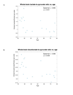 |
Hyperpolarized 13C MRI reveals age-related changes in lactate metabolism in the human brain
Biranavan Uthayakumar1,2, Casey Y Lee1,2, Nadia Bragagnolo1,2, Hany Soliman3, Albert P Chen4, Ruby Endre5, William J Perks2, Chris Heyn6, Sandra E Black2, and Charles Cunningham7
1Medical Biophysics, University of Toronto, Toronto, ON, Canada, 2Sunnybrook Health Sciences Centre, Toronto, ON, Canada, 3Radiation oncolocy, Sunnybrook Health Sciences Centre, Toronto, ON, Canada, 4GE Healthcare, Toronto, ON, Canada, 5Physical sciences, Sunnybrook Health Sciences Centre, Toronto, ON, Canada, 6Radiology, Sunnybrook Health Sciences Centre, Toronto, ON, Canada, 7Medical Biophysics, Sunnybrook research institute, Toronto, ON, Canada
In this study, hyperpolarized 13C MRI was used to investigate age-related changes to lactate and bicarbonate production in the brain in a healthy aging population. A whole-brain parcellation method was used to investigate regional changes. A global reduction in the production of both 13C-lactate and 13C-bicarbonate was observed vs. age, with certain regions showing increased rates of change in comparison to the rest of the brain. Our results suggest an age-related change in the underlying metabolic processes, and paves the way for future analysis of pathological aging as seen in disorders like Alzheimer's disease.
|
|
|
0676.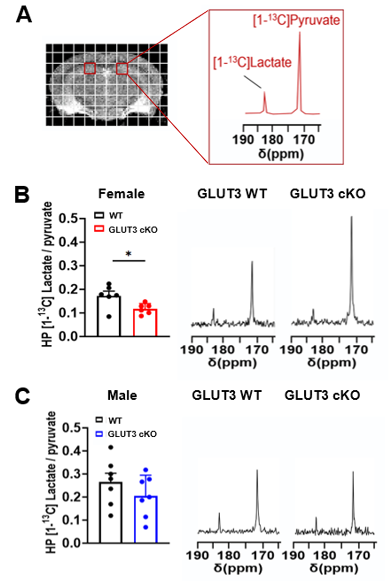 |
Hyperpolarized [1-13C]pyruvate detects brain glucose metabolism and sex-specific vulnerability in glucose transporter deficient mice
Caroline Guglielmetti1,2, Huihui Li3, Lydia M. Le Page1,2, Lauren Y. Shields3, Jeffrey C. Rathmell4, Ken Nakamura3, and Myriam M. Chaumeil1,2
1Department of Physical Therapy and Rehabilitation Science, University of California San Francisco, San Francisco, CA, United States, 2Department of Radiology and Biomedical Sciences, University of California San Francisco, San Francisco, CA, United States, 3Gladstone Institute of Neurological Disease, San Francisco, CA, United States, 4Vanderbilt Center for Immunobiology, Department of Pathology, Microbiology, and Immunology, Vanderbilt University Medical Center, Nashville, TN, United States
We used hyperpolarized 13C magnetic resonance spectroscopic imaging (HP 13C MRSI), fluorine-18 fluorodeoxyglucose (18F-FDG) positron emission tomography (PET) imaging and T2-weighted MRI to detect brain glucose metabolism in mice harboring deletion of the glucose transporter 3 (GLUT3) in CA1 hippocampal neurons. GLUT3 deletion induced memory impairment in males and females, highlighting the importance of glucose uptake by neurons. HP [1-13C]lactate-to-pyruvate ratios and brain volumes were decreased in female GLUT3 deficient mice, but not in males, indicating sex-specific vulnerability. No changes were detected using 18F-FDG PET imaging, highlighting the potential of HP [1-13C]pyruvate to detect downstream alterations in brain glucose metabolism.
|
|
0677.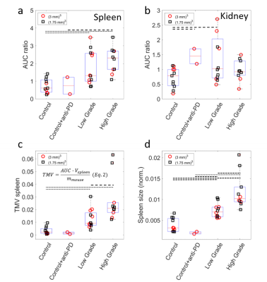 |
Characterization of glycolytic phenotypes using hyperpolarized 13C-MR and [18F]FDG PET in endogenous T-cell lymphomas in mice
Frits H.A. van Heijster1, Jason G. Skinner1, Tim Wartewig2, Christian Hundshammer1, Martin Grashei1, Geoffrey J. Topping1, Erik Hameister2, Jürgen Ruland2, and Franz Schilling1
1Technical University Munich, Nuclear Medicine, Klinikum rechts der Isar, München, Germany, 2Technical University Munich, TranslaTUM, Center for Translational Cancer Research, München, Germany
High- and low- glycolytic phenotypes of murine T-cell lymphoma are characterized using hyperpolarized MR spectroscopy/imaging and [18F]FDG-PET. Differences in pyruvate-to-lactate conversion are found within tumor groups, where PET imaging did not show this distinction. Using tumor metabolic volumes derived from PET imaging on the other hand, it’s possible to distinguish between low- and high-grade tumors. The complementary information of the two modalities gives a more complete view of the characteristics of the glycolytic phenotypes in T-cell lymphoma.
|
||
|
0678.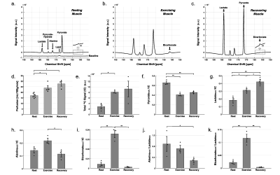 |
Hyperpolarized 13C MRI Detects In-Vivo Effect of Exercise on Pyruvate Metabolism in Human Skeletal Muscle
Jun Chen1, Junjie Ma1, Crystal E Harrison1, James Ratnakar1, Zungho Zun2, Jeff Liticker1, Galen D Reed3, Avneesh Chhabra4, Thomas Jue5, Craig R Malloy1,3,6, and Jae Mo Park1,4,7
1AIRC, UT Southwestern Medical Center, Dallas, TX, United States, 2The Developing Brain Institute, Children’s National Hospital, Washington, DC, United States, 3GE, Chicago, IL, United States, 4Radiology, UT Southwestern Medical Center, Dallas, TX, United States, 5Biochemistry and Molecular Medicine, UC Davis, Davis, CA, United States, 6Internal Medicine, UT Southwestern Medical Center, Dallas, TX, United States, 7Electrical and Computer Engineering, University of Texas at Dallas, Richardson, TX, United States
Pyruvate dehydrogenase (PDH) and lactate dehydrogenase (LDH) are essential for ATP production in skeletal muscle. However, directly PDH flux in exercising human muscle has been challenging and never assessed. This study was to demonstrate the feasibility of assessing PDH activation and changes in pyruvate metabolism in human skeletal muscle after the onset of exercise using hyperpolarized [1-13C]pyruvate. During moderate flexion-extension exercise, total HP 13C signals (tC), [1-13C]lactate/tC, and [13C]bicarbonate/tC increased significantly compared to resting state. This study demonstrates that PDH flux in skeletal muscle increases rapidly after the onset of exercise and decreases during recovery.
|
|
 |
0679.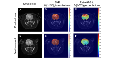 |
Hyperpolarized δ-[1-13C]gluconolactone detects response to chemotherapy in brain tumors in vivo
Georgios Batsios1, Celine Taglang1, Anne Marie Gillespie1, Peder Larson1, Sabrina M Ronen1, and Pavithra Viswanath1
1Radiology and Biomedical Imaging, UCSF, San Francisco, CA, United States
Glucose metabolism via the pentose phosphate pathway (PPP) is typically upregulated in tumors, including gliomas. We previously showed that hyperpolarized δ-[1-13C]gluconolactone metabolism via the PPP to 6-phospho-[1-13C]gluconate (6PG) differentiates tumor from contralateral normal brain in preclinical glioma models. Here, we examined the ability of hyperpolarized δ-[1-13C]gluconolactone to probe response to temozolomide, which is a key chemotherapeutic drug for glioma patients. Our studies in live cells and rats bearing orthotopic gliomas indicate that 6PG production from hyperpolarized δ-[1-13C]gluconolactone serves as an early biomarker of response to temozolomide, a finding that has the potential to improve treatment response monitoring for glioma patients.
|
|
0680.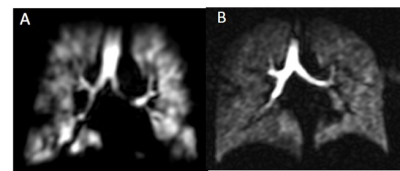 |
Hyperpolarised xenon ventilation MRI in difficult asthma; initial experience in a clinical setting
Helen Marshall1, Grace T Mussell1, Laurie J Smith1, Alberto M Biancardi1, Paul JC Hughes1, Andrew J Swift1, Smitha Rajaram1, Alison M Condliffe1, Guilhem J Collier1, Chris S Johns1, Nick D Weatherley1, Ian Sabroe2, and Jim M Wild1
1University of Sheffield, Sheffield, United Kingdom, 2Sheffield Teaching Hospitals, Sheffield, United Kingdom
The ability of hyperpolarised gas MRI to translate into real-world clinical practice is unknown. 129Xe ventilation images were acquired as part of routine care in patients referred from a difficult asthma service, and evaluated by a multi-disciplinary team. Evidence of airways obstruction on MRI can support the use of further treatment, for example in those with normal spirometry and high symptom burden. Well preserved ventilation on MRI alongside poor spirometry and/or symptom control may suggest coexisting breathing control issues or laryngeal disorders. 129Xe MRI can provide additional unique and valuable information in the evaluation of clinical presentations of asthma.
|
||
|
0681.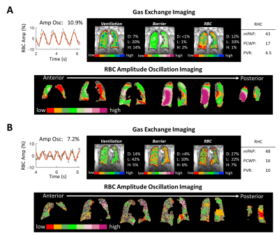 |
Extension of a Diagnostic Model for Pulmonary Hypertension with Hyperpolarized 129Xe Magnetic Resonance Imaging and Spectroscopy
Elianna Ada Bier1, Fawaz Alenezi2, Junlan Lu3, Joseph G Mammarappallil4, Bastiaan Driehuys4, and Sudarshan Rajagopal2
1Biomedical Engineering, Duke University, Durham, NC, United States, 2Division of Cardiology, Department of Medicine, Duke Univeristy, Durham, NC, United States, 3Medical Physics Graduate Program, Duke University, Durham, NC, United States, 4Radiology, Duke University, Durham, NC, United States
129Xe dynamic spectroscopy combined with 129Xe gas exchange MRI can detect both pre- and postcapillary pulmonary hypertension (PH). However, reliance on whole lung spectroscopy limits this technique’s ability to detect spatially heterogeneous impacts of PH. For example, the algorithm cannot identify combined pre- and postcapillary PH (CpcPH) and does not currently account for out-of-proportion PH in patients with parenchymal disease. Here we use the recently introduced method of imaging signal oscillations from 129Xe in red blood cells to gain additional insights into 26 subjects who have also undergone right heart catheterization to determine PH status within 2 months of imaging.
|
|
|
0682.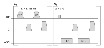 |
Pulmonary acinar structure and function assessed by hyperpolarized 129Xe stimulated echo NMR
Agilo Luitger Kern1,2, Marcel Gutberlet1,2, Frank Wacker1,2, Jens Hohlfeld2,3,4, and Jens Vogel-Claussen1,2
1Institute of Diagnostic and Interventional Radiology, Hannover Medical School, Hannover, Germany, 2Biomedical Research in Endstage and Obstructive Lung Disease Hannover (BREATH), German Center for Lung Research (DZL), Hannover, Germany, 3Department of Clinical Airway Research, Fraunhofer Institute for Toxicology and Experimental Medicine, Hannover, Germany, 4Department of Respiratory Medicine, Hannover Medical School, Hannover, Germany
A pulse sequence for hyperpolarized 129Xe NMR based on accumulated stimulated echoes has been implemented. Pairs of 90° pulses are repeatedly irradiated at the frequency of 129Xe in tissue-plasma and the decaying signal transferred to the gas phase is subsequently acquired. A study with healthy volunteers and patients of chronic obstructive pulmonary disease (COPD) has been performed to assess the method’s sensitivity for disease. A significant reduction of initial stimulated echo signal and trend for faster decay are observed in COPD. The proposed method has been demonstrated to be highly sensitive for emphysema and hyperinflation and shows promising diagnostic potential.
|
|
|
0683. |
Hyperpolarized 129Xe MRI Ventilation Texture Features to Characterize Long-haul COVID-19 Survivors
Harkiran K Kooner1, Marrissa J McIntosh1, Maksym Sharma1, Alexander M Matheson1, Yasal Rajapaksa1, Inderdeep Dhaliwal2, Michael Nicholson2, and Grace Parraga1
1Robarts Research Institute, Western University, London, ON, Canada, 2Department of Medicine, Western University, London, ON, Canada
Persistent, long-term COVID-19 symptoms and pulmonary function abnormalities, beyond the acute infectious pulmonary disease phase, is now recognized in certain patients and referred to as long-haul or long COVID. We used hyperpolarized 129Xe MRI ventilation defect percent (VDP) and texture analysis to evaluate and characterize second-order 129Xe MRI ventilation texture features in a pilot study of participants with long-haul COVID-19. We observed statistically significant differences in 129Xe MRI VDP and ventilation texture features between COVID-19 survivors and volunteers who were not infected. Second-order 129Xe MRI ventilation texture features dichotomized long-haul COVID-19 and volunteers in the absence of qualitative VDP differences.
|
|
|
0684.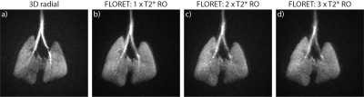 |
Preclinical Hyperpolarized 129Xe Ventilation Imaging Using 3D Spiral (FLORET) Encoding
Brice J Albert1, Peter J Niedbalski1, and Zackary I Cleveland1,2,3,4
1Center for Pulmonary Imaging Research, Cincinnati Children's Hospital Medical Center, Cincinnati, OH, United States, 2Imaging Research Center, Cincinnati Children's Hospital Medical Center, Cincinnati, OH, United States, 3Department of Pediatrics, University of Cincinnati Medical Center, Cincinnati, OH, United States, 4Department of Biomedical Engineering, University of Cincinnati, Cincinnati, OH, United States
Center-out trajectories are often used in preclinical HP gas MRI to reduce the impact of physiological motion and magnetization decay on image quality. Recently, implementation of 3D spiral (FLORET) imaging for human 129Xe ventilation imaging demonstrated higher accuracy in detecting ventilation abnormalities than traditional sequences. Here we show FLORET sequences provide superior SNR, consume less xenon, and reduce scan time by more than five times when used to image ventilation in mice.
|
The International Society for Magnetic Resonance in Medicine is accredited by the Accreditation Council for Continuing Medical Education to provide continuing medical education for physicians.