Oral
Novel & Multicontrast Approaches
ISMRM & SMRT Annual Meeting • 15-20 May 2021

| Concurrent 4 | 14:00 - 16:00 | Moderators: Elena Kleban |
 |
0083.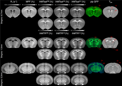 |
Comparison of inhomogeneous Magnetization Transfer (ihMT), R1 and MPF for myelin specific imaging
Andreea Hertanu1,2, Lucas Soustelle1,2, Arnaud Le Troter1,2, Julie Buron1,2,3, Julie Le Priellec3, Myriam Cayre3, Pascale Durbec3, Gopal Varma4, David C. Alsop4, Olivier M. Girard1,2, and Guillaume Duhamel1,2
1Aix Marseille Univ, CNRS, CRMBM, Marseille, France, 2APHM, Hôpital Universitaire Timone, CEMEREM, Marseille, France, 3Aix Marseille Univ, CNRS, IBDM, Marseille, France, 4Division of MR Research, Radiology, Beth Israel Deaconess Medical Center, Harvard Medical School, Boston, MA, United States
We conducted a comparative study of ihMT metrics variably weighted in T1D, with R1 (1/T1) and MPF (macromolecular proton fraction) for myelin specific imaging. These MRI methods were compared with histology by fluorescence microscopy using transgenic plp-GFP (proteolipid protein-Green Fluorescent Protein) mice.
|
|
 |
0084.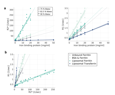 |
Uncovering the specificity of quantitative MRI to different molecular forms of iron in the brain.
Shir Filo1, Rona Shaharabani1, and Aviv Mezer1
1The Edmond and Lily Safra Center for Brain Science, The Hebrew University of Jerusalem, Jerusalem, Israel
The main iron compounds, ferritin and transferrin, are distributed heterogeneously across the brain and are often implicated in neurodegenerative diseases. While quantitative MRI has been linked to brain tissue’s microstructure, non-invasive discrimination between iron forms still remains a challenge. We propose an in vivo approach for assessing brain iron forms, based on the dependency of R1 on R2*. We establish this approach in phantoms and validate it against histology. In the in vivo human brain, the dependency of R1 on R2*, rather than each parameter by itself, predicts the inhomogeneous distribution of iron-binding proteins with age and across brain regions.
|
|
|
0085.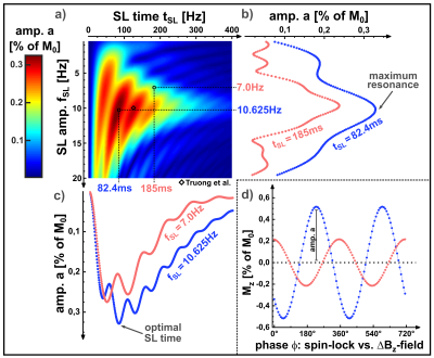 |
Improved spin-lock based detection of ultra-low-field electro-magnetic oscillations for direct fMRI
Maximilian Gram1,2, Markus Dippold2, Daniel Gensler1,3, Martin Blaimer4, Peter Nordbeck1,3, and Peter Michael Jakob2
1Department of Internal Medicine I, University Hospital Würzburg, Würzburg, Germany, 2Experimental Physics 5, University of Würzburg, Würzburg, Germany, 3Comprehensive Heart Failure Center (CHFC), University Hospital Würzburg, Würzburg, Germany, 4Fraunhofer Institute for Integrated Circuits IIS, Würzburg, Germany
BOLD-based fMRI is currently the method of choice for the spatially resolved investigation of neuronal activity, although this technique only allows indirect measurements of activity by evaluating hemodynamic effects. Recently, a spin-lock-based technique for the direct measurement of neuro-electro-magnetic oscillations was presented, which impressively demonstrated imaging of the alpha activity. In the corresponding publication, however, elementary parameters of the spin-lock preparation were specified ad hoc. In the present work it is shown that the choice of these preparation parameters is of essential importance. For example, a clever choice of the spin-lock time can improve and stabilize the signal detection.
|
|
|
0086.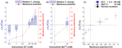 |
Noninvasive Detection of Changes in Membrane Potential with MR Measurements
Kyeongseon Min1, Sungkwon Chung2, Phan Tan Toi3,4, Jongho Lee1, Seung‐Kyun Lee3,4,5, and Jang-Yeon Park3,4
1Laboratory for Imaging Science and Technology, Department of Electrical and Computer Engineering, Seoul National Univeristy, Seoul, Korea, Republic of, 2Department of Physiology, Samsung Biomedical Research Institute, Sungkyunkwan University School of Medicine, Suwon, Korea, Republic of, 3Department of Biomedical Engineering, Sungkyunkwan University, Suwon, Korea, Republic of, 4Department of Intelligent Precision Healthcare Convergence, Sungkyunkwan University, Suwon, Korea, Republic of, 5Center for Neuroscience Imaging Research, Institute for Basic Science, Suwon, Korea, Republic of
In this study, the relationships between changes in cell membrane potential and MR measurements of T1, T2, and qMT parameters were examined using a neuroblastoma cell line (SH-SY5Y) as an in vitro model. When the membrane potential was depolarized from -37.7 mV to −7.5 mV, the changes in MR parameters were: T1, +4.29%; T2, +21.2%; pool size ratio (PSR), −12.4%. Contrarily, when the membrane potential was hyperpolarized to −43.0 mV, the changes in MR parameters were: T1, −3.40%; T2, −10.0%; PSR, +6.97%. These observations are expected to be utilized to noninvasively detect changes in the membrane potential of cells.
|
|
|
0087.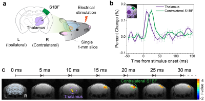 |
High temporospatial resolution MR imaging of neuronal activity in vivo
Phan Tan Toi1,2,3, Hyun Jae Jang4, Jeehyun Kwag4, and Jang-Yeon Park1,2,3
1Department of Biomedical Engineering, Sungkyunkwan University, Suwon, Korea, Republic of, 2Center for Neuroscience Imaging Research, Institute for Basic Science, Suwon, Korea, Republic of, 3Department of Intelligent Precision Healthcare Convergence, Sungkyunkwan University, Suwon, Korea, Republic of, 4Department of Brain and Cognitive Engineering, Korea University, Seoul, Korea, Republic of
Advanced non-invasive functional imaging methods have been widely used, but with certain limitations in either temporal or spatial information. There has long been a demand for a noninvasive imaging method capable of capturing neuronal activity with high temporal and spatial resolution. Here, we demonstrate a novel imaging method (called DIANA-fMRI) for directly detecting neuronal activity with high temporal (=5ms) and spatial (=0.22mm) resolution. DIANA-fMRI was capable of capturing sensory responses in mice at 9.4T with statistically significant signal changes (~0.1-0.2%). Temporally sequential DIANA responses were also confirmed along the thalamocortical pathway, together with further validation by electrophysiological experiments.
|
|
0088.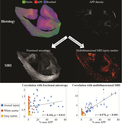 |
Diffuse axonal injury has a specific multidimensional MRI signature in traumatically injured corpus callosum
Dan Benjamini1, Diego Iacono2, Michal Komlosh1, Daniel Perl2, David Brody2, and Peter Basser1
1National Institute of Child Health and Human Development, Bethesda, MD, United States, 2Uniformed Services University of the Health Sciences, Bethesda, MD, United States
Can microscopic diffuse axonal injury lesions following trauma be seen non-invasively? We report that simultaneously integrating multiple MRI dimensions - T1, T2, and diffusion - can be targeted to image microscopic damage. Corpora Callosa derived from eight subjects that sustained TBI and three healthy control brain donors underwent post-mortem ex-vivo MRI at 7T. Multidimensional-, diffusion tensor-, and quantitative T1- and T2-MRI data were acquired and processed, along with corresponding pathohistological data. Although invisible using the conventional MRI modalities, multidimensional MRI provided images of the microscopic injuries, suggesting that it can be used for the detection of microscopic axonal injury.
|
||
|
0089.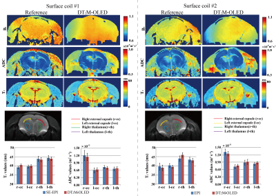 |
Single-shot simultaneous diffusion and T2 mapping based on overlapping-echo detachment planar imaging
Lingceng Ma1, Xinran Chen1, Jian Wu1, Lijun Bao1, Shuhui Cai1, Congbo Cai1, and Zhong Chen1
1Department of Electronic Science, Xiamen University, Xiamen, China
A single-shot simultaneous diffusion and T2 mapping method is developed based on overlapping-echo detachment planar imaging (DT2M-OLED) incorporating with deep-learning reconstruction. The method makes it possible to realize simultaneous diffusion and T2 mapping in around one hundred milliseconds for the first time, and owns an advantage in resisting motion artifacts. The accuracy of the proposed method was verified by numerical experiment and in vivo rat brain experiment. Dynamic diffusion and T2 mapping on rat recovering from deep anesthesia was also conducted to test the capability of the proposed method in real-time imaging.
|
|
|
0090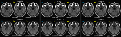 |
Deep learning based acceleration of multi-contrast MRI examinations by acquiring contrast and sharing inter-contrast structure information Video Permission Withheld
Sudhanya Chatterjee1, Suresh Emmanuel Joel1, Sajith Rajamani1, Shaik Ahmed1, Uday Patil1, Ramesh Venkatesan1, and Dattesh Dayanand Shanbhag1
1GE Healthcare, Bangalore, India
We propose a method to accelerate multi-contrast MRI examination for a subject. We accelerate typically longer scans of MR exam such as FLAIR-T1, FLAIR-T2 by only acquiring its contrast information and sharing structure information from a reference scan from the same exam which is fully sampled (typically with the lowest acquisition time e.g. T2FSE). The resulting view-shared images have systematic artifacts which are then removed by a deep learning module trained on more than 200 cases. We observed high quality reconstruction (SSIM>0.9) for both healthy control and pathology cases with acceleration factors of 2x and 3x for FLAIR-T1 and FLAIR-T2.
|
|
0091.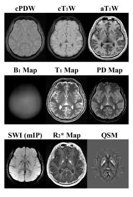 |
MULTI-Parametric MR imaging with fLEXible modular design (MULTIPLEX)
Yongquan Ye1, Jingyuan Lv1, Yichen Hu1, Zhongqi Zhang2, Jian Xu1, and Weiguo Zhang1
1UIH America, Inc., Houston, TX, United States, 2United Imaging Healthcare, Shanghai, China
We have developed a GRE based multi-parametric MR imaging method with flexible modular design, namely MULTIPLEX. Featuring a design of dual-TR, dual-FA and multi-echo, one single MULTIPLEX scan can provide multiple imaging contrasts and quantitative mappings with 3D high resolution within clinically friendly duration, including T1W, PDW, augmented T1W (aT1W), SWI and T1/T2*/PD/QSM maps, as well as optional MRA images.
|
||
0092.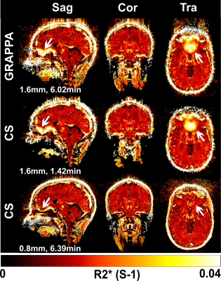 |
R2* mapping of the whole brain with 0.8 mm isotropic resolution at 7T in less than 7 minutes
Arun Joseph1,2,3, Tobias Kober4,5,6, and Tom Hilbert4,5,6
1Advanced Clinical Imaging Technology, Siemens Healthcare AG, Bern, Switzerland, 2Translational Imaging Center, Sitem-Insel, Bern, Switzerland, 3Departments of Radiology and Biomedical Research, University of Bern, Bern, Switzerland, 4Advanced Clinical Imaging Technology, Siemens Healthcare AG, Lausanne, Switzerland, 5Department of Radiology, Lausanne University Hospital and University of Lausanne, Lausanne, Switzerland, 6LTS5, École Polytechnique Fédérale de Lausanne (EPFL), Lausanne, Switzerland
T2*-weighted imaging is an important diagnostic tool to evaluate normal and pathological tissues due to its sensitivity to iron deposition. This comes with high sensitivity to magnetic field inhomogeneities within a voxel, resulting in susceptibility artifacts. These artefacts are largely reduced at higher resolutions; high-resolution protocols however lead to clinically unfeasible scan times with multi-echo gradient echo sequences. Here, we propose a compressed sensing multi-echo GRE acquisition for 7T to obtain 0.8 mm isotropic R2* maps of the whole brain in <7 minutes and with substantially reduced artefacts. Preliminary qualitative and quantitative validations are performed on healthy subjects.
|
The International Society for Magnetic Resonance in Medicine is accredited by the Accreditation Council for Continuing Medical Education to provide continuing medical education for physicians.