Oral
Current Trends in MRI Contrast Mechanisms
ISMRM & SMRT Annual Meeting • 15-20 May 2021

| Concurrent 3 | 12:00 - 14:00 | Moderators: Anahita Fathi Kazerooni |
 |
0241.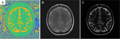 |
Heavily T2-weighted Imaging with Phase-Based RF Modulated GRE Imaging
Soudabeh Kargar1, Daiki Tamada1, Ruvini Navaratna1, Jayse Merle Weaver1, and Scott B Reeder1
1Radiology, University of Wisconsin - Madison, Madison, WI, United States
Heavily T2-weighted imaging is used for a variety of fluid sensitive imaging such as MRCP. However, these fast spin-echo-based methods suffer from long acquisition duration. In this work we apply a novel strategy using RF phase modulated gradient echo (GRE) with small RF phase increments to encode T2 information into the phase of the signal. In this work, we optimize this strategy for long T2 species to achieve heavily T2-weighted imaging, including the introduction of a novel cross-product strategy to highlight signal in tissues with long T2 and suppress signal in tissues with shorter T2 values.
|
|
0242. |
Deuterium metabolic imaging (DMI) of Water, Glucose and Lactate using spectroscopic multi-echo bSSFP: A higher Signal to Noise Approach
Dana C. Peters1, Stefan Markovic2, Qingjia Bao2, Dina Preise2, Keren Sasson2, Lilach Agemy2, Avigdor Scherz2, and Lucio Frydman2
1Radiology and Biomed Eng., Yale University, New Haven, CT, United States, 2Weizmann Institute of Science, Rehovot, Israel
Deuterium metabolic imaging (DMI) maps the individual in vivo fate of 2H-enriched metabolites. Upon injecting 2H6,6’-glucose, DMI images a 2H-water peak, and a small but diagnostic 2H3,3’-lactate signature, highlighting tumors and their aberrant metabolism. DMI faces major sensitivity challenges, that can be alleviated by a multi-echo balanced SSFP approach. When suitably tuned, multi-echo bSSFP yields good spectral isolation of all metabolites, and thanks to the relatively large T2/T1 ratios of deuterated compounds, several-fold increases in SNR vs. chemical shift imaging are then obtained. This is demonstrated in phantoms, and in in vivo mice studies of orthotopic pancreatic tumors.
|
||
0243.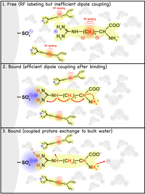 |
Detection of ionic bonding using IMMOBILISE MRI and theoretical description of the relayed NOE transfer mechanism
Yang Zhou1, Peter van Zijl2,3, Chongxue Bie2,3,4, Jiadi Xu2,3, Xin Liu1, and Nirbhay N. Yadav2,3
1Institute of Biomedical and Health Engineering, Shenzhen Institutes of Advanced Technology, Shenzhen, China, 2The Russell H. Morgan Department of Radiology, The Johns Hopkins University School of Medicine, Baltimore, MD, United States, 3F.M. Kirby Research Center for Functional Brain Imaging, Kennedy Krieger Institute, Baltimore, MD, United States, 4Department of Information Science and Technology, Northwest University, Xian, China
Saturation transfer MRI has previously been applied to study transient molecular binding using the IMMOBILISE MRI approach, yet the multiple mechanisms of magnetization transfer during such binding are still under investigation. We studied electrostatic-interaction-mediated molecular binding of small substrates (choline and arginine) to ion-exchange column media. A theoretical model using relayed NOEs to exchangeable protons in the bound substrate is proposed to quantitatively describe the saturation transfer during such binding.
|
||
0244.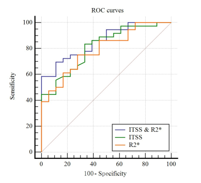 |
The combination of ITSS and R2* in quantitatively and automatically evaluating histological grade of HCC using ESWAN: A feasibility study
Dahua Cui1, Ailian Liu1, Hongkai Wang2, Mingrui Zhuang2, and Qingwei Song1
1The First Affiliated Hospital of Dalian Medical University, Dalian, China, 2Dalian University of Technology, Dalian, China
The aim of this study was to investigate the feasibility of intratumoral susceptibility signal intensities (ITSS) combined with R2* values obtained from T2 star-weighted angiography (ESWAN) in quantitatively and automatically evaluating histological grading of hepatocellular carcinoma (HCC). The results showed that the combination of quantitative ITSS and R2* was feasible to evaluate the histological grading of HCC performance (AUC = 0.856, P < 0.0001, sensitivity of 88.89%, specificity of 69.44%) automatically.
|
||
0245.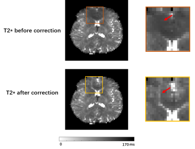 |
Improved T2' Mapping in Simultaneous Neurometabolic and Oxygenation Imaging Experiments
Tianxiao Zhang1, Rong Guo2,3, Yudu Li2,3, Yibo Zhao2,3, Zhi-Pei Liang2,3, and Yao Li1
1School of Biomedical Engineering, Shanghai Jiao Tong University, Shanghai, China, 2Beckman Institute for Advanced Science and Technology, University of Illinois at Urbana-Champaign, Urbana, IL, United States, 3Department of Electrical and Computer Engineering, University of Illinois at Urbana-Champaign, Urbana, IL, United States
Simultaneous MRSI and T2* mapping has been demonstrated recently using SPICE. With a T2 map, T2' values could be calculated, which reflects tissue oxygenation. However, the accuracy of T2* and T2 measurements suffers from system imperfections. In this work, we improved T2' mapping by overcoming signal dephasing in T2* mapping and estimation bias in T2 mapping. The signal dephasing in T2* caused by B0 inhomogeneity was corrected utilizing high-resolution field map and pre-learned subspaces, and the estimation bias in T2 caused
by B1+ inhomogeneity was corrected with a dictionary-based estimation. The proposed method provided improved T2' mapping in SPICE experiments.
|
||
0246.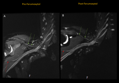 |
Evaluation of an Iron-Oxide Nanoparticle Contrast Agent for Vascular Suppression in Magnetic Resonance Neurography
Sophie Queler1, Ek Tsoon Tan1, Martin Prince2, John Carrino1, and Darryl Sneag1
1Radiology and Imaging, Hospital for Special Surgery, New York, NY, United States, 2Weill Cornell Medicine, New York, NY, United States
In this study, we investigated the use of ferumoxytol, an iron-oxide nanoparticle, for vascular signal suppression in 3 Tesla magnetic resonance neurography of the brachial plexus. A 3D, T2-weighted STIR sequence was prospectively acquired of 19 normal brachial plexi in 10 volunteers (1 unilateral; 9 bilateral) before and after ferumoxytol infusion. Independent assessment of anonymized exams by two radiologists demonstrated overall improved vascular suppression as well as improved visualization of the suprascapular nerve with increased diagnostic confidence. Improvements in nerve-, fat-, and blood-to-muscle contrast were supported by signal simulations.
|
||
|
0247.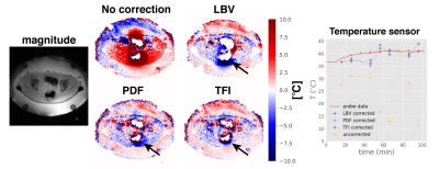 |
Susceptibility artifact correction in MR thermometry for monitoring of mild RF hyperthermia using total field inversion
Christof Boehm1, Marianne Goeger-Neff2, Hendrik T. Mulder3, Benjamin Zilles2, Lars H. Lindner2, Gerard C. van Rhoon3, Dimitrios C. Karampinos4, and Mingming Wu1
1Technical University of Munich, Munich, Germany, 2Department of Medicine III, University Hospital, LMU Munich, Munich, Germany, 3Erasmus MC Cancer Institute, Rotterdam, Netherlands, 4Department of Diagnostic and Interventional Radiology, School of Medicine, Technical University of Munich, Munich, Germany
Motion-induced susceptibility changes induce field variations, leading to large errors during MR thermometry based on the linear proton resonance frequency shift. These artefacts aggravate temperature quantification in the face of both the long treatment duration and the mild temperature change during mild RF hyperthermia treatments. We show with the help of simulations, a phantom heating experiment, volunteer scans and mild hyperthermia treatment of a patient with cervical cancer and a sarcoma patient how to correct for this artefact source by methods known from quantitative susceptibility mapping. The recently introduced total field inversion shows advantages over the background field removal methods.
|
|
0248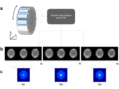 |
Fast MR thermometry based on propeller echo‐planar time‐resolved imaging with dynamic encoding (PEPTIDE) Video Permission Withheld
Zhehong Zhang1, Fair Merlin2, Fuyixue Wang3,4, Zijing Dong3,5, Wending Tang1, Menghan Li1, Danna Wei6, Kawin Setsompop2,7, and Kui Ying1
1Department of Engineering Physics, Tsinghua University, Beijing, China, 2Department of Radiology, School of Medicine, Stanford University, Stanford, CA, United States, 3Athinoula A. Martinos Center for Biomedical Imaging, Massachusetts General Hospital, Charlestown, MA, United States, 4Harvard-MIT Health Sciences and Technology, MIT, Cambridge, MA, United States, 5Department of Electrical Engineering and Computer Science, MIT, Cambridge, MA, United States, 6Department of Biomedical Engineering, School of Medicine, Tsinghua University, Beijing, China, 7Department of Electrical Engineering, Stanford University, Stanford, CA, United States
Echo‐planar time‐resolved imaging (EPTI) is a multi-shot EPI technique capable of rapidly obtaining distortion‐free and blurring‐free time-resolved multi-contrast images across the EPI readout. PROPELLER is an extension to EPTI, which incorporates PROPRELLER-like acquisition, to enable shot-to-shot motion toleration. In this work, PEPTIDE was applied to the MR thermometry application, where an image reconstruction framework that leveraged sparsity across blades of PEPTIDE rawdata was proposed. The potential in using PEPTIDE to provide distortion- and blurring-free temperature mapping at high temporal resolution was then demonstrated via simulated human brain PEPTIDE data with various temperature profiles.
|
||
0249.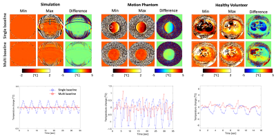 |
Motion-robust, multi-slice, real-time MR PRFS Thermometry for MR-guided ultrasound thermal therapy in abdominal organs
Kisoo Kim1, Chris Diederich2, and Eugene Ozhinsky1
1Department of Radiology & Biomedical Imaging, University of California, San Francisco, San Francisco, CA, United States, 2Department of Radiation Oncology, University of California, San Francisco, San Francisco, CA, United States
MR temperature monitoring during ultrasound thermal therapy of abdominal organs is challenging due to respiratory motion. It is especially important for hyperthermia therapy, which requires accurate temperature measurements to ensure therapeutic heating within a narrow temperature window (37-45 °C). In this study we developed a motion-robust, multi-slice, real-time MR thermometry sequence and reconstruction pipeline for the monitoring of temperature in hyperthermia or thermal ablation therapy in abdominal organs. This technique was implemented in RTHawk and evaluated in simulated acquisitions, phantom experiments with a custom-made motion phantom, and in-vivo healthy volunteer experiment without heating.
|
||
0250.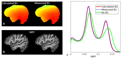 |
Calibrationless B1 Mapping for Accurate Macromolecular Proton Fraction Mapping Using Relaxometry Constraints
Alexey Samsonov1
1University of Wisconsin-Madison, Madison, WI, United States
Macromolecular proton fraction (MPF) is an established myelin marker with confirmed clinical relevance, but with sensitivity to technological variations such as B1 field errors. We propose a method to derive B1 map for MPF correction from MPF data itself. The method is based on standard two-pool MT formalism enhanced with improved relaxometry constraints.
|
The International Society for Magnetic Resonance in Medicine is accredited by the Accreditation Council for Continuing Medical Education to provide continuing medical education for physicians.