Oral
Quantitative MSK MRI
ISMRM & SMRT Annual Meeting • 15-20 May 2021

| Concurrent 2 | 18:00 - 20:00 | Moderators: Dimitrios Karampinos & Ashley Williams |
0843.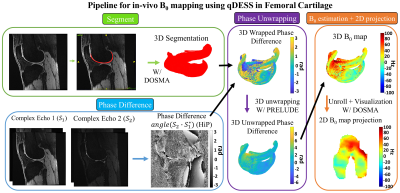 |
A Method for Measuring B0 Field Inhomogeneity using Quantitative DESS (qDESS)
Marco Barbieri1, Akshay S. Chaudhari1,2, Catherine J. Moran1, Garry E. Gold1,3, Brian A. Hargreaves1,3,4, and Feliks Kogan1
1Department of Radiology, Stanford University, Stanford, CA, United States, 2Department of Biomedical Data Science, Stanford University, Stanford, CA, United States, 3Department of Bioengineering, Stanford University, Stanford, CA, United States, 4Department of Electrical Engineering, Stanford University, Stanford, CA, United States
Quantitative T2 mapping is a valuable tool for studying OA changes. qDESS is a rapid sequence that provides accurate T2 measurements and SNR-efficient morphological imaging. B0 mapping is an auxiliary scan acquired to correct field inhomogeneity-induced errors using techniques such as WASSR and 2-GRE. This work proposes a method for B0 mapping that exploits the phase difference between the two echoes acquired with qDESS. The experiments with phantom and in-vivo simultaneous bilateral knee acquisitions showed that the B0 maps obtained with the qDESS method were in good agreement with those obtained using the WASSR method and the 2-GRE method.
|
||
0844.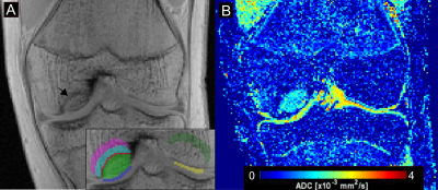 |
Analysis of Diffusion Changes in Patients with Juvenile Osteochondritis Dissecans (JOCD) of the Knee at 3T
Abdul Wahed Kajabi1,2,3, Stefan Zbyn1,3, Cyrus M. Nouraee1, Kai D. Ludwig1,3, Casey P. Johnson1,4, Steen Moeller1, Mark A. Tompkins5, Bradley J. Nelson5, Gregory J. Metzger1, Cathy S. Carlson4, and Jutta M. Ellermann1,3
1Center for Magnetic Resonance Research, University of Minnesota, Minneapolis, MN, United States, 2Research Unit of Medical Imaging, Physics and Technology, University of Oulu, Oulu, Finland, 3Department of Radiology, University of Minnesota, Minneapolis, MN, United States, 4Department of Veterinary Clinical Sciences, University of Minnesota, Minneapolis, MN, United States, 5Department of Orthopaedic Surgery, University of Minnesota, Minneapolis, MN, United States
This 3T study evaluates 46 juvenile osteochondritis dissecans (JOCD) lesions of 41 skeletally immature patients using clinical, morphological MRI and diffusion-weighted MRI (DWI). In this study, apparent diffusion coefficient (ADC) values were able to differentiate between healed and not yet healed JOCD lesions, and distinguish between the operative and nonoperative treatment groups. DWI provides noninvasive, quantitative assessment of disease status which may help inform clinical management of JOCD. Further follow-up studies are needed to evaluate the potential of DWI for the prediction of lesion healing in JOCD patients.
|
||
0845.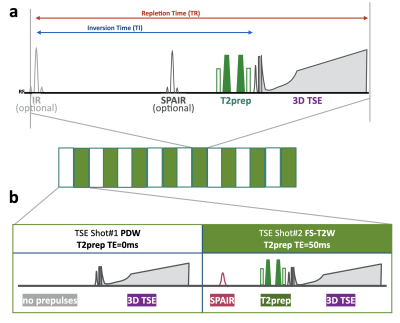 |
Simultaneous anatomical, pathological and T2 quantitative knee imaging with 3D submillimeter isotropic resolution using MIXTURE
Takayuki Sakai1,2, Masami Yoneyama3, Atsuya Watanabe4,5, Daichi Murayama1, Shigehiro Ochi1, Shuo Zhang6, and Tosiaki Miyati7
1Radiology, Eastern Chiba Medical Center, Tonage, Japan, 2Division of Health Sciences, Graduate School of Medical Sciences, Kanazawa University, Kanazawa, Japan, 3Philips Japan, Tokyo, Japan, 4General Medical Services, Chiba University Graduate School of Medicine, Chiba, Japan, 5Orthopaedic Surgery, Eastern Chiba Medical Center, Chiba, Japan, 6Philips Healthcare, Hamburg, Germany, 7Faculty of Health Sciences, Institute of Medical, Pharmaceutical and Health Sciences, Kanazawa University, Kanazawa, Japan
We propose a new sequence called MIXTURE (Multi-Interleaved X-prepared TSE with inTUitive RElaxometry). MIXTURE is a 3D TSE that can set arbitrary echo times using the T2 preparation pulses, and enables several image contrasts (such as PDW, T2W) and T2 mapping by acquiring at least two echo time images. In this study, we evaluated the clinical feasibility of morphological and pathological MRI with two contrasts images and quantitative MRI with T2 mapping using MIXTURE.
|
||
0846.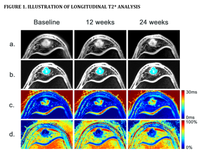 |
Association Between UTE-MRI T2* Relaxation Times and Symptoms During Exercise Therapy for Patellar Tendinopathy
Stephan J. Breda1, Robert-Jan de Vos2, Dirk Poot1, Gabriel Krestin1, Juan A. Hernandez-Tamames1, and Edwin Oei1
1Radiology & Nuclear Medicine, Erasmus University Medical Center, Rotterdam, Netherlands, 2Orthopaedics, Erasmus Univerity Medical Center, Rotterdam, Netherlands
Patellar tendinopathy (PT) is an overuse injury of the patellar tendon in athletes involving typical degenerative changes to the patellar tendon. The association of MRI-assessed structural changes with symptoms is largely unknown. UTE-MRI was implemented to study longitudinal changes in T2* within the patellar tendon in athletes performing exercise therapy for PT. We found that T2* relaxation times in the degenerative tissue of the patellar tendon were associated with symptom severity and that decreased T2* was associated with clinical improvement.
|
||
0847.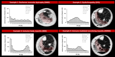 |
Differences in distribution of MRI-based fat fraction in lower limb skeletal muscles of six different neuromuscular disorders
Harmen Reyngoudt1,2, Pierre-Yves Baudin1,2, Ericky C.A. Araujo1,2, Pierre G. Carlier3, and Benjamin Marty1,2
1NMR Laboratory, Neuromuscular Investigation Center, Institute of Myology, Paris, France, 2NMR Laboratory, CEA/DRF/IBFJ/MIRCen, Paris, France, 3CEA, DRF, Service Hospitalier Frédéric Joliot, Orsay, France
Quantitative MRI fat-water separation techniques such as Dixon are often used to evaluate disease progression in muscle of neuromuscular diseases. Although the mean fat fraction (FF) value per region of interest (ROI) is a valuable objective MRI biomarker, it does not reflect the variation of FF within this ROI. In this study we analyzed 6582 muscle ROIs in leg and thigh of 6 different neuromuscular diseases and analyzed, besides the mean FF, other statistical metrics such as median, standard deviation, kurtosis and skewness. The differences in FF distribution might reveal additional information about the individual patient’s disease evolution.
|
||
0848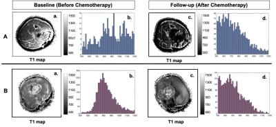 |
Tumor T1 for early chemotherapeutic response evaluation in patients with osteosarcoma with correlation to histological necrosis Video Permission Withheld
Esha Baidya Kayal1, Nikhil Sharma1, Raju Sharma2, Sameer Bakhshi3, Devasenathipathy Kandasamy2, and Amit Mehndiratta1,4
1Centre for Biomedical Engineering, Indian Institute of Technology Delhi, New Delhi, India, 2Radio diagnosis, All India Institute of Medical Sciences Delhi, New Delhi, India, 3Department of Medical Oncology, Dr. B.R. Ambedkar Institute-Rotary Cancer Hospital (IRCH), All India Institute of Medical Sciences Delhi, New Delhi, India, 4Department of Biomedical Engineering, All India Institute of Medical Sciences Delhi, New Delhi, India
The spin relaxation time (T1) of the water protons, an intrinsic property of tissue, can be a useful marker of therapeutic response in osteosarcoma. T1 values were estimated in whole tumor volume before and after completion of chemotherapy and histogram analysis was performed to characterize tumor T1 and its changes in the course of chemotherapy. Results showed mean and skewness of T1 relaxation time in tumor may be useful as non-invasive imaging markers of chemotherapeutic response in osteosarcoma.
|
||
0849.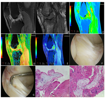 |
The clinical value of MRI in quantitatively evaluating anterior cruciate ligament mucoid degeneration
Guangtao Fan1, Yudan Li1, Fenglin Xue2, Yilong Huang1, Yanlin Li3, Guoliang Wang3, Tianfu Qi1, Lisha Nie4, and Bo He1
1Department of Imaging, the First Affiliated Hospital of Kunming Medical University, Kunming, China, 2Department of Pathology, the First Affiliated Hospital of Kunming Medical University, Kunming, China, 3Department of Sports Medicine, the First Affiliated Hospital of Kunming Medical University, Kunming, China, 4GE Healthcare, Kunming, China
The study aims to explore the clinical application value of MRI to quantitatively assess the anterior cruciate ligament mucoid degeneration (ACL-MD).The results indicated MRI Values of T1, T2 and T2*(relaxation time) show potential for diagnosis of ACL-MD and T2* may deliberately has the highest diagnostic efficacy.
|
||
0850.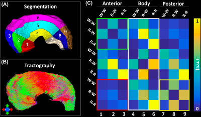 |
Diffusion Tensor Imaging and Fiber Tractography in Porcine Meniscus
Jikai Shen1, Qi Zhao1, Yi Qi1, Gary Cofer1, G. Allan Johnson1, and Nian Wang2
1Duke University, Durham, NC, United States, 2Radiology and Imaging Sciences, Indiana University, Indianapolis, IN, United States
Recently, diffusion MRI and tractography in musculoskeletal system has used to investigate the tissue microstructure, local collagen fiber alignment, and the 3D collagen network. To the best of our knowledge, nondestructively probing the local collagen fiber direction and 3D collagen fiber architecture is still limited. The water diffusion properties derived from DTI were quantified at different areas of meniscus using the proposed automatic segmentation method. Combining tractography and automatic segmentation, we were able to observe the structural connections among different areas of the meniscus.
|
||
0851.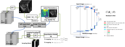 |
Achieving Rapid and Accurate Relaxometry of Whole Knee Joint using Self-Supervised Deep Learning
Fang Liu1, Georges El Fakhri1, Martin Torriani1, Richard Kijowski2, and Miho Tanaka3
1Radiology, Massachusetts General Hospital and Harvard Medical School, Boston, MA, United States, 2New York University School of Medicine, New York, NY, United States, 3Orthopaedic Surgery, Massachusetts General Hospital and Harvard Medical School, Boston, MA, United States
The purpose of this work was to develop and evaluate a model-guided self-supervised deep learning MRI reconstruction framework called REference-free LAtent map eXtraction (RELAX) for rapid quantitative relaxometry of the whole knee joint. This approach incorporated end-to-end CNN mapping to perform image-to-parameter domain transform. A concept of cyclic loss was utilized to enforce data fidelity and eliminate the explicit need for full-sampled training references. This approach was demonstrated in accelerated T1/T2 mapping of the whole knee joint and proven to outperform state-of-the-art reconstruction methods. The result suggests that RELAX allows accelerated relaxometry without training with reference data.
|
||
 |
0852. |
3T-Chemical Shift Encoded MRI with Ultra-Short Echo Time Acquisition for Bone Quality Assessment: Preliminary Results in the Hip.
Dimitri MARTEL1, Benjamin LEPORQ2, Stephen HONIG3, and Gregory CHANG1
1Radiology, NYU Langone Health, New york, NY, United States, 2Université de Lyon; CREATIS CNRS UMR 5220, Inserm U1206, INSA-Lyon, UCBL Lyon 1, Villeurbanne, France, 3Osteoporosis Center, Hospital for Joint Diseases, NYU Langone Health, New york, NY, United States Osteoporosis (OP) is a disease associated with low bone mass and deterioration of bone microarchitecture leading to bone fragility and increased fracture risk, especially in the proximal femur. Therefore, we have developed a chemical-shift encoded acquisition performed with a spiral k-space sampling to acquire ultrashort echo-time and longer echo time in the echo train. This study aims to determine if uTE acquisition can be performed for fat/water separation and if additional information can be provided through cortical bone imaging. |
The International Society for Magnetic Resonance in Medicine is accredited by the Accreditation Council for Continuing Medical Education to provide continuing medical education for physicians.