Oral
Muscle MRI
ISMRM & SMRT Annual Meeting • 15-20 May 2021

| Concurrent 4 | 16:00 - 18:00 | Moderators: Pierre Carlier & Erin Englund |
0579.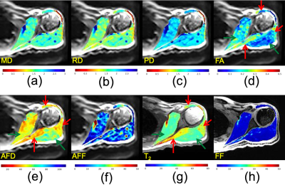 |
Diffusion MRI Fiber Diameter for Muscle Denervation Assessment
Ek T Tan1, Kelly C Zochowski1, Kenneth Serrano1, and Darryl B Sneag1
1Radiology and Imaging, Hospital for Special Surgery, New York, NY, United States
Diffusion imaging may provide intracellular muscle denervation characterization complimentary to T2- and fat fraction (FF) mapping. Non-invasive muscle diameter assessment was performed using multi-shell diffusion MRI with a cylindrical model. The apparent fiber diameter (AFD) was obtained using a dictionary method mapping restricted and free fractions to AFD. 16 subjects with suspected muscle denervation involving the brachial plexus and 4 healthy control subjects were imaged. The denervated AFD was smaller than non-denervated and healthy controls by 11 µm and 23 µm respectively. AFD in healthy controls were 67-101 µm, which compare well against histology values from the literature (50-90 µm).
|
||
0580.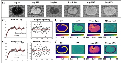 |
On the reconstruction of MR-fingerprinting with water and fat separation for quantitative skeletal muscle imaging
Benjamin Marty1,2, Fabian Balsiger3, Pierre-Yves Baudin1,2, Lopez Alfredo1,2, Ericky CA Araujo1,2, and Harmen Reyngoudt1,2
1Neuromuscular Investigation Center, NMR Laboratory, Institute of Myology, Paris, France, 2NMR Laboratory, CEA, DRF, IBFJ, MIRCen, Paris, France, 3Support Center for Advanced Neuroimaging, Institute for Diagnostic and Interventional Neuroradiology, Inselspital, Bern University Hospital, University of Bern, Bern, Switzerland
Our objective was to determine the influence of the reconstruction parameters on fat fraction and water T1 estimated in the skeletal muscle using MR fingerprinting with water and fat separation, to propose an optimized reconstruction procedure maximizing the accuracy and precision of the MRI variables, and evaluate it on in vivo data. We show that aliasing artefacts generate a bias in the estimates that could be mitigated by introducing a simple correction factor. Then, the accuracy and precision were improved by reconstructing the frames using a single radial spoke, and by using 10 SVD components for dictionary matching.
|
||
 |
0581.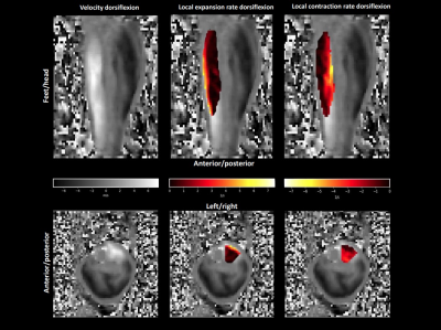 |
4D Phase Contrast MRI detects heterogeneous strain rate patterns along the length of the Tibialis Anterior muscle during dynamic contractions
Thom T. J. Veeger1, Gustav J. Strijkers2, Valentina Mazzoli3, Hans C. van Assen1, Jurriaan H. de Groot4, Lukas M. Gottwald5, Aart J. Nederveen5, Hermien E. Kan1,6, and Melissa T. Hooijmans2
1Radiology, Leiden University Medical Center, Leiden, Netherlands, 2Biomedical Engineering & Physics, Amsterdam Movement Sciences, Amsterdam University Medical Center, Location AMC, Amsterdam, Netherlands, 3Radiology, Stanford University, Stanford, CA, United States, 4Rehabilitation Medicine, Leiden University Medical Center, Leiden, Netherlands, 5Radiology and Nuclear Medicine, Amsterdam Movement Sciences, Amsterdam University Medical Center, Location AMC, Amsterdam, Netherlands, 6Duchenne Center Netherlands, Veenendaal, Netherlands
We assessed strain rate distributions within the Tibialis Anterior muscle by acquiring whole lower leg 4D PC-MRI during dynamic exercise, with and without load. Our data revealed a spatially heterogeneous strain rate pattern along the length of the Tibialis Anterior muscle during movement cycle. During dorsiflexion, the smallest negative strain rates were observed (in first approximation) along the fiber in the most proximal segment and the largest positive strain rates (in first approximation) across the fiber in the most distal segment, while plantarflexion showed the expected opposite. No effect of load was detected on strain rates.
|
|
0582.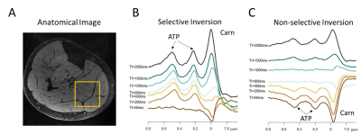 |
Characterization of cross-relaxation in human skeletal muscle using downfield 1H MRS at 7T
Sophia Swago1, Abigail Cember2, Brianna Moon1, Puneet Bagga3, Neil Wilson4, Mark A. Elliott2, Hari Hariharan2, Ravinder Reddy2, and Walter Witschey2
1Department of Bioengineering, University of Pennsylvania, Philadelphia, PA, United States, 2Department of Radiology, University of Pennsylvania, Philadelphia, PA, United States, 3St. Jude Children's Research Hospital, Memphis, TN, United States, 4Siemens Medical Solutions USA Inc, Malven, PA, United States
Water suppression limits the detection of non-labile proton downfield resonances in 1H magnetic resonance spectroscopy (MRS) due to cross-relaxation with water, and the cross-relaxation properties of these resonances has yet to be quantified in human skeletal muscle. We use spectrally-selective excitation in an inversion recovery experiment to compare the apparent T1 relaxation time of downfield resonances in skeletal muscle under selective and nonselective inversion conditions at 7T. Nonselective inversion significantly prolongs the longitudinal relaxation rate of resonances found 8.0, 8.2, and 8.5 ppm. This change is larger for the resonances at 8.2 and 8.5 ppm, indicating a stronger cross-relaxation effect.
|
||
0583.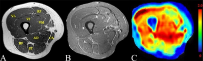 |
Decreased muscular perfusion in dermatomyositis: initial results detected by Inflow-based vascular-space-occupancy MR imaging
Yuankui Wu1, Xiaomin Liu1, Jun Hua2,3, Xiaodan Li1, Haimei Cao1, Yingjie Mei4, and Yikai Xu1
1Department of Medical Imaging, Nanfang Hospital, Southern Medical University, Guangzhou, China, 2Neurosection, Division of MRI Research, Department of Radiology, Johns Hopkins University School of Medicine, Baltimore, MD, United States, 3F.M. Kirby Research Center for Functional Brain Imaging, Kennedy Krieger Institute, Department of Radiology, Johns Hopkins University School of Medicine, Baltimore, MD, United States, 4Philips healthcare, Guangzhou, China
Dermatomyositis (DM) is a chronic autoimmune microangiopathy. Accurate quantification of muscular microcirculation can assist in early diagnosis and improve clinical outcomes. Inflow-based vascular-space-occupancy (iVASO) is a novel perfusion technique without the need for exogenous contrast agents. This study aimed to determine the potential diagnostic value of iVASO-MRI for patients with DM. The results showed decreased arteriolar muscular blood volume (MBVa) in DM patients, which worsens with the progression of disease, and the diminished MBVa in morphologically-normal appearing muscles. This suggests that iVASO-derived MBVa has the potential to be used as a biomarker for early diagnosis of DM.
|
||
0584.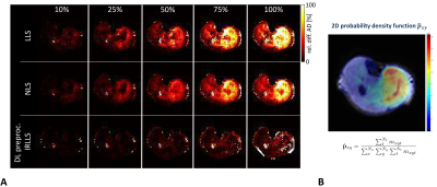 |
Spontaneous Muscular Activities and Estimating their Influence on Derived Diffusion Tensor Parameters
Martin Schwartz1,2, Petros Martirosian1, Günter Steidle1, Bin Yang2, and Fritz Schick1
1Section on Experimental Radiology, University Hospital of Tuebingen, Tuebingen, Germany, 2Institute of Signal Processing and System Theory, University of Stuttgart, Stuttgart, Germany
Estimation of diffusion tensor parameters in healthy subjects and patients suffering from neuromuscular disease can be markedly affected by a high rate of spontaneous muscular activities during the MR examination. Therefore, a concept for realistic simulation of spontaneous muscular activities in diffusion tensor imaging and the estimation of their influence on derived parameters is given in this work. The degradation of the derived parameters depends strongly on the robustness of the chosen approach for tensor estimation.
|
||
|
0585. |
Quantitative muscle MRI in monitoring disease progression and nusinersen treatment effects in spinal muscular atrophy
Louise Otto1, Martijn Froeling2, Ruben van Eijk1,3, Renske Wadman1, Inge Cuppen4, Danny van der Woude5, Bart Bartels5, Fay-Lynn Asselman1, Jeroen Hendrikse2, and Ludo van der Pol1
1Department of Neurology, UMC Utrecht Brain Center, University Medical Center, Utrecht, Utrecht, Netherlands, 2Department of Radiology, University Medical Center Utrecht, the Netherlands, Utrecht, Netherlands, 3Biostatistics & Research Support, Julius Center for Health Sciences and Primary Care, University Medical Center Utrecht, Utrecht, Netherlands, 4Department of Neurology and Child Neurology, UMC Utrecht Brain Center, University Medical Center, Utrecht, Utrecht, Netherlands, 5Department of Child Development and Exercise Center, University Medical Center Utrecht, the Netherlands, Utrecht, Netherlands
Quantitative MRI of muscles allows to measure disease progression or to assess therapeutic effects in neuromuscular diseases. We executed two studies on patients with spinal muscular atrophy, treated and untreated, with a protocol consisting of DIXON, T2 mapping and DTI on a 3T MR scanner. In treatment-naïve adult patients we demonstrated that qMRI was able to measure subclinical disease progression. In young children with SMA, quantitative MR parameters of the DIXON and DTI sequence showed ongoing fatty infiltration and normalization of thigh muscle microstructure during the first year of nusinersen treatment.
|
|
0586.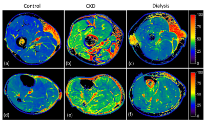 |
Multimodal MR Assessment of Skeletal Muscle in Patients with Chronic Kidney Disease and Dialysis
Can Wu1,2, Qi Peng3, William Paredes4, Moriel Vandsburger5, and Matthew K. Abramowitz4
1Department of Medical Physics, Memorial Sloan Kettering Cancer Center, New York, NY, United States, 2Philips Healthcare, Andover, MA, United States, 3Department of Radiology, Albert Einstein College of Medicine and Montefiore Medical Center, Bronx, NY, United States, 4Department of Medicine, Albert Einstein College of Medicine and Montefiore Medical Center, Bronx, NY, United States, 5Department of Bioengineering, University of California, Berkeley, Berkeley, CA, United States
Chronic kidney disease (CKD) is associated with reduced skeletal muscle mass, strength, and function, but a quantitative approach to systematically assess changes in skeletal muscle is lacking. The purpose of this study was to develop a multimodal MR method for quantitative assessment of skeletal muscle in patients with CKD compared to normal controls. The study revealed significant changes of T1ρ, intra- and extra-myocellular lipid ratio, ADC, and FA in CKD or dialysis patients. In addition, there was significant correlation between T1ρ and DTI biomarkers. These findings may provide new insights into the impaired skeletal muscle function in CKD patients.
|
||
 |
0587.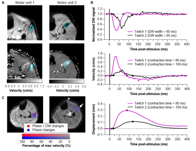 |
The muscle twitch profile assessed with Motor Unit Magnetic Resonance Imaging (MUMRI)
Linda Heskamp1, Matthew Birkbeck1,2,3, Roger Whittaker1, Ian Schofield1, and Andrew Blamire1
1Newcastle University Translational and Clinical Research Institute (NUTCRI), Newcastle University, Newcastle upon Tyne, United Kingdom, 2Newcastle Biomedical Research Centre, Newcastle University, Newcastle upon Tyne, United Kingdom, 3Northern Medical Physics and Clinical Engineering, Freeman Hospital, Newcastle upon Tyne NHS Foundation Trust, Newcastle upon Tyne, United Kingdom
Motor units (MUs) play a fundamental role in muscle physiology and disease. Contraction of muscle fibres belonging to a MU induces signal voids on diffusion weighted (DW) images enabling us to image these MUs (MUMRI). We demonstrated that MUMRI can also extract the twitch profile of single MUs. Computational modelling showed that the attenuation of net magnetisation and the cumulative phase changes increase with twitch magnitude. Therefore, we can measure the MU time profile by altering the timing of electrical nerve stimulation relative to the diffusion-encoding gradients. We applied this experimentally and measured in-vivo contraction times of human MUs.
|
|
|
0588.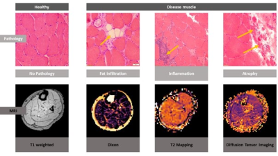 |
Quantitative muscle-MRI correlates with histopathology in skeletal muscle biopsies – a pilot study
Lara Schlaffke1, Robert Rehmann1, Anja Schreiner1, Marlena Rohm1, Johannes Forsting1, Martijn Froeling2, Martin Tegenthoff1, Matthias Vorgerd1, and Anne-Katrin Güttsches1
1Neurology, University Clinic Bergmannsheil Bochum gGmbH, Bochum, Germany, 2Radiology, UMC Utrecht, Utrecht, Netherlands
Skeletal muscle biopsy is the gold-standard in the diagnosis of inflammatory and hereditary muscle disorders. Ten patients who underwent muscle biopsy for diagnostic purposes were examined by qMRI (Fat-fraction, water T2-time and diffusion). The fat fraction, the severity of degenerative and inflammatory parameters and the amount of type 1/2-fibers were determined in all samples. The amount of fat tissue correlated significantly between histopathology and qMRI. EPG Water T2-time correlated with the in the histopathologic analysis. The study provides the basis for qMRI methods in the follow up of patients with neuromuscular disorders, especially in the context of emerging treatment strategies.
|
The International Society for Magnetic Resonance in Medicine is accredited by the Accreditation Council for Continuing Medical Education to provide continuing medical education for physicians.