Oral
Bone, Cartilage & Joint MRI
ISMRM & SMRT Annual Meeting • 15-20 May 2021

| Concurrent 4 | 14:00 - 16:00 | Moderators: Martijn Froeling & Valentina Mazzoli |
0519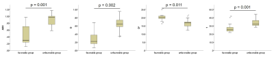 |
Predicting delayed union in osteoporotic vertebral fractures in the acute phase with intravoxel incoherent motion Video Permission Withheld
Hiroyuki Takashima1,2, Tsuneo Takebayashi3, Yasuhisa Abe3, Rui Imamura1, Hiroshi Oguma3, Izaya Ogon2, Yoshihiro Akatsuka1, and Toshihiko Yamashita2
1Division of Radiology and Nuclear Medicine, Sapporo Medical University Hospital, Sapporo, Japan, 2Department of Orthopaedic Surgery, Sapporo Medical University School of Medicine, Sapporo, Japan, 3Department of Orthopaedic Surgery, Sapporo Maruyama Orthopedic Hospital, Sapporo, Japan
Previous studies have reported that the disorder of intramedullary perfusion in the vertebral fracture (VF) delays the bone union process. However, few reports evaluating VF using intravoxel incoherent motion (IVIM) exist. The IVIM parameters between favorable and unfavorable VF prognosis were compared, and we investigated whether possible to evaluate for the VF prognosis. ADC, D, D*, and f in VF as IVIM parameters were measured, and the IVIM parameters between the favorable and unfavorable prognosis groups was compared. The IVIM parameters were significantly different between groups. Therefore, it is concluded that the IVIM analysis enables the prediction of VF prognosis.
|
||
0520.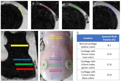 |
Quantification of Synovial Fluid using Magnetic Resonance Fingerprinting Multicomponent Imaging in Articular Cartilage of Knee
Seung Eun Lee1, Joon-Yong Jung1, and Dongyeob Han2
1Seoul St. Mary’s Hospital, Seoul, Korea, Republic of, 2Siemens Healthineers, Seoul, Korea, Republic of
The morphologic MR imaging is limited in identifying sub-voxel sized cartilage defect due to partial volume averaging. We assessed the feasibility of synovial fluid fraction (SFF) map generated by multicomponent approach using MRF-derived relaxation maps to characterize sub-voxel sized cartilage defect. In ex vivo experiment, we proved that SFF map can quantify synovial fluid fraction in sub-voxel sized cartilage defects. In clinical study, we demonstrated that SFF map can complement morphologic imaging in cartilage segmentation and volumetric assessment.
|
||
0521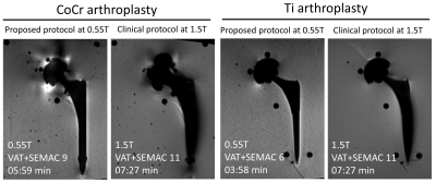 |
Advanced Low-Field MRI of Hip Arthroplasty Implants: First Experience at 0.55 T Video Permission Withheld
Iman Khodarahmi1, Inge Manuela Brinkmann2, Dana Lin1, Mary Bruno1, Patricia Johnson1, Florian Knoll1, Mahesh Bharath Keerthivasan2, Hersh Chandarana1, and Jan Fritz1
1Department of Radiology, New York University School of Medicine, New York, NY, United States, 2Siemens Medical Solutions USA Inc., Malvern, PA, United States
Next-generation, advanced low-field MRI holds promise to improve metal artifact reduction MRI of hip arthroplasty implants due to inherently lower susceptibility effects. Using titanium-on-ceramic and metal-on-metal cobalt-chromium total hip arthroplasty implant phantoms, we compared the degree of metal artifacts and signal-to-noise ratios of MR images obtained with modified 0.55T prototype and clinical 1.5T MRI systems. The 0.55T SEMAC MR images with 6-9 encoding steps invariably demonstrated superior, near-complete metal artifact reduction. Our preliminary results suggest clinically viable sequence acquisition times of ≤ 6-min with advanced 0.55T MRI.
|
||
0522.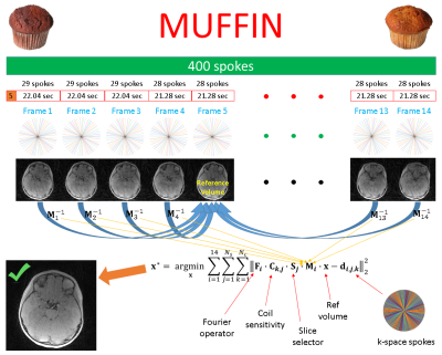 |
Skull MRI with MUFFIN: MUlti-Frame Forward-modeled Image Numismatics
Cihat Eldeniz1, Udayabhanu Jammalamadaka1, Gary B. Skolnick2, Paul K. Commean1, Kamlesh B. Patel2, and Hongyu An1
1Mallinckrodt Institute of Radiology, Washington University in St. Louis, St. Louis, MO, United States, 2Division of Plastic and Reconstructive Surgery, Washington University in St. Louis, St. Louis, MO, United States
Computed tomography (CT) is the reference method for skull imaging, but can cause cancer due to ionizing radiation. Magnetic resonance imaging (MRI) is safer, but the prolonged scan time increases the chance of motion, especially for pediatric patients. Sedation helps reduce motion significantly, but is associated with risks. A sedation-free MRI scheme that is robust to motion is therefore highly desirable. Here, we proposed such a method by making use of a radial acquisition scheme that is inherently robust to motion. The robustness was further boosted by a forward-modeled motion-corrected reconstruction. The results show the promise of the method.
|
||
0523.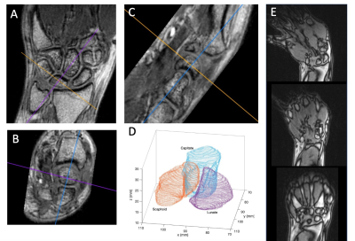 |
Assessing the Viability of Carpal Bone Kinematic Profiles Extracted from 4D MRI
Kevin Matthew Koch1, Mohammad Zarenia2, V. Emre Arpinar2, L Tugan Muftuler3, Alyssa Joy Schnorenberg 4, Joshua Leonardis4, Brooke Slavens4, and Andrew S Nencka2
1Medical College of Wisconsin, Milwaukee, WI, United States, 2Radiology, Medical College of Wisconsin, Milwaukee, WI, United States, 3Neurosurgery, Medical College of Wisconsin, Milwaukee, WI, United States, 4University of Wisconsin, Milwaukee, Milwaukee, WI, United States
A unique approach to analysis of wrist mechanics is analyzed. In this approach, 4D dynamic MRI is utilized to track unconstrained movement of individual wrist carpal bones. Through a boundary-based slab-to-volume registration approach, fiducial points identified or computed on high resolution static images are tracked using the dynamic time series to generate time-domain signals indicative of independent and relative carpal bone movement. After rudimentary processing of 12 derived signals, each computed on 3 asymptomatic control subjects, correlation analysis is utilized to elucidate the utility of these metrics in establishing a normative kinematic profile of the healthy wrist.
|
||
0524.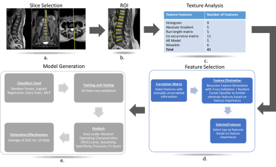 |
2D Texture Analysis based approach for detection of Osteoporosis on 1.5T on T1-weighted MR images
Preety Krishnan1, Tejas J Shah2, Akshay Godkhindi2, Rupsa Bhattacharjee3, Stanley Kovil Pichai3, Ajay Krishnan1, Bharat Dave1, and Indrajit Saha3
1Stavya Spine Research Institute, Ahmedabad, India, 2MR, Philips Innovation Campus, Bangalore, India, 3Philips India Limited, Gurgaon, India
Given the prevalence and disease burden of osteoporosis, it is critical to detect it as early as possible. This is challenging not only because the disease is typically asymptomatic but also due to known limitations of the gold standard method of DEXA. The aim of this study was to determine if alternative approach of 2D texture analysis in L1-L5 lumbar spine on T1W images can be used to detect osteoporosis. It is demonstrated that such an approach can indeed be used to clinically detect osteoporosis with an AUC of 0.8.
|
||
0525.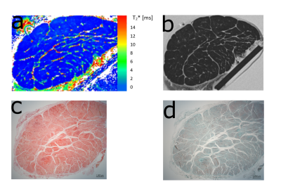 |
Transverse Relaxation Anisotropy of Tendons Studied by MR Microscopy
Benedikt Hager1,2,3, Markus M. Schreiner4, Sonja M. Walzer4, Lena Hirtler5, Vladimir Mlynarik1, Martin Zalaudek1, Andreas Berg6, Xeni Deligianni7,8, Oliver Bieri7,8, Reinhard Windhager4, Vladimir Juras1, and Siegfried Trattnig1
1Department of Biomedical Imaging and Image-guided Therapy, High Field MR Centre, Medical University of Vienna, Vienna, Austria, 2CD Laboratory for Clinical Molecular MR Imaging, Vienna, Austria, 3Austrian Cluster for Tissue Regeneration, Ludwig Boltzmann Institute for Experimental and Clinical Traumatology, Vienna, Austria, 4Department of Orthopedics and Trauma-Surgery, Medical University of Vienna, Vienna, Austria, 5Center for Anatomy and Cell Biology, Division of Anatomy, Medical University of Vienna, Vienna, Austria, 6Center for Medical Physics and Biomedical Engineering, Medical University of Vienna, Vienna, Austria, 7Division of Radiological Physics, Department of Radiology, University of Basel Hospital, Basel, Switzerland, 8Department of Biomedical Engineering, University of Basel, Allschwil, Switzerland
In this study, we analyzed the T2* anisotropy and mono- vs. bi-exponentiality of T2* decay of Achilles and patellar tendons in vitro with a variable echo time sequence, ultrashort echo times and microscopic resolution and compared the results with histological findings. A total of four human Achilles tendons and four patellar tendons were measured at their maximum and minimum dipolar interaction (0°, 55°). In addition, one Achilles tendon and one patellar tendon were measured at 11 fiber-to-field angles (0,10,20,30,40,50,60,70,80,90°) each in order to study the change in T2* values at these angles.
|
||
 |
0526.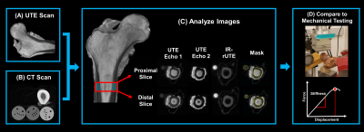 |
Measures of bone water and porosity are associated with whole-bone stiffness and mineral density in the human femur
Brandon Clinton Jones1,2, Hyunyeol Lee1, Shaowei Jia1,3, Anna Feng1, Snehal S Shetye4, Hee Kwon Song1, Felix Werner Wehrli1, and Chamith Sudesh Rajapakse1,4
1Radiology, University of Pennsylvania, Philadelphia, PA, United States, 2Bioengineering, University of Pennsylvania, Philadelphia, PA, United States, 3Biomedical Science and Medical Engineering, Beihang University, Beijing, China, 4Orthopaedic Surgery, University of Pennsylvania, Philadelphia, PA, United States
UTE measures of cortical bone water were evaluated in 15 cadaveric proximal femora. Pore water content, total water content, and porosity index were all negatively associated with whole-bone stiffness obtained in a sideways fall loading configuration and with volumetric bone mineral density. In contrast, bound water content was not found to be related to stiffness or mineral density. This data suggest that bone water measures may provide useful information on cortical bone mechanical competence.
|
|
0527.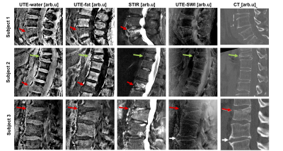 |
Simultaneous assessment of vertebral fractures and edema of the thoracolumbar spine on water-fat and SW images derived from a single-TE UTE scan
Sophia Kronthaler1, Christof Boehm1, Peter Börnert2, Ulrich Katscher2, Kilian Weiss3, Marcus R. Makowski1, Benedikt J. Schwaiger1, Alexandra S. Gersing1, and Dimitrios C. Karampinos1
1Department of Diagnostic and Interventional Radiology, School of Medicine, Technical University of Munich, Munich, Germany, 2Philips Research Laboratory, Hamburg, Germany, 3Philips Healthcare, Hamburg, Germany
CT and MR imaging are often both performed in patients with vertebral fractures or degenerative changes, with CT aiming at the characterization of osseous changes and the MR focusing on bone marrow edema. CT is associated with radiation exposure and therefore it is desirable to assess both soft-tissue and osseous components in one MR examination. The present work develops a methodology for simultaneously extracting susceptibility weighted imaging (SWI) and single point Dixon imaging based on a single-TE ultrashort echo time (UTE) scan to simultaneously assess vertebral fractures and degenerative bone changes in the thoracolumbar spine with a single MR sequence.
|
||
0528.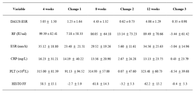 |
Quantification of bone marrow edema in RA by using high-speed T2-corrected multiecho acquisition of 1H magnetic resonance spectroscopy
Wenzhao Yuan1, Yiwu Lei1, Cheng Tang1, Fang Qin1, Jing Wen1, Chenhui Li2, Min Ling1, Jiang Huang1, Huiting Zhang3, and Liling Long1
1The First Affiliated Hospital of Guangxi Medical University, Nanning, China, 2Siemens Healthcare Ltd., Guangzhou, China, 3Siemens Healthcare Ltd., Wuhan, China The purpose of this study was to determine whether high-speed T2-corrected multiecho (HISTO) sequences can quantify bone marrow edema (BME) in the capitate bone in rheumatoid arthritis (RA), and whether the HISTO fat fraction (FF) reflects therapeutic effectiveness. The results showed that the HISTO sequence can measure the bone marrow FF of the wrist joint in RA patients. We also found that the HISTO sequence may help quantify BME in RA and help monitor the effectiveness of RA treatment. |
The International Society for Magnetic Resonance in Medicine is accredited by the Accreditation Council for Continuing Medical Education to provide continuing medical education for physicians.