Oral
fMRI: Applications with Clinical Relevance & Beyond
ISMRM & SMRT Annual Meeting • 15-20 May 2021

Oral
fMRI: Applications with Clinical Relevance & Beyond
| Concurrent 5 | 16:00 - 18:00 | Moderators: Baxter Rogers & Yu-Feng Zang |
0155.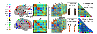 |
Functional network degeneration is associated with blood neurofilament light and cognitive decline in autosomal dominant Alzheimer disease
Muriah D Wheelock1, Patricia Mansfield2, Jeremy F Strain1, Beau M Ances1, Oliver Preische3, John C Morris1, Randall J Bateman1, Mathias Jucker3, Tammie L.S. Benzinger1, Adam T Eggebrecht1, and Brian A Gordon1
1Washington University in St. Louis, St. Louis, MO, United States, 2St. Louis University, St. Louis, MO, United States, 3University of Tubingen, Tubingen, Germany
Research suggests that serum neurofilament light (NfL), an indirect measure of neuronal cell death, is associated with volumetric and white matter changes, and is predictive of cognitive decline in Alzheimer disease (AD). We report that NfL is associated with default mode network (DMN) functional connectivity as well as DMN connectivity with control networks. DMN connectivity with control networks is additionally associated with concurrent cognition. Hierarchical regression demonstrates NfL, DMN, and Aβ-amyloid each contribute to predicting cognition. These findings suggest NfL is an indirect marker of functional network degeneration and both NfL and DMN connectivity are distinct biomarkers of AD progression.
|
||
 |
0156.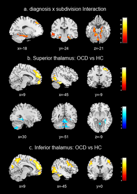 |
Mapping functional connectivity of thalamus subdivisions in obsessive-compulsive disorder
Lingxiao Cao1, Hailong Li1, Jing Liu1, Xue Li2, Suming Zhang1, Xinyu Hu1, Qiyong Gong1, and Xiaoqi Huang1
1Huaxi MR Research Center (HMRRC), Functional and molecular imaging Key Laboratory of Sichuan Province, Department of Radiology, West China Hospital, Sichuan University, Chengdu, China, 2Sichuan University, Chengdu, China
Using connectivity-based parcellation technique, we segmented thalamus into two distinct subdivisions comprising superior thalamus and inferior thalamus based on their similarity in resting-state functional connectivity properties. We then compared the functional connectivity profiles of each thalamic subdivisions between the obsessive-compulsive disorder (OCD) patients and healthy control (HC), and revealed the disturbances of the superior/inferior thalamo-cortical and inferior thalamo-cerebellar circuitry in OCD patients. These findings suggested that thalamus subdivisions play different role in motor, cognitive, affective processes in OCD, which may underlie the pathophysiology of the disorder.
|
|
0157.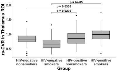 |
Abnormal cerebrovascular reactivity in human immunodeficiency virus-infected patients with or without smoking: a resting-state fMRI study
Lincoln Kartchner1, Linda Chang2, Thomas Ernst2, Huajun Liang2, Yuangi Shang2, and Peiying Liu1
1Department of Radiology, Johns Hopkins University School of Medicine, Baltimore, MD, United States, 2Department of Diagnostic Radiology and Nuclear Medicine, University of Maryland School of Medicine, Baltimore, MD, United States
Human immunodeficiency virus (HIV) infection is associated with neurodegeneration, but its effect on cerebrovascular function is not well understood. In a cohort of 160 participants, we investigated the effects of HIV and smoking on cerebrovascular reactivity (CVR) measured with standard resting-state blood oxygenation level dependent (BOLD)-fMRI. Across four participant groups (HIV+/HIV- x smokers/nonsmokers), both HIV-infection and smoking status altered CVR, but their effects were different across brain regions. Furthermore, lower nadir CD4 predicted lower thalamus CVR.
|
||
0158.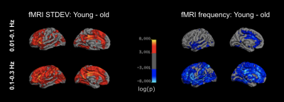 |
Resting-State fMRI Frequency Change During Brain Aging
Xiaole Zhong1 and J. Jean Chen1,2
1Rotman Research Institute, Baycrest Health Sciences, Toronto, ON, Canada, 2Department of Medical Biophysics, University of Toronto, Toronto, ON, Canada
Frequency features of the resting-state functional magnetic resonance imaging (rs-fMRI) signal can be crucial metrics that reveal patterns of brain aging. However, how the frequency shift during brain aging is still unclear. In this study, we examined the peak frequency and standard deviation of the rs-fMRI signal in healthy aging adults, divided into (0-0.1 Hz) and (0.1-0.3 Hz) bands. We found that in older adults, rs-fMRI fluctuation amplitude is lower but fluctuation frequency is higher in older adults, and these effects depend on the fMRI frequency range.
|
||
0159.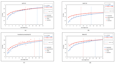 |
The effect of scan length on reliability of resting-state fMRI in patients with drug-resistant epilepsy (DRE) in awake and under anesthesia
Faezeh Vedaei1,2, Mahdi Alizadeh1, Sara Thalheimer1, Victor Romo3, Feroze Mohamed4, and Chengyuan Wu1
1Department of Neurosurgery, Thomas Jefferson University, Philadelphia, PA, United States, 2Department of Bioengineering, Temple University, Philadelphia, PA, United States, 3Department of Anesthesiology, Thomas Jefferson University, Philadelphia, PA, United States, 4Department of Radiology, Thomas Jefferson University, Philadelphia, PA, United States
Resting-state fMRI suffers from poor test-retest reliability because of between-subject and within-subject variability. Scan duration is one the main factors affects the reliability of rs-fMRI studies. We showed that under anesthesia, the time needed to optimize ICC of rs-fMRI metrics including ALFF, fALFF, functional connectivity, and ReHo is lower compare with awake state. The optimum scan duration that satisfies good reliability is 14-20 min and 8-17 min in awake and under anesthesia, respectively. Also, variability of ICCs is lower under anesthesia than in awake.
|
||
0160.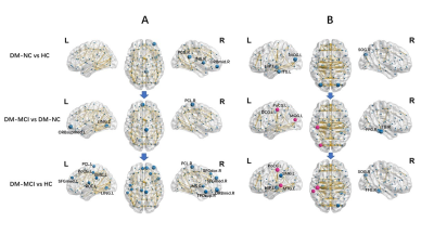 |
Disrupted Topological Organization of Structural and Functional Brain Connectomes in Type-2 Diabetes Patients
Ying Xiong1, Qiang Zhang2, and Wenzhen Zhu1
1Radiology, Tongji Hospital Tongji Medical College Huazhong University of Science and Technology, Wuhan, China, 2Neurology, Tongji Hospital Tongji Medical College Huazhong University of Science and Technology, Wuhan, China
The T2DM patients with cognitive impairment showed altered global efficiency(Eg), local efficiency(Eloc), clustering-coefficient(Cp), shortest-path-length(Lp) as well as nodal efficiencies in both structural and functional networks, compared to those with normal cognition and healthy controls. Some network metrics were correlated with neuropsychological assessments and disease severity. The disrupted topological organization of structural and functional connectomes (measured by Eg, Eloc, Cp and Lp) were found in T2DM with cognitive impairment, while these topological properties in T2DM with normal cognition were preserved equally to controls. The structural and functional connectomes research shows potential feasibility in characterizing intrinsic alterations of diabetic encephalopathy.
|
||
0161. |
Neuroimaging and obesity in adults with pre-diabetes or diabetes: results from the UK Biobank
Christopher R. Kouyoumdjian1, Kayley Marchena2,3, Masud Hussain3, and Bradley J. MacIntosh2,3
1University of Toronto, Scarborough, ON, Canada, 2Department of Medical Biophysics, University of Toronto, Toronto, ON, Canada, 3Sunnybrook Research Institute, Toronto, ON, Canada
Glycated haemoglobin and body mass index (BMI) are implicated in neurodegeneration and diabetes. Diabetes-related changes in T2* estimates and fractional amplitude of low frequency fluctuations (fALFFs) are less understood. We investigated these neuroimaging estimates within the thalamic nuclei, hippocampi, and caudate nuclei of the UK Biobank-derived pre-diabetes and diabetes adult groups. Multiple regression analyses elucidated BMI as the robust predictor of hippocampal T2* estimates in both subgroups. These metrics extracted from two pulse sequences were helpful in understanding the brain and BMI associations, which is important in the assessment of diabetes and pre-diabetes as a risk factor for neurodegenerative disorders.
|
||
0162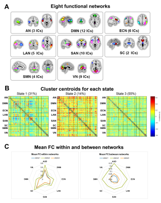 |
Altered brain functional network dynamics in obsessive-compulsive disorder Video Permission Withheld
Lekai Luo1, Qian Li1, Wanfang You1, Yuxia Wang1, Yanchun Yang2, Qiyong Gong1, and Fei Li1
1Huaxi MR Research Centre (HMRRC), Department of Radiology, West China Hospital of Sichuan University, Chengdu, China, 2Psychiatry, West China Hospital of Sichuan University, Chengdu, China
Dynamic functional connectivity (dFC) and dynamic topological analyses were used to investigate the whole brain resting-state dynamic property abnormalities in patients with obsessive-compulsive disorder (OCD). Our results provide evidence of clinically relevant aberrant dynamic brain activity in OCD. Increased functional segregation among networks and impaired functional flexibility in connections among brain regions in default mode network (DMN) and salience network (SAN) may play important roles in the neuropathology of OCD.
|
||
0163.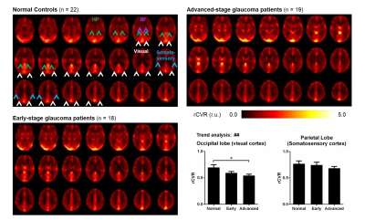 |
Cerebrovascular reactivity changes in glaucoma patients using resting-state fMRI
Russell W. Chan1,2, Ji Won Bang2, Vivek Trivedi2, Peiying Liu3, Gadi Wollstein2, Joel S. Schuman2, and Kevin C. Chan1,2,4
1Neuroscience Institue, New York University Grossman School of Medicine, New York, NY, United States, 2Department of Ophthalmology, New York University Grossman School of Medicine, New York, NY, United States, 3Department of Radiology, Johns Hopkins University School of Medicine, Baltimore, MD, United States, 4Department of Radiology, New York University Grossman School of Medicine, New York, NY, United States
Cerebrovascular reactivity (CVR) is the response of cerebral blood vessels to vasoactive stimuli. Dampened CVR can precede and contribute to neuropathology. However, CVR assessments in glaucoma patients have been lacking at the whole-brain scale. Here, we applied relative CVR (rCVR) mapping using resting-state fMRI to investigate vascular reserve changes in glaucoma patients. Our results show that visual cortical rCVR decreases with severity and is coupled with clinical ophthalmic assessments. Interestingly, rCVR in both basal forebrain and hippocampus increase with severity indicating their involvements in glaucoma. Together, resting-state fMRI derived rCVR can potentially be used for studying, diagnosing and monitoring glaucoma.
|
||
0164.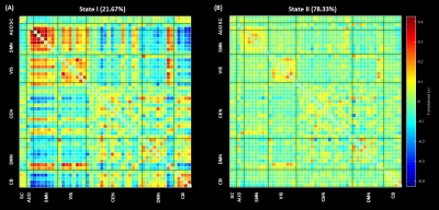 |
Altered dynamic functional connectivity in subjects with cerebral glioma
Siqi Cai1,2, Zhifeng Shi3, Yuchao Liang4, Chunxiang Jiang1,2, Shihui Zhou1,2, and Lijuan Zhang*1
1Shenzhen Institutes of Advanced Technology, Chinese Academy of Sciences, Shenzhen, China, 2University of Chinese Academy of Sciences, Beijing, China, 3Huashan Hospital of Fudan University, Shanghai, China, 4Neurosurgery, Beijing Tiantan Hospital of Capital Medical University, Beijing, China
This study investigated the impact of cerebral glioma on the dynamic properties of functional connectivity (dFC). Cerebral gliomas induce alteration in dFC featuring more frequent strengthened connectivity and state transition between strong and sparse functional connectivity, indicating an extensive disturbance of functional segregation. As alterations in the dFC were reported to be associated with the progression of brain diseases, dFC variation induced by glioma may entail additional information to interpret the clinical profiles that are not ascribed to the lesion topology and serve as a new biomarker for the tumor characterization of glioma.
|
The International Society for Magnetic Resonance in Medicine is accredited by the Accreditation Council for Continuing Medical Education to provide continuing medical education for physicians.