Oral
Imaging Metabolites: CEST, MT & MRS
ISMRM & SMRT Annual Meeting • 15-20 May 2021

| Concurrent 6 | 12:00 - 14:00 | Moderators: Puneet Bagga & Elena Vinogradov |
0715.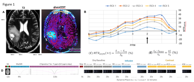 |
Dynamic GlucoCEST MRI: results in primary brain tumors at 3 Tesla
REGIS OTAVIANO FRANCA BEZERRA1, Hae Won Lee2, Gustavo Kaneblai3, Eduardo Figueiredo3, Mitsuharu Miyoshi4, Thomas Doring3, Claudia da Costa Leite5, Giovanni Guido Cerri2, and Frederico Perego Costa6
1RADIOLOGY, HOSPITAL SÍRIO-LIBANÊS, SAO PAULO, Brazil, 2Radiology, Hospital Sírio-Libanês, Sao Paulo, Brazil, 3General Eletric, Sao Paulo, Brazil, 4General Eletric, Tokyo, Japan, 5RADIOLOGIA, HOSPITAL SÍRIO-LIBANÊS, SAO PAULO, Brazil, 6Oncology, Hospital Sírio-Libanês, Sao Paulo, Brazil
This study shows the glucoCEST effect in 15 patients with primary malignant brain tumors compared with normal white matter(NWM) at 3-Tesla-MRI. The procedure included a full Z-spectra CEST and a dynamic glucoCEST. The signal % at 2 ppm calculated using mean asymmetric magnetization transfer ratio values were significantly higher in the whole cancer compared to NWM. Similarly, the dynamic glucoCEST presented as mean area-under-the-curve values were significantly different in whole cancer in comparison to normal tissue. This method, using clinical field strength of 3Tesla, was capable of identifying distinct patterns of glucose metabolismand also heterogeneity in the cancer metabolism
|
||
0716.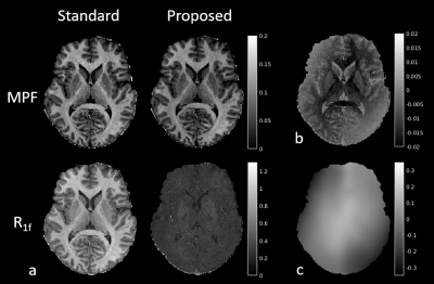 |
Confounding of Macromolecular and Paramagnetic Tissue Content in Quantitative MTI Remedied by Explicit Estimation of Bound Pool Relaxation
Alexey Samsonov1 and Aaron S. Field1
1Radiology, University of Wisconsin-Madison, Madison, WI, United States We study the effect of macromolecular proton fraction (MPF) and R1 interdependency in quantitative MT experiments. We hypothesize that the two-pool model with properly calibrated relaxation constraints on the bound proton pool can separate interdependency of both metrics, potentially improving the specificity of both, specifically, MPF to macromolecular content and R1 to paramagnetic ions. The simulation and in vivo results support feasibility of such refinement. |
||
0717.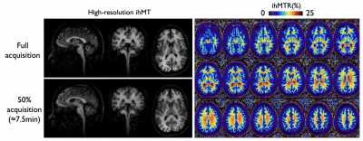 |
Improved volumetric inhomogeneous magnetization transfer (ihMT) using a CSF-suppressed FSE sequence (FLAIR-ihMT)
Manuel Taso1, Fanny Munsch1, Olivier M Girard2, Guillaume Duhamel2, David C Alsop1, and Gopal Varma1
1Division of MRI research, Department of Radiology, Beth Israel Deaconess Medical Center, Harvard Medical School, Boston, MA, United States, 2CRMBM, Aix-Marseille Univ, CNRS, Marseille, France 3D myelin imaging using inhomogeneous magnetization transfer (ihMT) has seen increased interest as sequence implementations evolve. This has mostly been based on prepared or pseudo-steady-state gradient echo sequences. However, Fast-Spin-Echo (FSE) sequences should have a theoretical SNR advantage over GRE. Here we report implementation of a CSF-suppressed FSE sequence for ihMT imaging (FLAIR-ihMT), exploring through simulations the effect of CSF suppression and providing first imaging results, highlighting the benefits of CSF suppression, potential for whole brain high-resolution imaging and translation down to the spinal cord. |
||
 |
0718. |
Inhomogeneous magnetization transfer in the healthy adult brain: reproducibility and correlation with MTR and myelin water imaging
Sarah Rosemary Morris1,2,3, Irene M. Vavasour1,4, Anastasia Smolina5,6, Erin MacMillan4,7, Guillaume Gilbert7, Michelle Lam2,4, Piotr Kozlowski1,2,4,8, Carl Michal2, Alan Manning2, Alex L. MacKay1,2,4, and Cornelia Laule1,2,4,8,9
1Radiology, University of British Columbia, Vancouver, BC, Canada, 2Physics & Astronomy, University of British Columbia, Vancouver, BC, Canada, 3International Collaboration on Repair Discoveries, Vancouver, BC, Canada, 4UBC MRI Research Centre, Vancouver, BC, Canada, 5Physics & Astronomy, McMaster University, Hamilton, ON, Canada, 6Hospital for Sick Children, Toronto, ON, Canada, 7MR Clinical Science, Philips Healthcare Canada, Markham, ON, Canada, 8International Collaboration on Repair Discoveries (ICORD), Vancouver, BC, Canada, 9Pathology & Laboratory Medicine, University of British Columbia, Vancouver, BC, Canada
We compared three myelin-sensitive MRI metrics (inhomogeneous magnetization transfer ratio (ihMTR), magnetization transfer ratio (MTR) and gradient and spin echo-derived myelin water fraction (MWF)) in 15 white matter regions of interest from 14 healthy adults at 3T. Reproducibility of ihMTR was also investigated. We found a moderately strong correlation between MWF and ihMTR but only a very weak correlation between MWF and MTR. Myelination rankings were similar for ihMTR and MWF but had no relation to MTR. Finally, we established an average ihMTR scan-rescan variability of 8.2% of the mean in each ROI.
|
|
0719.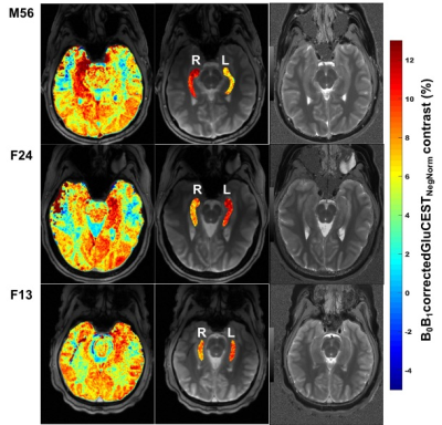 |
GluCEST as an in vivo biomarker for monitoring abnormal glutamate dehydrogenase activity in Hyperinsulinism/Hyperammonemia syndrome at 7.0T
Ravi Prakash Reddy Nanga1, Elizabeth A Rosenfeld2, Deepa Thakuri1, Mark Elliott1, Ravinder Reddy1, and Diva D De Leon2
1Radiology, University of Pennsylvania, Philadelphia, PA, United States, 2Endocrinology and Diabetes, Children’s Hospital of Philadelphia, Philadelphia, PA, United States
Hyperinsulinism/Hyperammonemia (HI/HA) syndrome is an orphan disease characterized by fasting and protein-induced hypoglycemia, hyperammonemia, and has high prevalence of epilepsy, developmental delays, and learning disabilities. Understanding the mechanism involved in brain phenotype remains limited. We have previously shown the application of glutamate weighted chemical exchange saturation transfer (GluCEST) imaging in small set of subjects to spatially map the glutamate levels of hippocampus. In this study we have expanded the human subject pool for studying the abnormal function of glutamate dehydrogenase (GDH) enzyme activity with HI/HA syndrome.
|
||
0720.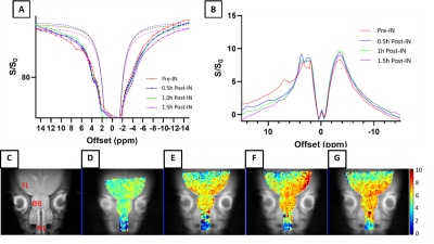 |
CEST Imaging of Nose-to-Brain Drug Delivery using Iohexol liposomes at 3T
Lok Hin LAW1, Peng XIAO1, Jianpan Huang1, Xiongqi HAN1, and Kannie WY CHAN1,2,3
1Department of Biomedical Engineering, City University of Hong Kong, Hong Kong, Hong Kong, 2Russell H. Morgan Department of Radiology and Radiological Science, Johns Hopkins University School of Medicine, Baltimore, MD, United States, 3City University of Hong Kong Shenzhen Research Institute, Shenzhen, China
Imaging-guided nose-to-brain drug delivery provide a non-invasive monitoring of drug delivery to brain, which increases effective dose via bypassing Blood-Brain-Barrier(BBB). Here, we investigated imaging of nanomedicine delivery via intranasal-administration using CEST-detectable mucus-penetrating-liposome(with 10%PEG). Liposomes were loaded with Iohexol(Ioh-Lipo) and CEST properties were examined both in-vitro and in-vivo by injecting into mouse nostril. Ioh-Lipo generated CEST contrast of 33.4% at 4.3 ppm in-vitro. which was also observed in nostril, olfactory-bulb and frontal-lobe after intranasal-administration at 3T. We demonstrated the liposomes detectability both in nostril and olfactory-bulb by CEST. The result demonstrates an approach for imaging-guided Nose-to-Brain Intranasal Liposomal Drug Delivery.
|
||
0721.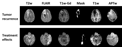 |
Peritumoral radiomics features from amide proton transfer-weighted MRI unveil the progressive pattern in early recurrent malignant gliomas
Shanshan Jiang1, Pengfei Guo2, Hye Young Heo1, Peter van Zijl1,3, and Jinyuan Zhou1
1Department of Radiology, Johns Hopkins University, Baltimore, MD, United States, 2Whiting School of Engineering, Johns Hopkins University, Baltimore, MD, United States, 3F.M. Kirby Research Center for Functional Brain Imaging, Kennedy Krieger Institute, Baltimore, MD, United States
Assessing post-treatment malignant gliomas early has remained one of the most critical dilemmas in neuro-oncology for three decades. Amide protein transfer weighted (APTw) MRI has been validated to accurately detect recurrent malignant gliomas in more and more studies. The peritumoral area, as the one of the most aggressive regions, has been seldom studied. Here, we explore radiomics features extracted from peritumoral areas on APTw images to unveil the progressive pattern in early recurrent malignant gliomas. Our results suggest that the use of APTw radiomic features can add important value to structural MRI to assess the treatment response.
|
||
 |
0722.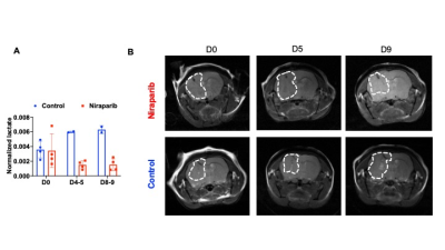 |
Deuterium metabolic imaging of tumor burden and response to therapy in mutant IDH gliomas in vivo
Celine Taglang1, Georgios Batsios1, Mers Tran1, Anne Marie Gillespie1, Hema Artee Luchman2, Russell O Pieper3, Sabrina M Ronen1, and Pavithra Viswanath1
1Radiology and Biomedical Imaging, University of California San Francisco, San Francisco, CA, United States, 2Cell Biology and Anatomy, University of Calgary and Hotchkiss Brain Institute, Calgary, AB, Canada, 3Neurological Surgery, University of California San Francisco, San Francisco, CA, United States
2H-magnetic resonance spectroscopy (MRS) recently emerged as a novel, non-invasive method of monitoring metabolic fluxes in high-grade glioblastomas in vivo. However, its utility for imaging low-grade gliomas and for assessing treatment response has not been examined. Here, we show that [6,6’-2H]-glucose metabolism to lactate serves to delineate tumor from contralateral normal brain in mice bearing orthotopic patient-derived low-grade glioma xenografts. Importantly, reduced lactate production from [6,6’-2H]-glucose informs on early response to therapy, at timepoints when volumetric alterations cannot be detected by anatomical imaging, pointing to the ability of [6,6’-2H]-glucose to assess pseudoprogression, which is a major challenge in glioma imaging.
|
|
0723.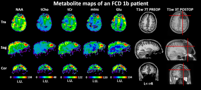 |
CRT-FID-MRSI at 7T for the high-resolution metabolic imaging of epilepsy: Preliminary results
Gilbert Hangel1, Philipp Lazen2, Matthias Tomschik1, Jonathan Wais1, Eva Hečková2, Lukas Hingerl2, Stephan Gruber2, Bernhard Strasser2, Gregor Kasprian3, Daniela Prayer3, Julia Furtner3, Christoph Baumgartner4, Johannes Koren4,
Robert Diehm5, Martha Feucht5, Christian Dorfer1, Ekaterina Pataraia6, Wolfgang Bogner2, Siegfried Trattnig2,7, and Karl Rössler1
1Department of Neurosurgery, Medical University of Vienna, Vienna, Austria, 2High Field MR Centre, Department of Biomedical Imaging and Image-guided Therapy, Medical University of Vienna, Vienna, Austria, 3Division of Neuroradiology and Musculoskeletal Radiology, Department of Biomedical Imaging and Image-guided Therapy, Medical University of Vienna, Vienna, Austria, 4Department of Neurology, Clinic Hietzing, Vienna, Austria, 5Department of Paediatrics and Adolescent Medicine, Medical University of Vienna, Vienna, Austria, 6Department of Neurology, Medical University of Vienna, Vienna, Austria, 7Christian Doppler Laboratory for Clinical Molecular MR Imaging, Vienna, Austria
We successfully implemented a fast high-resolution 3D-MRSI protocol covering the whole brain at 7T in a preliminary study of 14 patients with pharmacoresistant epilepsy. With an isotropic resolution of 3.4 mm acquired in 15:30 min, we detected focal metabolic alterations in thirteen patients. From all metabolites, tCr, Glu, mIns and NAA and especially tCho appear as the most relevant markers for detection of focal metabolic alterations in epilepsy. Especially concerning cortical developmental alterations as a cause of epilepsy, our findings may have the potential to differentiate metabolic fingerprints for FCD subclasses.
|
||
0724.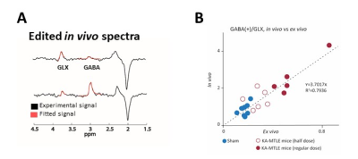 |
In vivo GABA increase as a biomarker of the epileptogenic zone: an unbiased metabolomics approach
Florence Fauvelle1,2, Vasile Stupar1,2, Jia Guo3, Wafae Labriji1, Chen Liu3, Alicia Plaindoux1, Emmanuel Luc Barbier1,2, Sophie Hamelin1, and Antoine Depaulis1
1Grenoble Institut Neurosciences, University Grenoble Alpes, La Tronche, France, 2IRMaGE, University Grenoble Alpes, La Tronche, France, 3Departement of Psychiatry, Columbia University, New York, NY, United States
New non invasive methods are required to better delimit the epileptogenic zone (EZ) during the pre-surgical exam of epileptic patients. By combining ex vivo NMR spectroscopy-based (MRS) untargeted metabolomics and in vivo MRS-based targeted metabolomics, we found that GABA was the most discriminant metabolite of the epileptogenic zone vs adjacent brain regions in a mouse model of mesio-temporal lobe epilepsy (MTLE). GABA appears therefore as a specific in vivo biomarker of EZ in MTLE.
|
The International Society for Magnetic Resonance in Medicine is accredited by the Accreditation Council for Continuing Medical Education to provide continuing medical education for physicians.