Oral
Hot Topics in Preclinical Models of CNS Disease
ISMRM & SMRT Annual Meeting • 15-20 May 2021

Oral
Hot Topics in Preclinical Models of CNS Disease
| Concurrent 6 | 18:00 - 20:00 | Moderators: Sanam Assili & Po-Wah So |
0427.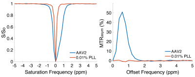 |
Viral-CEST: Exploiting AAV capsids as endogenous CEST agents for tracking of viral cell transduction
Mark Velasquez1, Laurel Nelson1, Bonnie Lam1, Kevin Godines1, Soo Hyun Shin1, and Moriel Vandsburger1
1Department of Bioengineering, University of California, Berkeley, Berkeley, CA, United States
Viral vectors, including adeno-associated viruses (AAV), are increasingly used for therapies that utilize somatic cell gene editing. Validation of successful delivery of gene editing machinery requires biopsy, which is a fundamental barrier to development of gene therapy. Research into chemical exchange saturation transfer (CEST)-MRI contrast agents has yielded numerous targeted and pH sensitive exogenous agents for diagnostic purposes. In this study we demonstrated that CEST-MRI can detect the abundant serine, threonine, and lysine residues on AAV capsid surfaces across serotypes that are responsible for targeted receptor binding; further that transduction of cells by AAV2 can be detected by CEST.
|
||
 |
0428.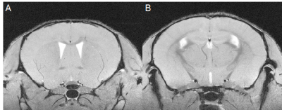 |
Grey matter atrophy measured in-vivo with 9.4T MRI in the cuprizone mouse model of demyelination
A. Max Hamilton1,2,3,4, Qandeel Shafqat1,2,3,4, Nils D. Forkert1,2,3, Ying Wu1,2,3,4, and Jeff F. Dunn1,2,3,4
1Department of Radiology, University of Calgary, Calgary, AB, Canada, 2Hotchkiss Brain Institute, University of Calgary, Calgary, AB, Canada, 3Department of Clinical Neurosciences, University of Calgary, Calgary, AB, Canada, 4Experimental Imaging Center Cumming School of Medicine, University of Calgary, Calgary, AB, Canada
Grey matter atrophy is a marker of progressive disability in multiple sclerosis (MS). To better study atrophy in MS, mouse models that display grey matter loss are needed. A possible candidate is the cuprizone mouse model, which exhibits demyelination, gliosis, and axonal injury. We used high-resolution MRI (37.5x37.5x250μm3) and atlas-based volumetrics to measure volumes of 62 structures in the brains of cuprizone mice following acute (6-weeks) and chronic (12-weeks) demyelination. We found no atrophy associated with acute demyelination but identified atrophy in 7 regions following chronic demyelination including the corpus callosum, internal capsule, striatum, and thalamus.
|
|
|
0429.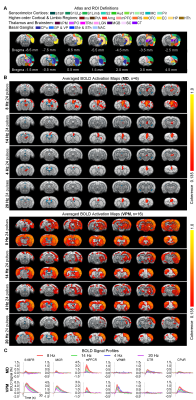 |
Optogenetic fMRI Reveals Distinct Response Characteristics of Sensory and Limbic Thalamic Spindle-like Activities
Xunda Wang1,2, Alex T. L. Leong1,2, and Ed X. Wu1,2
1Laboratory of Biomedical Imaging and Signal Processing, The University of Hong Kong, Hong Kong SAR, China, 2Department of Electrical and Electronic Engineering, The University of Hong Kong, Hong Kong SAR, China
Spindle-like activities constitute one of the most critical brain-wide oscillatory activities for memory consolidation. Spindle-like activities with different temporal characteristics have been associated with heterogeneous distribution patterns. Studies postulated that such heterogeneous distribution and differences in temporal characteristics of spindle-like activities were determined by differences in corresponding spindle-generation thalamo-cortical circuits. However, no direct evidence has been shown. In this study, we demonstrate distinct brain-wide targets but similar temporal-characteristics dependent cross-modal recruitment property of limbic and sensory thalamically-evoked spindle-like activities. Our work provides direct evidence that spindle-like activities initiated from distinct thalamic nuclei can recruit distinct brain-wide targets
|
|
0430.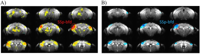 |
Polarity of BOLD fMRI as a function of balance between excitation and inhibition
Kostiantyn Cherkas1, G. H. Im1, and S.G. Kim2
1Cener for Neuroscience Imaging Research (CNIR), Institute for Basic Science (IBS), Suwon 16419, Republic of Korea, Suwon, Korea, Republic of, 2Cener for Neuroscience Imaging Research (CNIR), Institute for Basic Science (IBS), Suwon 16419, Republic of Korea, Department of Biomedical Engineering, Sungkyunkwan University, Suwon, Korea, Suwon, Korea, Republic of
To investigate neural source of positive and negative BOLD signals, we modulated a balance of excitatory and inhibitory activity (EI) within the same somatosensory area to determine whether the polarity of evoked BOLD response is reversed. We measured BOLD fMRI at 15.2T and calcium photometry of ketamine/xylazine-anesthetized mice in response to frequency dependent whisker pad stimulation. At 4 Hz, positive BOLD and increased calcium activity were observed in the barrel cortex, whereas at 20 Hz stimulation, negative BOLD and decreased calcium activity were detected. This indicates that the BOLD polarity closely links to the EI balance.
|
||
0431.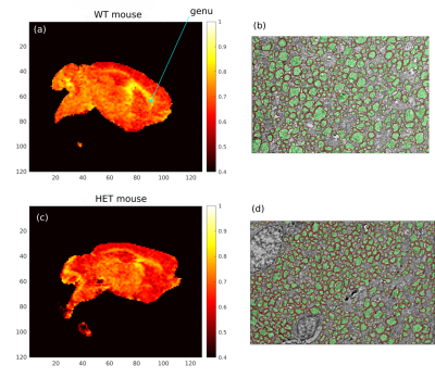 |
Validation of MRI Measurements of Myelination Changes in an Absence Epilepsy Mouse Model
Gustavo Chau Loo Kung1, Juliet Knowles2, Lijun Ni2, John Huguenard2, Michelle Monje2, and Jennifer McNab3
1Bioengineering Department, Stanford University, Stanford, CA, United States, 2Neurology Department, Stanford University, Stanford, CA, United States, 3Radiology Department, Stanford University, Stanford, CA, United States
Maladaptive myelination may contribute to both the predisposition to seizures and cognitive impairment in diseases such as absence epilepsy. To support this hypothesis, we performed MRI microstructural measurements in ex vivo mouse brains from the Scn8amed+/- model of absence epilepsy and validated them against matching Electron Microscopy (EM) quantifications. Our MRI g-ratio results strongly agree with these measurements and both show a statistically significant decrease in g-ratio of the genu in seizure mice. Future work will look to continue the current analysis in longitudinal studies to probe the previously unknown effects of dynamic myelination on the progression of absence epilepsy.
|
||
0432. |
fMRI connectivity mapping in the awake mouse brain reveals state-dependent network reconfiguration
Neha Atulkumar Singh1, Daniel Gutierrez-Barragan1, Elizabeth de Guzman1, Mauro Uboldi2, Ludovico Coletta1, and Alessandro Gozzi1
1Functional Neuroimaging Laboratory, Istituto Italiano di Tecnologia, Rovereto, Italy, 2Ugo Basile S.r.L., Gemonio, Italy
Resting state fMRI mapping in the mouse is typically carried out under light anesthesia, preventing a full characterization of how the ensuing functional architecture compares to awake conditions. Leveraging a novel protocol for fMRI connectivity mapping in awake mice, we provide a fine-grained description of the network structure and dynamic organization of brain-wide functional connectivity in this species. Notably, by comparing network features across brain states, we identify a robust set of state-dependent network changes, including a distinctive dynamic signature of consciousness. These results open the way to the implementation of awake rsfMRI in the mouse.
|
||
|
0433.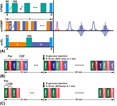 |
Sensitive imaging schemes for dynamic glucose enhanced (DGE) MRI to detect glucose uptake and clearance in mouse brain at 3T
Jianpan Huang1, Joseph H. C. Lai1, Xiongqi Han1, Zilin Chen1, Yang Liu1, Peng Xiao1, Lin Chen2,3, Jiadi Xu2,3, and Kannie W. Y. Chan1,3,4
1Department of Biomedical Engineering, City University of Hong Kong, Hong Kong, China, 2F.M. Kirby Research Center for Functional Brain Imaging, Kennedy Krieger Research Institute, Baltimore, MD, United States, 3Russell H. Morgan Department of Radiology and Radiological Science, The Johns Hopkins University School of Medicine, Baltimore, MD, United States, 4City University of Hong Kong Shenzhen Research Institute, Shenzhen, China
We developed a high-sensitivity dynamic glucose enhanced (DGE) MRI acquisition and post-processing scheme for sensitive monitoring glucose uptake and clearance in both brain parenchyma and cerebrospinal fluid (CSF) at 3T. By investigating Carr-Purcell-Meiboom-Gill sequence (CPMG), on-resonance variable delay multi-pulse (onVDMP) and on-resonance spin-locking (onSL), a high-sensitivity DGE MRI scheme composed of CPMG method for monitoring parenchyma and onVDMP method for monitoring CSF was proposed. We incorporated the multilinear singular value decomposition (MLSVD) based denoising method in post-processing, which enables the detection of DGE signals from the brain parenchyma and CSF at low concentration of D-glucose (12.5% w/w) injection.
|
|
0434. |
Quantitative neuroimaging biomarkers using 3D UTE MRI and ferumoxytol
Codi Gharagouzloo1, Praveen Kulkarni2, Joshua Leaston1, Kevin Johnson3, Jonathan Polimeni4, Ju Qiao5, Misung Han6, Peder Larson6, and Craig Ferris2
1Imaginostics, Inc., Cambridge, MA, United States, 2Center for Translational Neuroimaging, Northeastern University, Boston, MA, United States, 3Medical Physics, University of Wisconsin–School of Medicine and Public Health, Madison, WI, United States, 4Martinos Center, Massachusetts General Hospital and Athinoula A. Martinos Center for Biomedical Imaging, Boston, MA, United States, 5Massachusetts General Hospital, Boston, MA, United States, 6University of California, San Francisco, San Francisco, CA, United States
New quantitative vascular neuroimaging biomarkers are made possible by the combination of optimized 3D UTE MRI and ferumoxytol for contrast enhancement. Here, we have shown that this method, QUTE-CE MRI, can be used to image both the vascular and perivascular space. The small vessel density biomarker demonstrated sensitivity to hyper-to-normal microvascularization from 8-24 months of age in an ApoE4 genetic knock-in rat model. Disruption in the blood brain barrier was detected in individual animals and groups after even a single mild hit to the head. Finally, feasibility of QUTE-CE MRI is demonstrated on clinical scanners for human imaging.
|
||
|
0435.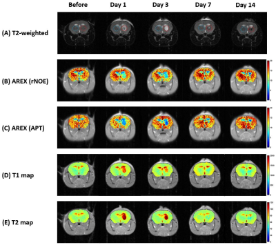 |
CEST MRI of temporal changes of hematoma in Intracerebral Hemorrhage (ICH) mouse at 3T
Joseph H. C. Lai1, Jiaxin Liu2, Jianpan Huang1, Yang Liu1, Zilin Chen1, Peng Xiao1, Gilberto K. K. Leung2, and Kannie W. Y. Chan1,3,4
1Department of Biomedical Engineering, City University of Hong Kong, Hong Kong, Hong Kong, 2Division of Neurosurgery, Department of Surgery, Li Ka Shing Faculty of Medicine, The University of Hong Kong, Hong Kong, Hong Kong, 3Russell H. Morgan Department of Radiology and Radiological Science, Johns Hopkins University School of Medicine, Baltimore, MD, United States, 4City University of Hong Kong Shenzhen Research Institute, Shenzhen, China
CEST MRI in hematoma could be challenging depending on the involvement of iron. This study examined the feasibility of CEST in monitoring ICH and its progression over two weeks. The AREX data supported that the iron-overloading pathology might not significantly attenuate CEST contrast as demonstrated both in vitro and in vivo at 3T. We observed the most significant decrease in rNOE (37%) and APT (47%) contrast in lesions were on day7 and day3, respectively, when compared to contralateral side. This could indicate neuropathologies related to lipid and amide, which could be valuable for ICH diagnosis and treatment planning at 3T.
|
|
|
0436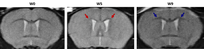 |
Abnormal Oxidative Metabolism in the Gray Matter of Cuprizone Mouse Model: An in-vivo NIRS-MRI Study Video Permission Withheld
Mada Hashem1,2,3,4,5, Ying Wu1,3,4,5, and Jeff F. Dunn1,2,3,4,5
1Department of Radiology, University of Calgary, Calgary, AB, Canada, 2Biomedical Engineering Graduate Program, University of Calgary, Calgary, AB, Canada, 3Hotchkiss Brain Institute, University of Calgary, Calgary, AB, Canada, 4Experimental Imaging Centre, University of Calgary, Calgary, AB, Canada, 5Cumming School of Medicine, University of Calgary, Calgary, AB, Canada
Non-invasive quantitative imaging of cerebral oxygen consumption is crucial to understand the involvement of oxidative metabolism in neurological diseases. We are applying a novel multimodal technique combining near-infrared spectroscopy and high-field MRI to study the mitochondrial status as well as oxygen delivery and consumption in the cortex of the cuprizone mouse model. In this study, multiple physiological parameters controlling oxidative metabolism were investigated in the cuprizone mice exhibiting demyelination. A mitochondrial impairment and a reduced oxygen consumption rate were found in the gray matter of cuprizone mice, emphasizing the association between abnormal oxidative metabolism and the observed demyelination.
|
The International Society for Magnetic Resonance in Medicine is accredited by the Accreditation Council for Continuing Medical Education to provide continuing medical education for physicians.