Oral
Diffusion in the Brain
ISMRM & SMRT Annual Meeting • 15-20 May 2021

| Concurrent 5 | 18:00 - 20:00 | Moderators: Tim Dyrby & Camilo Jaimes |
0205.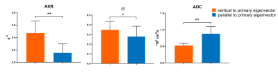 |
Characterization of Apparent Exchange Rate in Human Brain White Matter
Zhaoqing Li1,2, Yi-Cheng Hsu3, and Ruiliang Bai1,2
1Interdisciplinary Institute of Neuroscience and Technology (ZIINT), School of Medicine, Zhejiang University, Hangzhou, China, HangZhou, China, 2College of Biomedical Engineering and Instrument Science, Zhejiang University, Hangzhou, China, HangZhou, China, 3MR Collaboration, Siemens Healthcare, Shanghai, China, ShangHai, China
Filter exchange imaging (FEXI) is a non-invasive method to measure water exchange among diffusion compartments by apparent exchange rate (AXR). However, in human white matter, it is still controversial whether the diffusion-encoding gradient direction will affect AXR measurement. In this study, we performed FEXI on human brain with 20 diffusion gradient directions to explore features of AXR in white matter. We found that AXR measured with diffusion direction perpendicular to fiber (~ 0.47 s-1) is significantly larger than that parallel to fiber (~ 0.15 s-1), suggesting FEXI at different directions might measure exchanges between different biological compartments in white matter.
|
||
0206.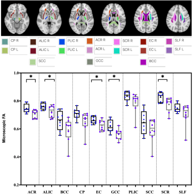 |
Evaluation of White Matter Microstructure in an HIV Population at Risk of Cerebral Small Vessel Disease using Microscopic Fractional Anisotropy
Md Nasir Uddin1, Abrar Faiyaz2, and Giovanni Schifitto1,3
1Department of Neurology, University of Rochester, Rochester, NY, United States, 2Electrical & Computer Engineering, University of Rochester, Rochester, NY, United States, 3Imaging Sciences, University of Rochester, Rochester, NY, United States
White matter microstructural abnormalities are well documented in HIV-infected individuals. DTI derived fractional anisotropy (FA) is frequently used to assess the abnormalities but it has some limitations. In this work, we compared FA and microscopic fractional anisotropy (μFA) in an HIV cohort, and our results indicate that μFA seems to provide better sensitivity than the conventional FA for detecting white matter microstructure changes associated with HIV infection, especially in the areas with crossing fibers.
|
||
0207.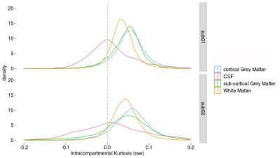 |
Human brain in vivo correlation tensor MRI on a clinical 3T system
Lisa Novello1, Rafael Neto Henriques2, Andrada Ianuş2, Thorsten Feiweier3, Noam Shemesh2, and Jorge Jovicich1
1Center for Mind/Brain Sciences - CIMeC, University of Trento, Rovereto, Italy, 2Champalimaud Research, Champalimaud Centre for the Unknown, Lisbon, Portugal, 3Siemens Healthcare GmbH, Erlangen, Germany
Resolving the underlying sources of kurtosis in biological systems is emerging as a promising strategy for non-invasive quantitative characterization of tissue microstructure. Recently, a novel framework termed Correlation Tensor Imaging (CTI), based on double-diffusion-encoding MRI, was shown to disentangle anisotropic, isotropic, and microscopic kurtosis sources in mouse brains. Here, we implemented CTI on a clinical 3T MRI system and scanned normal humans for the first time. The ensuing CTI-driven non-Gaussian inter/intra-compartmental estimates are promising and agree with expectations: positive intra-compartmental kurtosis for gray and white matter, larger for gray than white matter, and around zero for cerebrospinal fluid.
|
||
0208.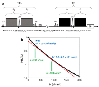 |
Feasibility of Filter-exchange Imaging (FEXI) in Measuring Different Exchange Processes in Human Brain
Zhaoqing Li1,2, Chaoliang Sun1,2, Yi-Cheng Hsu3, Hui Liang4, Peter Basser5, and Ruiliang Bai1,2
1Interdisciplinary Institute of Neuroscience and Technology (ZIINT), School of Medicine, Zhejiang University, Hangzhou, China, HangZhou, China, 2College of Biomedical Engineering and Instrument Science, Zhejiang University, Hangzhou, China, HangZhou, China, 3MR Collaboration, Siemens Healthcare, Shanghai, China, ShangHai, China, 4Department of Neurology, First Affiliated Hospital, School of Medicine, Zhejiang University, Hangzhou, China, HangZhou, China, 5Section on Quantitative Imaging and Tissue Sciences, NICHD, National Institutes of Health, Bethesda, MD, USA, Bethesda, MD, United States
In this study, we aim to explore the feasibility of Filter-exchange imaging (FEXI) in measuring different water exchange processes in human brain by modulating the diffusion filter (bf) and detection (b) blocks. We found the apparent exchange rate (AXR) estimated from a FEXI protocol with bf=250s/mm2 are significantly larger than those with bf=900s/mm2. Besides, the filter efficiency of FEXI with bf=250s/mm2 shows a strong correlation with vascular density estimated as the fraction of water exhibiting intravoxel incoherent motion (IVIM). Collectively, our current results demonstrate that FEXI targeting the vascular water could help characterize the intra-to-extravascular water exchange process.
|
||
0209.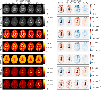 |
Is the inversion time important? A study of the reciprocal influence of inversion time and b-value on diffusion and longitudinal relaxation in MRI
Tomasz Pieciak1,2, Maryam Afzali3, Fabian Bogusz1, Dominika Ciupek1, Derek K. Jones3, and Marco Pizzolato4,5
1AGH University of Science and Technology, Kraków, Poland, 2LPI, ETSI Telecomunicación, Universidad de Valladolid, Valladolid, Spain, Valladolid, Spain, 3Cardiff University Brain Research Imaging Centre (CUBRIC), School of Psychology, Cardiff University, Cardiff, United Kingdom, 4Department of Applied Mathematics and Computer Science, Technical University of Denmark, Kongens Lyngby, Denmark, 5Signal Processing Lab (LTS5), École polytechnique fédérale de Lausanne (EPFL), Lausanne, Switzerland
The nervous tissue microstructure can be characterized by sensitizing the MRI signal to diffusion. The advent of multi-parametric sequences enabled the collection of diffusion data at different echo and inversion times. While the link between diffusion and transverse relaxation has undergone several investigations, in this work we characterize the relationship between the diffusion and longitudinal relaxation, and quantify the reciprocal influence of b-values and inversion time on the quantification of T1 and of diffusion metrics, interpreting our findings on in vivo data in the light of numerical simulations, and showing evidence that longitudinal relaxation locally modifies the diffusion contrast.
|
||
 |
0210.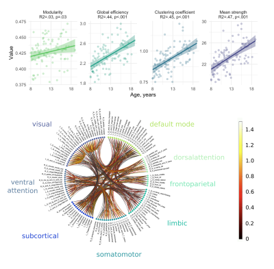 |
Unique insights into visual network development over childhood and adolescence from microstructure informed tractography
Simona Schiavi1, Sila Genc2, Maxime Chamberland2, Chantal M.W. Tax2,3, Erika P. Raven4, Alessandro Daducci1, and Derek K Jones2,5
1Department of Computer Science, University of Verona, Verona, Italy, 2CUBRIC, School of Psychology, Cardiff University, Cardiff, United Kingdom, 3Image Sciences Institute, University Medical Center Utrecht, Utrecht, Netherlands, 4Department of Radiology, Bernard and Irene Schwartz Center for Biomedical Imaging, New York University School of Medicine, New York, NY, United States, 5Mary MacKillop Institute for Health Research, Australian Catholic University, Melbourne, Australia
We employed the Convex Optimization Modeling for Microstructure Informed Tractography (COMMIT) approach to construct microstructure-informed connectomes and study the distinct patterns of age-related development in structural whole-brain network and sub-networks using global graph metrics. Whole brain analyses showed that with the new edge-weighting, the shortest-path length between all pairs of nodes decreases with age and thus efficiency increases. This reduction in shortest-path length is probably driven by previously reported age-related increases in the intra-axonal signal fraction. Sub-networks analyses revealed unique visual network characteristics over development and confirmed previously observed maturational pattern of posterior regions across childhood and adolescence.
|
|
 |
0211.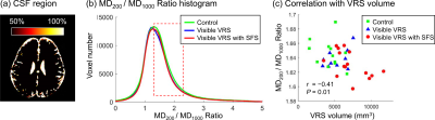 |
Infant cerebrospinal fluid dynamics assessed by low b-value diffusion tensor imaging and association with visible Virchow–Robin spaces
Xianjun Li1, Congcong Liu1, Mustafa Salimeen1, Miaomiao Wang1, Mengxuan Li1, Chao Jin1, Xiaocheng Wei1, and Jian Yang1
1Department of Radiology, The First Affiliated Hospital of Xi’an Jiaotong University, Xi'an, China
Cerebrospinal fluid (CSF) and interstitial fluid exchange via Virchow Robin space (VRS). It is reasonable to infer that VRS may be related to CSF dynamics. To investigate the association between CSF dynamics and VRS on infants, this work assessed CSF dynamics on infants by DTI with b values of 200 and 1000 s/mm2 and segmented VRS on T2WI images. Results suggest that the ratio between diffusivities derived from low and high b-value DTI could provide complementary information for assessing infant cerebrospinal fluid dynamics. There may exist potential association between Virchow–Robin space volume and cerebrospinal fluid dynamics.
|
|
|
0212.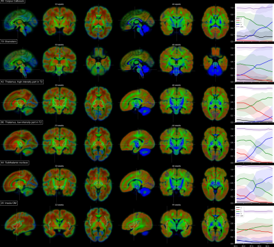 |
3t++ temporally consistent 3 tissue HARDI decomposition of neonatal brain tissue
Maximilian Pietsch1,2, Daan Christiaens2,3, Jana Hutter2,4, Lucilio Cordero-Grande2, Anthony N. Price2,4, Emer Hughes2, A. David Edwards2, Joseph V. Hajnal2,4, Serena J. Counsell2, Jonathan O'Muircheartaigh1,2,5,6, and J-Donald Tournier2,4
1Forensic & Neurodevelopmental Sciences, King's College London, London, United Kingdom, 2Centre for the Developing Brain, School of Biomedical Engineering and Imaging Sciences, King’s College London, Kings Health Partners, St. Thomas Hospital, London, SE1 7EH, UK, King's College London, London, United Kingdom, 3Department of Electrical Engineering (ESAT/PSI), KU Leuven, Leuven, Belgium, 4Biomedical Engineering Department, School of Biomedical Engineering and Imaging Sciences, King's College London, London, United Kingdom, 5Department of Perinatal Imaging & Health, School of Biomedical Engineering and Imaging Sciences, King's College London, London, United Kingdom, 6MRC Centre for Neurodevelopmental Disorders, King's College London, London, United Kingdom
We decompose neonatal HARDI signal into four components that together capture the orientation dependency and temporal dependency of signal in neonatal white and grey matter and CSF. We show that the voxel-wise volume fractions of the associated components exhibit distinct sigmoid-shaped time dependencies between 33 to 44 weeks gestational age and clearly delineate anatomical structures in developing white and grey matter, allowing detailed characterisation of microstructural properties of developing white and grey matter.
|
|
0213.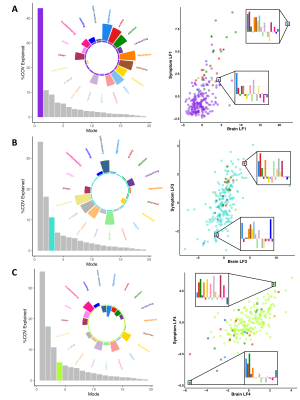 |
Redefining the architecture of white matter damage in paediatric concussions and their relationship with symptoms
Guido I. Guberman1, Sonja Stojanovski2,3, Alain Ptito1, Danilo Bzdok4, Anne Wheeler2,3, and Maxime Descoteaux5
1Department of Neurology and Neurosurgery, McGill University, Montreal, QC, Canada, 2Neuroscience and Mental Health Program, Hospital for Sick Children, Toronto, ON, Canada, 3Department of Physiology, University of Toronto, Toronto, ON, Canada, 4Department of Biomedical Engineering, McGill University, Montreal, QC, Canada, 5Department of Computer Science, Université de Sherbrooke, Sherbrooke, QC, Canada
Concussion heterogeneity remains a major challenge for clinical and scientific research. Yet, most studies continue to employ group comparison approaches that assume consistent one-to-one relationships between brain structure and symptoms. To parse concussion heterogeneity, we employed a double-multivariate approach using diffusion MRI of children with histories of concussion. Multivariate white-matter structural features captured more information about symptoms than all individual features. Results also revealed how different white-matter abnormalities led to similar symptom profiles. Lastly, multivariate features significantly predicted adverse psychiatric outcomes. These results suggest that concussion heterogeneity arising from complex structure/symptom relationships can be well captured by our double-multivariate approach.
|
||
|
0214.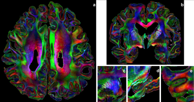 |
Chenonceau: an entire ex vivo human brain 11.7T anatomical and diffusion MRI dataset at the mesoscopic scale
Alexandros Popov1, Raïssa Yebga Hot1, Justine Beaujoin1, Ivy Uszynski1, Fawzi Boumezbeur1, Fabrice Poupon1, Christophe Destrieux2, and Cyril Poupon1
1NeuroSpin (CEA), Paris, France, 2Université de Tours, Tours, France
In this study, we present the Chenonceau dataset : a novel 11.7T MRI dataset of the entire human brain, combining an ultra-high resolution anatomical scan at 100μm with diffusion scans at 200μm using strong diffusion sensitizations. To obtain this dataset, a unique acquisition protocol has been established : a fixed ex vivo brain has been cut into blocks compatible in size with a Bruker 11.7T MRI system. The blocks were scanned individually over an extended period of time. The collection of datasets was then registered back to a reference blockface acquired at 3T, thus resulting in a unique high resolution brain dataset.
|
The International Society for Magnetic Resonance in Medicine is accredited by the Accreditation Council for Continuing Medical Education to provide continuing medical education for physicians.