Oral
Multiple Sclerosis
ISMRM & SMRT Annual Meeting • 15-20 May 2021

| Concurrent 6 | 12:00 - 14:00 | Moderators: Cristina Granziera & Susie Huang |
0483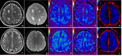 |
Differential Changes in Brain Viscoelastic Properties Observed with MR Elastography in MS and NMOSDs Video Permission Withheld
Ling Fang1, Matthew C. Murphy2, Qiuxia Luo1, Xiaodong Chen3, Linqi Zhang1, Bingjun He1, Jun Chen2, Jonathan M. Scott2, Meng Yin2, Kevin J. Glaser2, Richard L. Ehman2, Wei Qiu3, and Jin Wang1
1Department of Radiology, The Third Affiliated Hospital of Sun Yat-sen University, Guangzhou, China, 2Department of Radiology, Mayo Clinic, Rochester, MN, United States, 3Department of Neurology, The Third Affiliated Hospital of Sun Yat-sen University, Guangzhou, China
Multiple sclerosis (MS) is a chronic inflammatory disease of the central nervous system (CNS) characterized by demyelination, axonal loss and neurodegeneration. Because of overlapping clinical and imaging features, it is a challenge to distinguish MS from Neuromyelitis optica spectrum disorders (NMOSDs) for which the treatment is different. 3D MR Elastography (MRE) is a potential method to evaluate brain tissue damage in autoimmune diseases of the CNS. By measuring the viscoelasticity of the centrum ovale with 3D MRE, we found significantly decreased damping ratio and loss modulus in MS compared with NMOSDs, suggesting possible diagnostic utility for 3D MRE in MS.
|
||
 |
0484.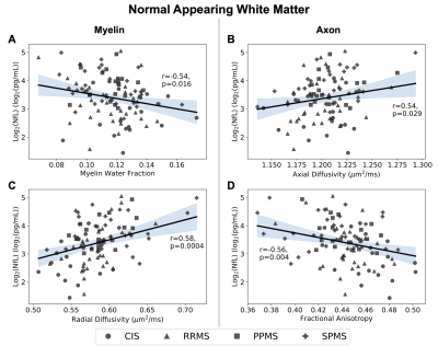 |
Correlations of serum neurofilament with myelin, axonal and volumetric imaging in multiple sclerosis
Jackie Yik1,2, Pierre Becquart3, Jasmine Gill3, Shannon H. Kolind1,2,4,5, Virginia Devonshire5, Ana-Luiza Sayao5, Alice Schabas5, Robert Carruthers5, Anthony Traboulsee5, G.R. Wayne Moore2,3, David K.B. Li4,5, Sophie Stukas3, Cheryl Wellington3,
Jacqueline A. Quandt3, Irene M. Vavasour2,4, and Cornelia Laule1,2,3,4
1Physics and Astronomy, University of British Columbia, Vancouver, BC, Canada, 2International Collaboration on Repair Discoveries, Vancouver, BC, Canada, 3Pathology and Laboratory Medicine, University of British Columbia, Vancouver, BC, Canada, 4Radiology, University of British Columbia, Vancouver, BC, Canada, 5Medicine, University of British Columbia, Vancouver, BC, Canada
Neurofilaments, particularly the light subunit (NfL), have become a biomarker of interest in multiple sclerosis (MS) and can be measured in blood serum after neuronal damage. NfL has been studied in MS prognosis and treatment monitoring, but little is known about the relationship between NfL and advanced quantitative MRI measures. This exploratory study characterizes the relationship between NfL and myelin water fraction and diffusion measures in different brain regions through regression models. We found NfL to correlate with myelin and axonal damage measures in the whole brain and normal appearing white matter but only to myelin water fraction in lesions.
|
|
|
0485.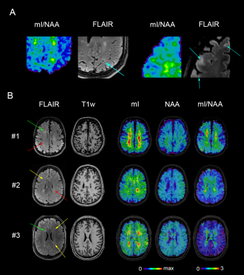 |
Disseminated brain pathology detected with high-resolution MRSI correlates with clinical disability in multiple sclerosis
Eva Heckova1, Alexandra Lipka1, Assunta Dal-Bianco2, Bernhard Strasser1, Gilbert Hangel1,3, Paulus Rommer2, Petra Hnilicová4, Ema Kantorová5, Lukas Hingerl1, Stanislav Motyka1, Fritz Leutmezer2, Stephan Gruber1, Siegfried Trattnig1,6,
and Wolfgang Bogner1
1High Field MR Centre, Department of Biomedical Imaging and Image-guided Therapy, Medical University of Vienna, Vienna, Austria, 2Department of Neurology, Medical University of Vienna, Vienna, Austria, 3Department of Neurosurgery, Medical University of Vienna, Vienna, Austria, 4Biomedical Center Martin, Jessenius Faculty of Medicine in Martin, Comenius University in Bratislava, Martin, Slovakia, 5Clinic of Neurology, Jessenius Faculty of Medicine in Martin, Comenius University in Bratislava, Martin, Slovakia, 6Christian Doppler Laboratory for Clinical Molecular MR Imaging, Vienna, Austria To enhance the detection of diffuse pathological alterations associated with multiple sclerosis, high-resolution MR spectroscopic imaging was performed at 7T, together with clinical MRI, in 68 MS patients with different levels of clinical disability and 20 healthy controls. Increased myo-inositol, a marker of neuroinflammation-induced astrogliosis, was found in white matter regions appearing normal on clinical MRI, even in subgroup of MS patients with no evidence of clinical disability. Myo-inositol/N-acetylaspartate ratio in the NAWM and cortical gray matter correlated with EDSS, suggesting that reactive astrogliosis and axonal injury play important role in the evolution of MS-related disability. |
|
|
0486.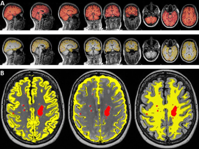 |
Neurometabolic changes in RRMS: comparison between fingolimod and injectables therapies
Oun Al-iedani1,2, Saadallah Ramadan2,3, Karen Ribbons2, Rodney Lea2, and Jeannette Lechner-Scott2,4,5
1School of Health Sciences, University of Newcastle, Newcastle, Australia, 2Hunter Medical Research Institute, Newcastle, Australia, 3Faculty of Health and Medicine, University of Newcastle, Newcastle, Australia, 4Department of Neurology, John Hunter Hospital, Newcastle, Australia, 5School of Medicine and Public Health, University of Newcastle, Newcastle, Australia
This novel study compares the neurometabolic effects of different DMTs on the MS brain. We evaluated volumetric and neurometabolic changes in RRMS patients on fingolimod(N=52), injectables(N=46) and HCs cohort(N=51). MRS was acquired from PCG and PFC. Compared to HCs, a significant reduction in NAA/tCr were detected in both locations and cohorts. Clinical parameters, MR-volumetrics and neurometabolic concentrations showed no statistically significant differences between RRMS cohorts. MRI metrics and neurometabolites from both locations, showed moderate correlations with cognition, fatigue and memory. This the first study demonstrating that fingolimod and injectable DMTs influence volumetric and neurometabolic profiles of MS-brain similarly.
|
|
0487.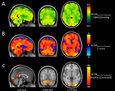 |
Grey Matter Cerebrovascular Reactivity in Multiple Sclerosis and its Changes with Immunomodulation: a Breath-Hold BOLD-MRI Study
Antonio Maria Chiarelli1, Daniele Mascali1, Nikolaos Petsas2, Carlo Pozzilli2, Richard Geoffrey Wise1, and Valentina Tomassini1
1Department of Neuroscience, Imaging and Clinical Sciences, University G. D'Annunzio of Chieti Pescara, Chieti Scalo, Italy, 2Department of Neurology and Psychiatry, Sapienza University, Rome, Italy
The cerebrovascular system is altered in MS. Using breath-hold BOLD-MRI, we tested whether pharmacological modulation of MS inflammation influences alteration in cerebrovascular reactivity (CVR). We found that CVR increased with immunomodulation and this increase was negatively correlated with pre-treatment CVR, suggesting that cerebrovascular alteration reflects disease activity. Moreover, lower CVR in the pre-treatment phase was associated with lower grey matter volume and this correlation was lost with immunomodulation, indicating an involvement of brain vasculature in neurodegeneration. Given the multiparametric characteristic of BOLD, further studies are warranted to clarify the vascular origin of our findings.
|
||
|
0488.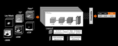 |
Toward Fully Automated Assessment of the Central Vein Sign Using Deep Learning
Till Huelnhagen1,2,3, Omar Al Louzi4, Mário João Fartaria1,2,3, Lynn Daboul4, Pietro Maggi5,6, Cristina Granziera7,8,9, Meritxell Bach Cuadra2,3,10, Jonas Richiardi2, Daniel S Reich4, Tobias Kober1,2,3, and Pascal Sati4,11
1Advanced Clinical Imaging Technology, Siemens Healthcare AG, Lausanne, Switzerland, 2Department of Radiology, Lausanne University Hospital and University of Lausanne, Lausanne, Switzerland, 3Signal Processing Laboratory (LTS 5), Ecole Polytechnique Fédérale de Lausanne (EPFL), Lausanne, Switzerland, 4Translational Neuroradiology Section, National Institute of Neurological Disorders and Stroke, National Institutes of Health (NIH), Bethesda, MD, United States, 5Department of Neurology, Lausanne University Hospital and University of Lausanne, Lausanne, Switzerland, 6Cliniques universitaires Saint-Luc, Université catholique de Louvain, Brussels, Belgium, 7Neurologic Clinic and Policlinic, Departments of Medicine, Clinical Research and Biomedical Engineering, University Hospital Basel and University of Basel, Basel, Switzerland, 8Translational Imaging in Neurology (ThINk) Basel, Department of Medicine and Biomedical Engineering, University Hospital Basel and University of Basel, Basel, Switzerland, 9Research Center for Clinical Neuroimmunology and Neuroscience (RC2NB) Basel, University Hospital Basel and University of Basel, Basel, Switzerland, 10Medical Image Analysis Laboratory (MIAL), Centre d'Imagerie BioMédicale (CIBM), University of Lausanne, Lausanne, Switzerland, 11Department of Neurology, Cedars-Sinai Medical Center, Los Angeles, CA, United States
The fraction of white matter lesions exhibiting the central vein sign (CVS) has shown promise as a biomarker in the diagnosis of multiple sclerosis. As manual CVS assessment is not clinically feasible, automated solutions have been proposed to perform this task. A deep-learning-based method called “CVSnet” demonstrated effective and accurate discrimination of MS from its mimics but required manual pre-selection. This work extends CVSnet to allow fully automated CVS assessment without manual interaction. High-quality, expert-reviewed segmentations of almost 6300 lesions were used for training and testing. The proposed method achieved accuracies between 75% and 80% in an unseen testing set.
|
|
|
0489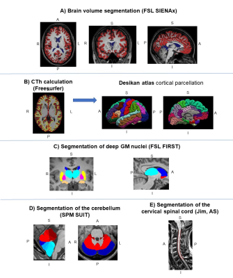 |
Damage of Different CNS Compartments Contributes to Explain Multiple Sclerosis Disability Milestones: A Multicenter Study Video Permission Withheld
Paola Valsasina1, Milagros Hidalgo de la Cruz1, Alessandro Meani1, Claudio Gobbi2,3, Antonio Gallo4, Chiara Zecca2,3, Alvino Bisecco4, Maria A. Rocca1,5,6, and Massimo Filippi1,5,6,7,8
1Neuroimaging Research Unit, Division of Neuroscience, IRCCS San Raffaele Scientific Institute, Milan, Italy, 2Multiple Sclerosis Center, Department of Neurology, Neurocenter of Southern Switzerland, Civic Hospital, Lugano, Switzerland, 3Faculty of Biomedical Sciences, Università della Svizzera Italiana, Lugano, Switzerland, 4Department of Advanced Medical and Surgical Sciences, and 3T MRI Center, University of Campania “Luigi Vanvitelli”, Naples, Italy, 5Neurology Unit, IRCCS San Raffaele Scientific Institute, Milan, Italy, 6Vita-Salute San Raffaele University, Milan, Italy, 7Neurorehabilitation Unit, IRCCS San Raffaele Scientific Institute, Milan, Italy, 8Neurophysiology Service, IRCCS San Raffaele Scientific Institute, Milan, Italy
Here, we assessed damage of cortex, deep grey matter, cerebellum and cervical cord to determine their relative contributions to the main clinical disability (EDSS) milestones in a multicentre cohort of 198 multiple sclerosis (MS) patients. The main determinants of EDSS=3.0 were cervical cord and thalamic atrophy, and brain lesion burden. The EDSS=4.0 milestone was better explained by cortical atrophy, together with cord and cerebellar damage, while the only predictor of EDSS=6.0 was cervical cord damage. This study is shading light on the differential weight of inflammatory and neurodegenerative processes leading to disability accumulation across various MS disease phases.
|
|
0490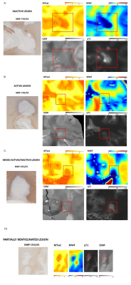 |
Multiparametric quantitative postmortem 3T-MRI of histopathological lesion types in multiple sclerosis Video Permission Withheld
Riccardo Galbusera1,2,3, Erik Bahn4, Matthias Weigel2,3,5, Po-Jui Lu1,2,3, Muhamed Barakovic1,2,3, Reza Rahmanzadeh1,2,3, Peter Dechent6, Antoine Lutti7, Govind Bhagavatheeshwaran8, Ludwig Kappos2,3, Wolfgang Brück4, Christine Stadelmann-Nessler4, and Cristina Granziera1,2,3
1Neurology Clinic and Policlinic, Departments of Medicine, Clinical Research and Biomedical Engineering, University Hospital Basel and University of Basel, Basel, Switzerland, Basel, Switzerland, 2Translational Imaging in Neurology (ThINk) Basel, Department of Biomedical Engineering, University Hospital Basel and University of Basel, Basel, Switzerland, Basel, Switzerland, 3Research Center for Clinical Neuroimmunology and Neuroscience (RC2NB) Basel, University Hospital Basel and University of Basel, Basel, Switzerland, Basel, Switzerland, 4Institute of Neuropathology, University Medical Center, Göttingen, Germany, Göttingen, Germany, 5Division of Radiological Physics, Department of Radiology, University Hospital Basel, Basel, CH, Basel, Switzerland, 6Department of Cognitive Neurology, MR-Research in Neurosciences, University Medical Center Göttingen, Göttingen, Germany, Göttingen, Germany, 7Centre for Research in Neuroscience - Department of Clinical Neurosciences, Laboratoire de recherche en neuroimagerie (LREN) University Hospital and University of Lausanne, Lausanne, Switzerland, Lausanne, Switzerland, 8National Institute of Neurological Disorders and Stroke, Bethesda, MD, USA, Bethesda, MD, United States
We have identified the imaging correlates of multiple sclerosis lesion subtypes by exploiting post-mortem multiparametric quantitative MRI and histopathological analysis. Remyelinated lesions showed distinct MRI characteristics compared to other MS lesions, and a remarkable resemblance to normal-appearing tissue properties. Our findings suggest that multiparametric quantitative MRI may well help to identify specific focal lesion types in vivo in MS patients.
|
||
0491.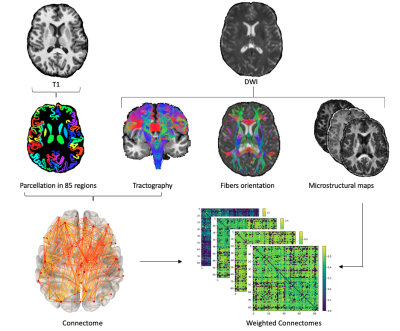 |
An investigation of the sensitivity of diffusion-based microstructure combined with network analysis in multiple sclerosis
Sara Bosticardo1, Simona Schiavi1, Sabine Schaedelin2, Po-Jui Lu3,4, Muhamed Barakovic2,3, Matthias Weigel2,3,5, Ludwig Kappos3,4, Jens Kuhle3,4, Alessandro Daducci1, and Cristina Granziera2,3,4
1Department of Computer Science, University of Verona, Verona, Italy, 2Departments of Medicine, Clinical Research and Biomedical Engineering, Neurology, University Hospital Basel and University of Basel, Basel, Switzerland, 3Department of Medicine and Biomedical Engineering, University Hospital Basel and University of Basel, Neurologic Clinic and Policlinic, Translational Imaging in Neurology (ThINk), Basel, Switzerland, 4Research Center for Clinical Neuroimmunology and Neuroscience, Basel, Switzerland, 5Department of Radiology, Division of Radiological Physics, University Hospital Basel, Basel, Switzerland
Graph measures derived from structural connectomes are widely used to study neurodegenerative diseases such as multiple sclerosis (MS). Usually, the connection strength is assessed by counting the number of streamlines connecting pairs of grey-matter regions. Here we used different ways to weight the edges to compare the sensitivity to MS structural disruptions of three diffusion-based microstructural models and their derived maps combined with network analysis. We found that the most sensitive are those whose derived maps are associated to intra-axonal signal fraction. Moreover, the segregation of the network appeared to be the most important in explaining clinical motor disability.
|
||
0492.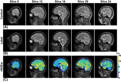 |
Relayed nuclear Overhauser effect (rNOE) imaging identifies multiple sclerosis: an initial human study
Jianpan Huang1, Jiadi Xu2,3, Joseph H. C. Lai1, Henry K. F. Mak4, Koon Ho Chan5, and Kannie W. Y. Chan1,3,6
1Department of Biomedical Engineering, City University of Hong Kong, Hong Kong, China, 2F.M. Kirby Research Center for Functional Brain Imaging, Kennedy Krieger Research Institute, Baltimore, MD, United States, 3Russell H. Morgan Department of Radiology and Radiological Science, The Johns Hopkins University School of Medicine, Baltimore, MD, United States, 4Department of Diagnostic Radiology, Li Ka Shing Faculty of Medicine, The University of Hong Kong, Hong Kong, China, 5Department of Medicine, Li Ka Shing Faculty of Medicine, The University of Hong Kong, Hong Kong, China, 6City University of Hong Kong Shenzhen Research Institute, Shenzhen, China
Multiple sclerosis (MS) is demyelinating disease of the central nervous system (CNS), which affects more than two million people globally. Here we applied our optimized pulsed-CEST MRI method to acquire relayed nuclear Overhauser effect weighted (rNOEw) images for detecting the pathology changes regarding myelin lipid/protein in human brain with neuromyelitis optica (NMO) and MS on clinical 3T scanner. We found that rNOEw signal of MS brains was significantly lower than that of NMO and NC brains. Our proposed rNOEw imaging method has great potential to assist MS diagnosis and specifically identify MS patients from NMO patients.
|
The International Society for Magnetic Resonance in Medicine is accredited by the Accreditation Council for Continuing Medical Education to provide continuing medical education for physicians.