Oral
Spinal Cord Imaging & More
ISMRM & SMRT Annual Meeting • 15-20 May 2021

| Concurrent 5 | 18:00 - 20:00 | Moderators: Alan Seifert & Seth Smith |
 |
0649.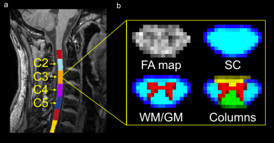 |
Variations of quantitative MRI metrics along the cervical spinal cord: multi-vendor, multi-center, multi-subject study
Jan Valošek1,2 and Julien Cohen-Adad2,3
1Department of Neurology, Faculty of Medicine and Dentistry, Palacký University Olomouc, Olomouc, Czech Republic, 2NeuroPoly Lab, Institute of Biomedical Engineering, Polytechnique Montreal, Montreal, QC, Canada, 3Functional Neuroimaging Unit, CRIUGM, University of Montreal, Montreal, QC, Canada
Quantitative MRI (qMRI) of the spinal cord (SC), such as diffusion-weighted and magnetic transfer imaging can be used in diagnosis of many different diseases. In this study, we established normative qMRI metrics along C2-C5 cervical SC levels for different regions-of-interest (spinal cord, white and gray matter, white matter columns) for 3 major MRI vendors (Siemens, Philips, GE) in large open-access dataset of ~250 healthy subjects. Metrics showed dependency on vertebral levels and confirmed different microstructural organization along SC. Moreover, differences in qMRI metrics were observed between individual vendors suggesting the influence of different MRI scanner configurations.
|
|
 |
0650.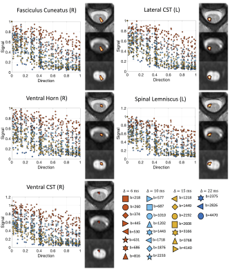 |
Exploring diffusion modeling for the human cervical spinal cord: an evaluation of 480 multicompartment models
Kurt G Schilling1, Qi Yang2, Vishwesh Nath3, Rutger Fick4, Kristin P O'Grady1, Adam W Anderson2, Bennett A Landman1, and Seth A Smith1
1Vanderbilt University Medical Center, Nashville, TN, United States, 2Vanderbilt University, Nashville, TN, United States, 3NVIDIA, Bethesda, MD, United States, 4TheraPanacea, Paris, France
A large number of models have been developed to describe the diffusion MRI signal as a sum of different neural compartments. However, development and optimization of these multicompartment models has largely focused on the brain. In this work, we apply and compare a combinatorially large number of biophysical models (N=480) in the in vivo human spinal cord, evaluating their ability to fit the signal and also predict unseen signal. We find that certain combinations of constraints and compartments better model the signal in the cervical spinal cord, and we give recommendations for future modeling of this structure with clinical acquisitions.
|
|
|
0651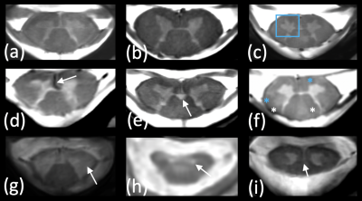 |
The good, the bad and the ugly : a retrospective study of image quality in human cervical spinal cord MRI at 7T Video Permission Withheld
Guillaume Frebourg1,2, Aurélien Massire1,2, Lauriane Pini1,2, Maxime Guye1,2, Bertrand Audoin2,3, Annie Veschueren2,4, Pierre-Hugues Roche5, and Virginie Callot1,2
1Aix-Marseille University, CNRS, CRMBM, Marseille, France, 2AP-HM, Hôpital de la Timone, CEMEREM, Marseille, France, 3AP-HM, Hôpital de la Timone, Neurology Dept., Marseille, France, 4AP-HM, Hôpital de la Timone, Neuromuscular Disease Dept., Marseille, France, 5AP-HM, Hôpital Nord, Neurosurgery Dept., Marseille, France
In the context of fast expansion of 7T clinical MR systems for neurological applications, a retrospective evaluation of cervical spinal cord image quality acquired on 41 patients and 25 healthy controls was performed. Several morphometric parameters were collected, and four MR modalities were rated. Altogether, this study provides guidance for both clinicians and physicists, regarding current sequence robustness and most frequent problems to be addressed.
|
|
|
0652.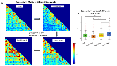 |
Detection of resting state correlations between white matter tracts in spinal cord using BOLD fMRI and their changes with injury
Anirban Sengupta1, Arabinda Mishra1, Feng Wang1, Li Min Chen1, and John C. Gore1
1Vanderbilt University Medical Center, Nashville, TN, United States
The study objective was to detect and quantify correlations between resting state BOLD signals in WM of spinal cord (SC) as potential indicator of functional connectivity, and evaluate changes that occur following injury. At first, BOLD activation was detected in response to tactile stimuli in certain WM regions in the SC of squirrel monkeys. Next, localized BOLD activity was observed during resting state in SC regions which resembled closely to WM tracts. There was a drop in resting state WM correlations after injury, followed by gradual recovery with time which mimics the pattern of SC functional recovery after an injury.
|
|
|
0653.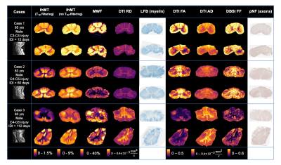 |
Correlating advanced MRI and histopathological measurements of axons and myelin in human traumatic spinal cord injury
Sarah Rosemary Morris1,2,3, Andrew Yung1,2,4, Valentin Prevost1,2,4, Shana George1, Andrew Bauman1,2,4, Piotr Kozlowski1,2,3,4, Farah Samadi1,5, Caron Fournier1,5, Lisa Parker6, Kevin Dong1, Femke Streijger1, Veronica Hirsch-Reinshagen1,5,6, G.R. Wayne Moore1,5,6,
Brian K Kwon1,7, and Cornelia Laule1,2,3,5
1International Collaboration on Repair Discoveries (ICORD), Vancouver, BC, Canada, 2Radiology, University of British Columbia, Vancouver, BC, Canada, 3Physics & Astronomy, University of British Columbia, Vancouver, BC, Canada, 4UBC MRI Research Centre, Vancouver, BC, Canada, 5Pathology & Laboratory Medicine, University of British Columbia, Vancouver, BC, Canada, 6Vancouver General Hospital, Vancouver, BC, Canada, 7Vancouver Spine Surgery Institute, Vancouver, BC, Canada
Using human spinal cord tissue donated to the International Spinal Cord Injury Biobank, we quantitatively correlated binarized histological stains for myelin and axons with myelin- and axon-sensitive advanced MRI metrics (myelin water fraction (MWF), inhomogeneous magnetization transfer (ihMT), diffusion tensor imaging (fractional anisotropy (FA), axial diffusivity (AD), radial diffusivity, RD), diffusion basis spectrum imaging (fibre fraction, FF)). MWF, ihMT and RD had significant, moderately strong correlations with Luxol fast blue staining for myelin phospholipids. FA, AD and FF did not have any significant correlation with phosphorylated neurofilament immunohistochemistry.
|
|
0654.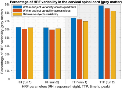 |
Variability of the hemodynamic response function in the healthy human cervical spinal cord at 3 Tesla
D Rangaprakash1 and Robert L Barry1
1Athinoula A. Martinos Center for Biomedical Imaging, Massachusetts General Hospital and Harvard Medical School, Charlestown, MA, United States
Functional MRI is an indirect measure of neural activity, being the convolution of the hemodynamic response function (HRF) and latent neural response. The HRF is variable across brain regions and individuals. However, resting-state spinal cord fMRI studies still largely ignore this variability, partly due to an incomplete understanding of HRF variability in the cord. To address this gap, we characterized within- and between-subjects HRF variability within the cervical spine (N=20). 6–9% HRF variability was observed in the gray matter, and 3–5% in the white matter. This is an important confound to be accounted for in future spinal cord fMRI studies.
|
||
0655.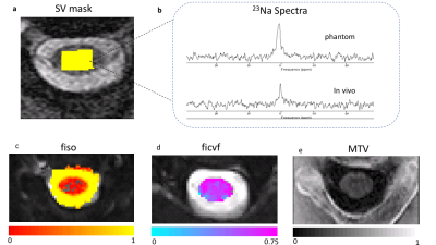 |
Associations between cervical cord sodium concentration, neuronal density and macromolecular tissue volume in spinal cord injury
Bhavana S Solanky1, Ferran Prados1,2, Francesco Grussu1,3, Marco Battiston1, Jon Stutters1, Selma Al-Ahmad4, Baris Kanber2, David Choi4, Jalesh Panicker5, and Claudia AM Gandini Wheeler-Kingshott1,6,7
1NMR Research Unit, Queen Square MS Centre, Department of Neuroinflammation, UCL Queen Square Institute of Neurology, Faculty of Brain Sciences, University College London (UCL), London, United Kingdom, 2Centre for Medical Image Computing, Department of Medical Physics and Biomedical Engineering, University College London (UCL), London, United Kingdom, 3Radiomics Group, Vall d’Hebron Institute of Oncology, Vall d’Hebron Barcelona Hospital Campus, Barcelona, Spain, 4National Hospital For Neurology and Neurosurgery, London, United Kingdom, 5Department of Uro-Neurology, The National Hospital for Neurology and Neurosurgery and UCL Queen Square Institute of Neurology, London, United Kingdom, 6Department of Brain & Behavioural Sciences, University of Pavia, Pavia, Italy, 7Brain Connectivity Centre Research Department, IRCCS Mondino Foundation, Pavia, Italy
Sodium retention as a consequence of spinal cord injury is thought to impair the regenerative ability of neurons but also reduce damage. Pilot studies suggest a possible increase in total sodium concentration (TSC) in spinal cord injury. Here we report increases in spinal cord TSC in cervical myelopathy patients relative to healthy controls. Given that the increase could be a consequence of intracellular accumulation of sodium or increases in extracellular sodium through enlarged extracellular space, the correlations of sodium with microstructure were investigated using neurite orientation dispersion and density imaging (NODDI) and macromolecular tissue volume imaging.
|
||
0656.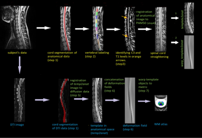 |
Atlas-based Quantification of DTI measures in Typically Developing Pediatric Spinal Cord
Shiva Shahrampour1, Benjamin De Leener2, Mahdi Alizadeh1, Devon Middleton1, Laura Krisa1, Adam Flanders1, Scott Faro1, Julien Cohen-Adad2, and Feroze Mohamed1
1Thomas Jefferson University, Philadelphia, PA, United States, 2Polytechnique Montreal, Montreal, QC, Canada
White matter microstructure, essential for efficient and coordinated transmission of neural communications, undergoes pronounced development during the first years of life. Hence, systematic evaluation of white matter microstructure in the normative pediatric spinal cord is critical for assessing early development and improving diagnosis of spinal cord related diseases.
|
||
0657.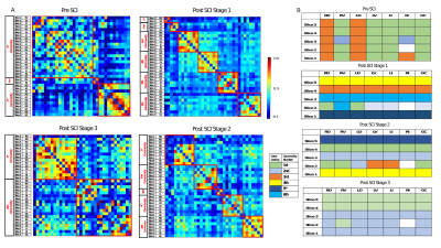 |
Detection of fine-scale functional networks in spinal cord and the effects of injury on intra- and inter-segmental networks
Anirban Sengupta1, Arabinda Mishra1, Feng Wang1, Li Min Chen1, and John C. Gore1
1Vanderbilt University Medical Center, Nashville, TN, United States
The objective of this study was to identify fine-scale resting state functional networks within the spinal cord gray matter of squirrel monkeys, and measure the changes in functional connectivity within the cord after a targeted injury. Independent Component Analysis of resting state fMRI data detected robust BOLD signals localized at the bilateral intermediate and gray-commissure regions of the spinal cord as well at the ‘4 horns’. A unilateral section of dorsal column tract at C5 segment of spinal cord damaged the inter-segmental connectivity more than intra-segmental connectivity, as observed through individual connectivity measures and community structures generated by graph-theory principles.
|
||
0658.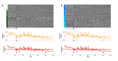 |
Visualization technique for assessment of spinal cord fMRI data quality
Kimberly J Hemmerling1,2 and Molly G Bright1,2
1Biomedical Engineering, McCormick School of Engineering, Northwestern University, Evanston, IL, United States, 2Physical Therapy & Human Movement Sciences, Feinberg School of Medicine, Northwestern University, Chicago, IL, United States
It is important to perform visual inspection of spinal cord imaging data throughout an fMRI analysis pipeline. The method presented here is an extension of an existing technique developed for the brain, adapted to spinal cord fMRI. We create a two-dimensional heatmap of the spinal cord derived from four-dimensional imaging data, which can be co-visualized with traces of motion and physiological signals, and identify examples of structured variations in the heatmap that may be attributed to these nuisance signals. Implementing this visualization of spinal cord fMRI data is a simple and fast method to examine data quality.
|
The International Society for Magnetic Resonance in Medicine is accredited by the Accreditation Council for Continuing Medical Education to provide continuing medical education for physicians.