Oral
Artificial Intelligence (Machine Learning & Deep Learning) Applications to Neuroradiology
ISMRM & SMRT Annual Meeting • 15-20 May 2021

Oral
Artificial Intelligence (Machine Learning & Deep Learning) Applications to Neuroradiology
| Concurrent 6 | 16:00 - 18:00 | Moderators: Mai-Lan Ho & Greg Zaharchuk |
0599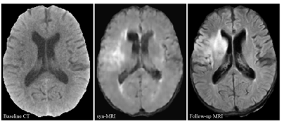 |
CT-to-MR image synthesis: A generative adversarial network-based method for detecting hypoattenuating lesions in acute ischemic stroke Video Permission Withheld
Na Hu1, Tianwei Zhang2, Yifan Wu3, Biqiu Tang1, Minlong Li1, Qiyong Gong1, Shi Gu2, and Su Lui1
1Huaxi MR Research Center (HMRRC), Department of Radiology, West China Hospital of Sichuan University, Chengdu, China, 2Department of Computer and Engineering, University of Electronic Science and Technology of China, Chengdu, China, 3Department of Bioengineering, University of Pennsylvania, Philadelphia, PA, United States
We aimed to develop a method of CT-to-MR image synthesis to assist in detecting hypoattenuating brain lesions in acute ischemic stroke. Emergency head CT images of 193 patients with suspected stroke and follow-up MR images were collected. A generative-adversarial-network model was developed for CT-to-MR image synthesis. With synthetic MRI compared to CT, sensitivity was improved by 116% in patient detection and 300% in lesion detection, and extra 75% of patients and 15% of lesions missed on CT were detected on synthetic MRI. Our method could be a rapid tool to improve readers’ detection of hypoattenuating lesions in AIS.
|
||
0600.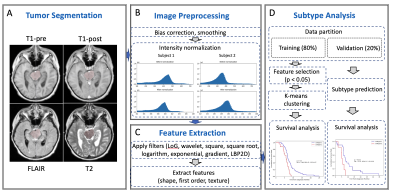 |
Identifying Diffuse Intrinsic Pontine Glioma (DIPG) Subtypes via Radiomic Approaches
Silu Zhang1, Zoltan Patay1, Bogdan Mitrea2, Angela Edwards1, Lydia McColl Makepeace1, and Matthew A. Scoggins1
1Diagnostic Imaging, St. Jude Children's Research Hospital, Memphis, TN, United States, 2Activ Surgical, Boston, MA, United States
Diffuse intrinsic pontine glioma (DIPG) is a pediatric brain tumor with very poor prognosis. In this study, we identify two subtypes of DIPG based on radiomic features. The two subtypes show a significant difference in survival rates. Subtype 1 has a mean progression-free survival (PFS) and overall survival (OS) of 8.9 and 12.7 months, respectively. Subtype 2 has a mean PFS and OS of 5.7 and 9.1 months, respectively. Our results suggest that shape features and intensity features extracted from FLAIR and T1-post contrast predict the prognosis of DIPG.
|
||
0601.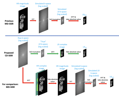 |
Deep learning super-resolution for sub-50-micron MRI of genetically engineered mouse embryos
Zihao Chen1,2, Yuhua Chen1,2, Ankur Saini3, William Devine3, Yibin Xie1, Cecilia Lo3, Debiao Li1,2, Yijen Wu3, and Anthony Christodoulou1
1Biomedical Imaging Research Institute, Cedars-Sinai Medical Center, Los Angeles, CA, United States, 2Department of Bioengineering, UCLA, Los Angeles, CA, United States, 3Department of Developmental Biology, University of Pittsburgh, Pittsburgh, PA, United States
Genetically engineered mouse models (GEMM) are indispensable in modeling human diseases. High resolution MRI with spatial resolution less than 100 μm has made incredible progress for phenotyping mouse embryos. However, it takes more than 10 hours' acquisition time to reach such high resolution, so reducing the scan time is of great need. Here we propose a deep learning based super-resolution approach for 3x3 super-resolution (SR) of mouse embryo images using raw k-space data. Our method can reduce the scan time by a factor of 9 while preserving the diagnostic details and shows better quantitative results than previous SR methods.
|
||
0602.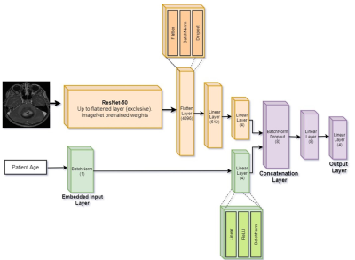 |
Classification of Pediatric Posterior Fossa Tumors using Convolutional Neural Network and Tabular Data
Moran Artzi1,2,3, Erez Redmard3, Oron Tzemach3, Jonathan Zeltser3, Omri Gropper4, Jonathan Roth2,5,6, Ben Shofty2,5,7, Danil A. Kozyrev5,7, Shlomi Constantini2,5,7, and Liat Ben-Sira2,8
1Sagol Brain Institute, Tel Aviv Sourasky Medical Center, Tel Aviv, Israel, 2Sackler Faculty of Medicine, Tel Aviv University, Tel Aviv, Israel, 3Sagol School of Neuroscience, Tel Aviv University, Tel Aviv, Israel, 4The Iby and Aladar Fleischman Faculty of Engineering, Tel Aviv University, Tel Aviv, Israel, 5Department of Pediatric Neurosurgery, Tel Aviv Sourasky Medical Center, Tel Aviv, Israel, 6The Gilbert Israeli Neurofibromatosis Center, Tel Aviv University, Tel Aviv, Israel, 7The Gilbert Israeli Neurofibromatosis Center, Tel Aviv Sourasky Medical Center, Tel Aviv, Israel, 8Division of Radiology, Tel Aviv Sourasky Medical Center, Tel Aviv, Israel
A fused architecture contrived of 2 neural networks, pre-trained ResNet-50 CNN and tabular based network is proposed for the classification of Posterior fossa tumors (PFT) types. The study included data for 158 MRI of healthy controls and pediatric patients with PFT. The input data were T1WI+C, FLAIR and diffusion MRI, and tabular data (subject's age). The best classification results obtained by the fused CNN + tubular data architecture and based on diffusion images, achieved cross-validation accuracy of validation=0.88±0.04, test=0.87±0.02. Overall, the proposed architecture achieved a ~16% improvement in accuracy for the test data compared to CNN method for this dataset.
|
||
0603.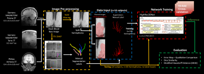 |
Deep Learning Segmentation of Lenticulostriate Arteries Using 3T and 7T 3D Black-Blood MRI
Samantha J Ma1,2, Mona Sharifi Sarabi2, Kai Wang2, Wenli Tan2, Huiting Wu3, Lei Hao3, Yulan Dong3, Hong Zhou3, Lirong Yan2, Yonggang Shi2, and Danny JJ Wang2
1Siemens Medical Solutions USA, Inc., Los Angeles, CA, United States, 2University of Southern California, Los Angeles, CA, United States, 3Department of Radiology, The First Affiliated Hospital of University of South China, Hunan, China
Given the inaccessibility of cerebral small vessels to existing clinical in vivo imaging technologies, early cerebral microvascular morphological changes in small vessel disease (SVD) can be difficult to evaluate. In this study, we trained a deep learning (DL)-based algorithm with 3T and 7T black-blood images on two vendor platforms to automatically segment lenticulostriate arteries (LSAs) in the brain. Our results show that black-blood imaging in conjunction with DL is a promising approach to enable quantitative morphometric analysis in patients with cerebral SVD.
|
||
0604.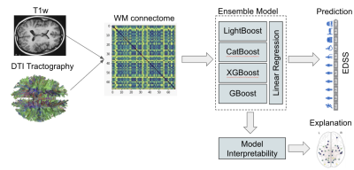 |
Disability Prediction in Multiple Sclerosis using Ensemble of Machine Learning Models and DTI Brain Connectivity
Berardino Barile1, Aldo Marzullo2, Claudio Stamile3, Françoise Durand-Dubief4, and Dominique Sappey-Marinier1,5
1CREATIS (UMR 5220 CNRS & U1206 INSERM), Université Claude Bernard Lyon 1, Villeurbanne, France, 2Department of Mathematics and Computer Science, University of Calabria, Rende, Italy, 3R&D Department, CGnal, Milan, Italy, 4Hôpital Neurologique, Hospices Civils de Lyon, Bron, France, 5MRI, CERMEP - Imagerie du Vivant, Bron, France
The Expanded Disability Status Scale (EDSS) monitors physical impairment in Multiple Sclerosis (MS). A Staking Ensemble model composed of 4 ML "boosting" models was used to predict EDSS using both white matter (WM) fiber-bundles and structural connectome data. This model provided excellent prediction results with an RMSE of 0.92 to 1.08. A counterfactual model was added to highlight the most important WM links and fiber-bundles in the prediction process. The accordance of the findings obtained with both data types confirmed the clinical interest of such methods for disability prediction using DTI data.
|
||
 |
0605.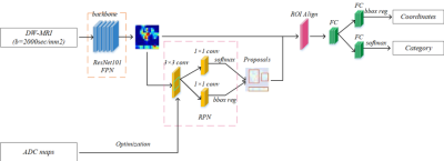 |
The feasibility of an optimized Faster R-CNN in detection and differentiation HT from PTMC Using high b-value DWI with RESOLVE
ChengLong Deng1,2, BingChao Wu1,2, QingJun Wang3, QingLei Shi4, Bei Guan1,2, DaCheng Qu5, and YongJi Wang*1,2,6
1Collaborative Innovation Center, Institute of Software, Chinese Academy of Sciences, Beijing, China, 2University of Chinese Academy of Sciences, Beijing, China, 3Department of Radiology, PLA 6th medical center, Beijing, China, 4MR Scientific Marketing, Siemens Healthcare, Beijing, China, 5School of Computer Science and Technology, Beijing Institute of Technology, Beijing, China, 6State Key Laboratory of Computer Science, Institute of Software, Chinese Academy of Sciences, Beijing, China
In this study, we optimized a Faster R-CNN algorithm through constraining anchor boxes generated by Region Proposal Network (RPN) based on prior knowledge, and evaluated the feasibility of the optimized model in detecting and differentiating Hashimoto's thyroiditis (HT) from papillary thyroid microcarcinomas (PTMC) based on high b-value (2000 sec/mm2) diffusion-weighted images that acquired with readout segmentation of long variable echo-trains (RESOLVE) sequence. The study indicated that our model based on high b-value (2000 sec/mm2) DWI images demonstrated great potential as a new inspection tool in the diagnosis of benign and malignant thyroid micronodules.
|
|
|
0606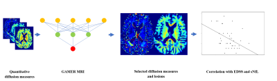 |
Identification of diffusion-based micro-structural measures most sensitive to multiple sclerosis focal damage using GAMER-MRI Video Permission Withheld
Po-Jui Lu1,2,3, Muhamed Barakovic1,2,3, Matthias Weigel1,2,3,4, Reza Rahmanzadeh1,2,3, Riccardo Galbusera1,2,3, Simona Schiavi5, Alessandro Daducci5, Francesco La Rosa6,7,8, Meritxell Bach Cuadra6,7,8, Robin Sandkühler9, Jens Kuhle2,3, Ludwig Kappos2,3, Philippe Cattin9,
and Cristina Granziera1,2,3
1Translational Imaging in Neurology (ThINk) Basel, Department of Biomedical Engineering, University Hospital Basel and University of Basel, Basel, Switzerland, 2Neurology Clinic and Policlinic, Departments of Medicine, Clinical Research and Biomedical Engineering, University Hospital Basel and University of Basel, Basel, Switzerland, 3Research Center for Clinical Neuroimmunology and Neuroscience (RC2NB) Basel, University Hospital Basel and University of Basel, Basel, Switzerland, 4Division of Radiological Physics, Department of Radiology, University Hospital Basel, Basel, Switzerland, 5Department of Computer Science, University of Verona, Verona, Italy, 6Signal Processing Laboratory (LTS5), Ecole Polytechnique Fédérale de Lausanne, Lausanne, Switzerland, 7Medical Image Analysis Laboratory, Center for Biomedical Imaging (CIBM), University of Lausanne, Lausanne, Switzerland, 8Department of Radiology, Lausanne University Hospital and University of Lausanne, Lausanne, Switzerland, 9Center for medical Image Analysis & Navigation, Department of Biomedical Engineering, University of Basel, Allschwil, Switzerland
We applied an attention-based convolutional neural network to select discriminating diffusion measures derived from mathematical models of multi-shell diffusion data in the classification of multiple sclerosis lesions. Further, we correlated the selected measures or their combinations with the Expanded Disability Status Scale (EDSS) and the serum level of neurofilament light chain (sNfL). Our results show that the combinations have stronger correlations with EDSS and sNfL than the individual measures. The proposed method might be useful for selecting the microstructural measures most discriminative of focal tissue damage and identifying the combination most related to clinical disability and neuroaxonal damage.
|
|
0607.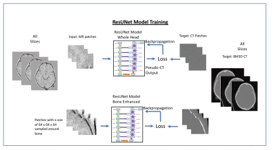 |
Deep Learning-based high-resolution pseudo-CT to detect cranial bone abnormalities for pediatric patients using MRI
Parna Eshraghi Boroojeni1, Yasheng Chen2, Paul K. Commean1, Cihat Eldeniz1, Udayabhanu Jammalamadaka1, Gary B. Skolnick3, Kamlesh B. Patel3, and Hongyu An1
1Mallinckrodt Institute of Radiology, Washington University in St. Louis, Saint louis, MO, United States, 2Department of Neurology, Washington University in St. Louis, Saint louis, MO, United States, 3Division of Plastic and Reconstructive Surgery, Washington University in St. Louis, Saint louis, MO, United States
Computed tomography (CT) scans are commonly used in pediatric patients with head trauma and craniosynostosis to identify skull fractures and sutures, respectively. However, the ionizing radiation associated with the CT scans increases the pediatric patients’ risk for cancer. We developed a deep learning-based method, which consists of two networks focusing on skull and head separately, to generate high-resolution pseudo-CT (pCT) from a radial MR scan. A Dice coefficient of 0.90 ± 0.02 was obtained in the bone.Moreover, a pCT mean absolute error (MAE) of 87.5 ± 4.4 HU was achieved.
|
||
|
0608.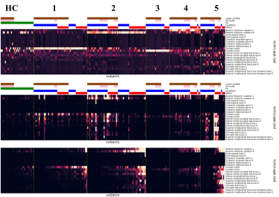 |
A unsupervised machine learning approach for classification of white matter hyperintensity patterns applied to Systemic Lupus Erythematosus.
Theodor Rumetshofer1, Francesca Inglese2, Jeroen de Bresser2, Peter Mannfolk3, Olof Strandberg4, Markus Nilsson1, Itamar Ronen2, Andreas Jönsen5, Linda Knutsson6,7, Tom Huizinga8, Gerda Steup-Beekman8, and Pia Sundgren1,9,10
1Clinical Science Lund / Diagnostic Radiology, Lund University, Lund, Sweden, 2Department of Radiology, Leiden University Medical Center, Leiden, Netherlands, 3Department of Medical Imaging and Physiology, Skåne University Hospital, Lund, Sweden, 4Clinical Memory Research Unit, Department of Clinical Sciences, Malmö, Lund University, Lund, Sweden, 5Department of Rheumatology, Lund University, Skåne University Hospital, Lund, Sweden, 6Department of Medical Radiation Physics, Lund University, Lund, Sweden, 7Russell H. Morgan Department of Radiology and Radiological Science, Johns Hopkins University School of Medicine, Baltimore, MD, United States, 8Department of Rheumatology, Leiden University Medical Center, Leiden, Netherlands, 9Department of Clinical Sciences/Centre for Imaging and Function, Skåne University Hospital, Lund, Sweden, 10Lund University BioImaging Center, Lund University, Lund, Sweden
White Matter Hyperintensities (WMH) are common clinical neuroimaging brain markers. However, WMH in Systemic Lupus Erythematosus (SLE) are non-specific. For this purpose, we developed and unsupervised machine learning approach based on individual WMH distribution to unveil hidden MRI phenotypes. Cluster analysis was performed on a two-site SLE dataset with significant different WMH burden and MRI acquisition protocols. The resulting MRI phenotypes show a clear lesion pattern on distinct WM tracts. This approach reduces the influence of the total WMH burden and MRI acquisition parameters and improves WMH characterization in SLE.
|
The International Society for Magnetic Resonance in Medicine is accredited by the Accreditation Council for Continuing Medical Education to provide continuing medical education for physicians.