Oral
Epilepsy & TBI: Damaged Brains
ISMRM & SMRT Annual Meeting • 15-20 May 2021

| Concurrent 5 | 12:00 - 14:00 | Moderators: David Abbott & Cornelia Laule |
 |
0037.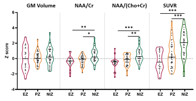 |
Simultaneous 18F-FDG-PET and 1H-MRSI in Temporal Lobe Epilepsy Reveals Metabolic Alterations Concordant with SEEG Epileptogenicity
Hui Huang1, Jia Wang1, Miao Zhang2, Wei Liu3, Lihong Tang1, Yibo Zhao4,5, Rong Guo4,5, Yudu Li4,5, Zhi-Pei Liang4,5, Yao Li1, Biao Li2, and Jie Luo1
1School of Biomedical Engineering, Shanghai Jiao Tong University, Shanghai, China, 2Department of Nuclear Medicine, Ruijin Hospital, Shanghai Jiao Tong University School of Medicine, Shanghai, China, 3Department of Neurosurgery, Ruijin Hospital, Shanghai Jiao Tong University School of Medicine, Shanghai, China, 4Department of Electrical and Computer Engineering, University of Illinois at Urbana Champaign, Urbana, IL, United States, 5Beckman Institute for Advanced Sciences and Technology, University of Illinois at Urbana Champaign, Urbana, IL, United States
Both PET and MRSI could provide metabolic information of the epileptogenic zone, which could add value to presurgical planning of epilepsy patients. This study investigated metabolic alternations in patients with temporal lobe epilepsy (TLE) across their brain regions with different epileptogenicity, as defined using stereo-EEG (SEEG). Our experimental results showed FDG hypometabolism and NAA decrease in epileptogenic zone and propagation zone. These findings may lay a foundation for further investigation of tissue damage associated with epileptogenicity using high-resolution metabolic imaging.
|
|
|
0038.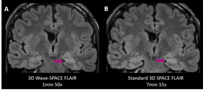 |
Highly Accelerated Wave-CAIPI 3D SPACE FLAIR Compared to Standard 3D SPACE FLAIR for Epilepsy Imaging at 3T
Augusto Lio M. Goncalves Filho1,2, Chanon Ngamsombat3, Stephen F. Cauley2, Wei Liu4, Daniel N. Splitthoff5, Wei-Ching Lo6, John E. Kirsch1, Pamela W. Schaefer1, Otto Rapalino1, Susie Y. Huang1,2, and John Conklin1,2
1Department of Radiology, Massachusetts General Hospital, Boston, MA, United States, 2Department of Radiology, Athinoula A. Martinos Center for Biomedical Imaging, Charlestown, MA, United States, 3Department of Radiology, Siriraj Hospital, Bangkok, Thailand, 4Siemens Shenzhen Magnetic Resonance Ltd., Shenzhen, China, 5Siemens Healthcare GmbH, Erlangen, Germany, 6Siemens Medical Solutions Inc., Boston, MA, United States
We performed a systematic comparison of highly accelerated Wave-CAIPI 3D SPACE FLAIR versus standard 3D SPACE FLAIR for the imaging evaluation of 77 patients with seizures or established epilepsy undergoing 3T MRI. There were no significant differences between the two sequences for the detection of lesions and overall diagnostic quality, despite a 2.5- to 4-fold decrease in acquisition time using Wave-CAIPI SPACE FLAIR. The application of highly accelerated 3D imaging using Wave-CAIPI technology may improve use of MRI resources while reducing motion artifacts and patient anxiety.
|
|
 |
0039.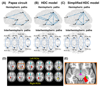 |
Seizure frequency in relation to effective connectivity of the hippocampal–diencephalic–cingulate in temporal lobe epilepsy
Yao-Chia Shih1,2,3, Fa-Hsuan Lin4,5, Aeden Kuek Zi Cheng1, Horng-Huei Liou6,7, and Wen-Yih Isaac Tseng3,7,8,9
1Department of Diagnostic Radiology, Singapore General Hospital, Singapore, Singapore, 2Duke-NUS Medical School, Singapore, Singapore, 3Institute of Medical Device and Imaging, College of Medicine, National Taiwan University, Taipei, Taiwan, 4Department of Medical Biophysics, University of Toronto, Toronto, ON, Canada, 5Physical Sciences Platform, Sunnybrook Research Institute, Toronto, ON, Canada, 6Department of Neurology, National Taiwan University Hospital and College of Medicine, Taipei, Taiwan, 7Graduate Institute of Brain and Mind Sciences, College of Medicine, National Taiwan University, Taipei, Taiwan, 8Department of Medical Imaging, National Taiwan University Hospital and College of Medicine, Taipei, Taiwan, 9Molecular Imaging Center, National Taiwan University, Taipei, Taiwan
To seek neural correlates of seizure recurrence, the structural equation modeling (SEM) and resting-state functional MRI were performed to evaluate intrinsic effective connectivity (iEC) within the Papez circuit, hippocampal–diencephalic–cingulate (HDC) model, and simplified HDC model in patients with left and right temporal lobe epilepsy. We verified that the simplified HDC model was the best model to estimate iEC and found associations between seizure frequency and aberrant iEC on the paths connecting to the mammillary body. Our findings could facilitate the discovery of potential epilepsy pathways and the development of novel targeted therapies for unilateral temporal lobe epilepsy.
|
|
0040. |
Mild Traumatic Brain Injury Predisposes to Thalamic Reticular Nucleus Impairment and Thalamocortical Dysrhythmia
Yi-Tien Li1,2, Duen-Pang Kuo2,3, Yun-Ting Lee2, Yung-Chieh Chen2,3, Hsiao-Wen Chung4, and Cheng-Yu Chen2,3,5,6
1Neuroscience Research Center, Taipei Medical University, Taipei, Taiwan, 2Translational Imaging Research Center, Taipei Medical University Hospital, Taipei, Taiwan, 3Department of Medical Imaging, Taipei Medical University Hospital, Taipei, Taiwan, 4Graduate Institute of Biomedical Electrics and Bioinformatics, Taipei, Taiwan, 5Department of Radiology, School of Medicine, College of Medicine, Taipei Medical University, Taipei, Taiwan, 6Research Center for Artificial Intelligence in Medicine, Taipei Medical University, Taipei, Taiwan
This study is the first to provide strong evidence that thalamocortical dysrhythmia (TCD) is involved in mild traumatic brain injury (mTBI) and plays a crucial role in protracted symptoms. The impaired cortical–thalamic tracts and thalamic reticular nuclei are recognized as two pathomechanisms of TCD in mTBI. TCD-induced thalamocortical disinhibition, such as within-thalamus hyperconnectivity, widespread low-frequency thalamocortical coherence, and thalamo-default-mode network disinhibition, are associated with patients’ prolonged symptoms, which were consistently presented at 1- and 2-year follow-ups. Our systematic analysis strengthens understanding of TCD involvement in mTBI and provides future directions for diagnosis, prognosis, and treatment of long-lasting symptoms in mTBI.
|
||
0041.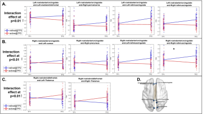 |
Improvements in neuropsychological functioning and recoveries in brain structures and functions in inactive professional fighters
Xiaowei Zhuang1,2, Lauren Bennett3, Virendra Mishra1, Zhengshi Yang1,2, Karthik Sreenivasan1,2, Aaron Ritter1, Charles Bernick1,4, and Dietmar Cordes1,2,5
1Lou Ruvo Center For Brain Health, Cleveland Clinic, Las Vegas, NV, United States, 2University of Nevada, Las Vegas, Las Vegas, NV, United States, 3Hoag Memorial Hospital Presbyterian, Newport Beach, CA, United States, 4UW Medicine, Seattle, WA, United States, 5University of Colorado Boulder, Boulder, CO, United States
Longitudinal changes in fighters’ cognitive performance and brain structural (cortical thickness) and functional (seeded functional connectivity) measures following their transitions to inactive fighting status were investigated and compared with fighters who remain active. A linear mixed effect model was applied for each measure. When fighters transitioned to inactive status, improvements in cognitive performances, structural thickness measures and related functional connectivity measures are evident. In contrast, in fighters who continue to compete in professional matches, neuropsychological performances and structural and functional brain measures are observed to remain largely stable or reflect subtle declines across time points.
|
||
0042.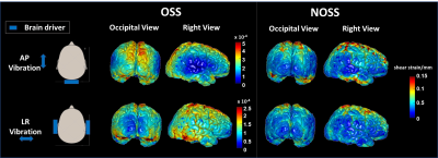 |
MRI-based assessment of regional patterns of cortical strain in the human brain resulting from non-impact dynamic mechanical loading
Ziying Yin1, Matthew C. Murphy1, Yi Sui1, Armando Manduca2, Richard L. Ehman1, and John III Huston1
1Radiology, Mayo Clinic, Rochester, MN, United States, 2Physiology and Biomedical Engineering, Mayo Clinic, Rochester, MN, United States
There is growing recognition of morbidity resulting from subconcussive repetitive head impact (RHI). Mechanical interactions between the skull-brain interface (e.g., transmission and tethering) contribute significantly to the response of the brain to head impact. Local cortical strain concentrations would likely reflect the nearby tethering interactions at the skull-brain interface. In this study, we have developed MR elastography (MRE)-based methods that enable in vivo visualization and quantification of 3D full-volume cortical strain in response to non-impact dynamic loading. We have found that the distribution of the cortical strain is region-dependent, constituting a possible mechanism for RHI vulnerability among individuals.
|
||
|
0043.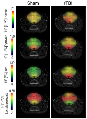 |
Brain metabolic impairment after mild repetitive traumatic brain injury can be measured by hyperpolarized [1-13C]pyruvate and [13C]urea
Caroline Guglielmetti1,2, Kai Qiao1,2, Brice Tiret1,2, Karen Krukowski1,3, Amber Nolan3,4, Susanna Rosi1,3,5,6, and Myriam M. Chaumeil1,2
1Department of Physical Therapy and Rehabilitation Science, University of California San Francisco, San Francisco, CA, United States, 2Department of Radiology and Biomedical Sciences, University of California San Francisco, San Francisco, CA, United States, 3Brain and Spinal Injury Center, University of California San Francisco, San Francisco, CA, United States, 4Department of Pathology, University of California San Francisco, San Francisco, CA, United States, 5Department of Neurological Surgery, University of California San Francisco, San Francisco, CA, United States, 6Weill institute for Neuroscience, University of California San Francisco, San Francisco, CA, United States
We used hyperpolarized 13C magnetic resonance spectroscopic imaging (HP 13C MRSI), T1- and T2-MRI to detect brain alterations in a mouse model of mild repetitive traumatic brain injury (rTBI). T1/T2-MRI did not detect brain damages. HP 13C MRSI detected metabolic changes in cortical areas, with decreased HP lactate/pyruvate and pyruvate dehydrogenase activity in rTBI. Interestingly, HP pyruvate and HP urea increased in rTBI, suggesting vascular and/or blood brain barrier alterations. Altogether, we demonstrated that HP 13C MRSI has potential to detect long-lasting metabolic alterations following rTBI and holds great potential for improving diagnosis and monitoring of rTBI in clinical practice.
|
|
|
0044.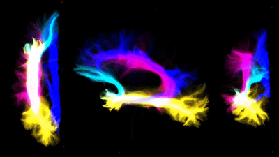 |
Assessing the impact of cerebro-cerebellar and long association fibers in Temporal Lobe Epilepsy: a tractography based study
Nicolò Rolandi1, Fulvia Palesi1,2, Francesco Padelli3, Isabella Giachetti3, Domenico Acquino3, Paul Summers4, Giancarlo Germani4, Gerardo Salvato1,5,6, Valeria Mariani5, Pina Scarpa5,6, Egidio D'Angelo1,2, Gabriella Bottini1,5,6, Laura Tassi5,
Paolo Vitali4,7, and Claudia Angela Michela Gandini Wheeler-Kingshott1,2,8
1Department of Brain and Behavioral Science, University of Pavia, Pavia, Italy, 2Brain Connectivity Center Research Deparment, IRCCS Mondino Foundation, Pavia, Italy, 3I.R.C.C.S. Istituto Neurologico Carlo Besta, Milano, Italy, 4Neuroradiology Unit, IRCCS Mondino Foundation, Pavia, Italy, 5Hospital Niguarda, Milano, Italy, 6Milan Center for Neuroscience, Milano, Italy, 7Department of Radiology, IRCCS Policlinico San Donato, Milano, Italy, 8NMR Research Unit, Queen Square MS Centre, Department of Neuroinflammation, UCL Queen Square Institute of Neurology, Faculty of brain Sciences, University College London (UCL), London, United Kingdom
Advance tractography were performed on cerebro-cerebellar and long association fibers, characterized with diffusion tensor imaging, diffusion kurtosis imaging and NODDI parameter maps. The aim of this work is investigate whether white matter alterations of specific tracts can be selectively related to disfunction of declarative long term memory. Our findings show how relationship between microstructural alterations and neuropsychological scores should be investigated taking account in specific area of white matter restriced to tracts or bundle.
|
|
|
0045.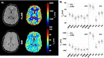 |
Evaluation of T1 and T2 from MR Fingerprinting as Markers for Predicting Patient Recovery in Mild Traumatic Brain Injury
TERESA GERHALTER1, Martijn Cloos2, Seena Dehkharghani1, Anna M. Chen1, Rosermary Peralta1, Fatemeh Adlparvar1, James S. Babb1, Tamara Bushnik3, Jonathan M. Silver4, Brian S. Im3, Stephen P. Wall5, Guillaume Madelin1, and Ivan Kirov1
1Center for Biomedical Imaging, Department of Radiology, New York University Grossman School of Medicine, NEW YORK, NY, United States, 2Centre for Advanced Imaging, The University of Queensland, Brisbane, Australia, 3Department of Rehabilitation Medicine, New York University Grossman School of Medicine, NEW YORK, NY, United States, 4Department of Psychiatry, New York University Grossman School of Medicine, NEW YORK, NY, United States, 5Ronald O. Perelman Department of Emergency Medicine, New York University Grossman School of Medicine, NEW YORK, NY, United States
We analyzed brain MRI data including clinical imaging and MR fingerprinting (MRF) of 22 mild traumatic brain injury (mTBI) patients measured ~1 month after injury and 18 healthy controls. T1 and T2 values in mTBI were not significantly different from controls’. However, increased T1 of three brain regions enabled the identification of non-recovered patients at 3-months (AUC=0.80-0.88). This suggests that T1 quantification is more sensitive to mTBI damage than T2, and is a potential candidate to predict patient outcome.
|
|
|
0046.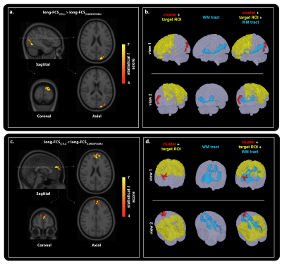 |
Alterations in Network Connectivity Within Special Forces Military Personnel: A Combined Resting-FMRI and DTI Study
Allen A Champagne1, Nicole S Coverdale2, Andrew Ross3, Christopher I Murray3, Isabelle Vallee4, and Douglas J Cook5
1School of Medicine, Queen's University, Kingston, ON, Canada, 2Center for Neuroscience Studies, Queen's University, Kingston, ON, Canada, 3Performance Phenomics, Toronto, ON, Canada, 4National Defence Headquarters, Ottawa, ON, Canada, 5Department of Surgery, Queen’s University, Kingston, ON, Canada
Chronic exposure to head trauma in Special Forces personnel may provide a mechanism for changes in connectivity making-up the architectural organization of functional hubs. Here, resting-state MRI and diffusion tensor imaging are integrated to highlight interdependent differences in functional and structural connectivity of Canadian military Special Operations Forces personnel, when compared to civilian. Changes in white matter integrity of fibers directly connecting functional nodes were observed, which may explain, at least in part, changes in functional markers within networks. These findings suggest a potential structural compensatory relationship between axonal injury and neural recruitment following head trauma from high-exposure military duties.
|
The International Society for Magnetic Resonance in Medicine is accredited by the Accreditation Council for Continuing Medical Education to provide continuing medical education for physicians.