Oral
MRI in Stroke: Oxygen, Metabolism & Tissue Function
ISMRM & SMRT Annual Meeting • 15-20 May 2021

Oral
MRI in Stroke: Oxygen, Metabolism & Tissue Function
| Concurrent 6 | 18:00 - 20:00 | Moderators: Alex Bhogal & Khin Tha |
 |
0869.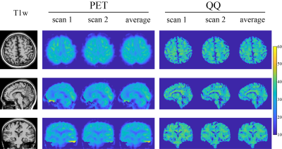 |
Cerebral oxygen extraction fraction (OEF): comparison of challenge-free gradient echo QSM+qBOLD (QQ) with 15O PET in healthy adults
Junghun Cho1, John Lee2, Hongyu An2, Manu S Goyal2, Yi Su3, and Yi Wang1
1Weill Cornell Medicine, New York, NY, United States, 2Washington University School of Medicine, Saint Louis, MO, United States, 3Banner Alzheimers Institute, Phoenix, AZ, United States
Cerebral oxygen extraction fraction (OEF) maps are critical to investigate salvageable tissue in ischemic stroke. We compare OEF maps obtained using quantitative susceptibility mapping plus quantitative blood oxygen level-dependent modeling (QSM+qBOLD=QQ) with the reference standard OEF maps obtained using 15O PET in 10 healthy adults. The whole brain and regional average OEF values were found to be substantially equivalent between the two methods, e.g. 32.8 ± 6.7 % on PET and 34.2 ± 2.6 % on QQ (p=0.002, TOST) for whole brain average. QQ estimates OEF maps from a single routine MRI sequence without burdensome gas inhalation or respiratory-control procedures.
|
|
0870.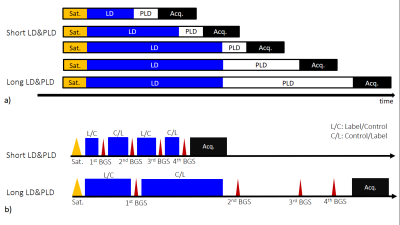 |
Pseudo-Continuous Arterial Spin Labeling using Multiple Label- and Post-Label Duration with Dynamically Optimized Background Suppression
Makoto Obara1, Osamu Togao2, Tatsuhiro Wada3, Chiaki Tokunaga3, Ryoji Mikayama3, Hiroshi Hamano1, Kim van de Ven4, Masami Yoneyama1, Tetsuo Ogino1, Yuta Akamine1, Yu Ueda1, Jihun Kwon1, and Marc Van Cauteren5
1Philips Japan, Tokyo, Japan, 2Department of Molecular Imaging & Diagnosis, Graduate School of Medical Sciences, Kyushu University, Fukuoka, Japan, 3Division of Radiology, Department of Medical Technology, Kyushu University Hospital, Fukuoka, Japan, 4Philips Healthcare, Best, Netherlands, 5Philips Healthcare, Tokyo, Japan
The arterial transit time (ATT) calculated from multiple time points pseudo continuous arterial spin labeling (pCASL) has recently attracted attention. To calculate ATT accurately, while ensuring reliable SNR, we propose multiple repetition time (multi-TR) scheme, that is TR is varied according to the label duration (LD) and post label delay (PLD) with dynamically optimized BGS and 3D acquisition. The multi points data is efficiently acquired and scan time is less than 3 minutes for whole brain coverage. We conducted a feasibility test. Reliable background suppression effectiveness for all time points and a significant SNR gain were confirmed in healthy volunteers.
|
||
 |
0871.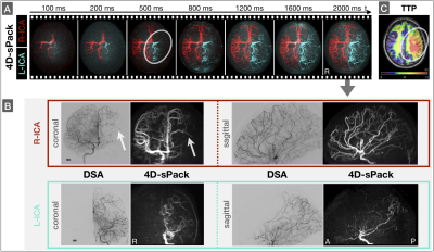 |
Clinical application of ASL-based non-invasive perfusion territory mapping and time-resolved angiography in cerebrovascular diseases
Stephan Kaczmarz1, Miriam Reichert1, Moritz Roman Hernandez Petzsche1, Andreas Hock2, Kilian Weiss3, Kim van de Ven4, Christine Preibisch1, Jan Kirschke1, Claus Zimmer1, Makoto Obara5, Michael Helle6, Nico Sollmann1,7, and Hans Liebl1
1School of Medicine, Department of Neuroradiology, Technical University of Munich (TUM), Munich, Germany, 2Philips Healthcare, Horgen, Switzerland, 3Philips Healthcare, Hamburg, Germany, 4Philips Healthcare, Best, Netherlands, 5Philips Japan, Tokyo, Japan, 6University Hospital Schleswig-Holstein Campus Kiel, Kiel, Germany, 7Department of Radiology, University Ulm Medical Center, Ulm, Germany
Imaging of blood supply patterns is of high clinical interest to improve diagnostics in cerebrovascular diseases (CVD). However, the only clinically available imaging method is invasive digital subtraction angiography (DSA). We present data from 17 participants, including 7 CVD patients, with non-invasive MRI-based perfusion territory mapping by super-selective arterial spin labeling (ss-ASL) and time-resolved angiography by 4D-sPack with automated planning. Our results successfully confirmed automated planning even in CVD patients. Collateral pathways were non-invasively depicted by ss-ASL and 4D-sPack. Moreover, congruent results with DSA, contrast-enhanced MR-angiography and perfusion MRI demonstrated great potential for future routine application of ss-ASL and 4D-sPack.
|
|
0872.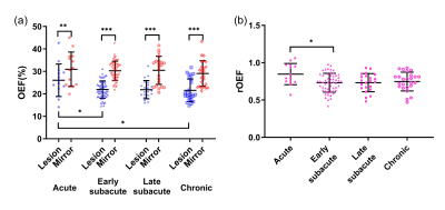 |
Application of MRI-based oxygen extraction fraction mapping in ischemic stroke
Di Wu1 and Shun Zhang1
1Tongji Hospital, Tongji Medical College, Huazhong University of Science and Technology, Wuhan, Hubei, China
This study aims to investigate spatiotemporal evolution of cerebral oxygen extraction fraction (OEF) in ischemic stroke with an MRI-based vascular-challenge-free method. The OEF of the infarcted area descends continuously from acute to chronic phase. Meanwhile, there exists tissue that is likely to be penumbra in the acute diffusion lesion, of which the OEF shows an increasing trend with time, suggesting timely reperfusion in this region. This MRI-based OEF mapping can precisely capture the heterogenous character of brain oxygen metabolism at different phases of ischemic stroke.
|
||
0873.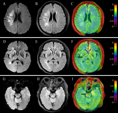 |
Prediction of Wallerian Degeneration in the Corticospinal Tract after Cerebral Ischemic Stroke: A Pilot APT and DWI Study
junxin wang1, yanwei Miao1, and Jiazheng Wang2
1Department of Radiology, the First Affiliated Hospital of Dalian Medical University, Dalian, China, 2Phillips healthcare, dalian, China
This study aimed to exploit the early Wallerian degeneration (WD) along the corticospinal tract following cerebral ischemic stroke using amide proton transfer weighted (APTw) and diffusion weighted imaging (DWI). This study included 34 patients with acute cerebral infarction and 21 healthy adult controls. Our data suggested that APTw and apparent diffusion coefficient (ADC) values was significantly increased on ipsilateral compared to contralateral and the control group in the posterior limb of internal capsule. It proves the feasibility of APTw and ADC in early detection of WD following cerebral ischemic stroke.
|
||
0874.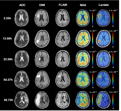 |
Assessment of Symptom Onset Time in Ischemic Stroke Patients Using Fast High-Resolution 3D 1H-MRSI
Zengping Lin1, Tianyao Wang2, Rong Guo3,4, Yudu Li3,4, Yibo Zhao3,4, Tianxiao Zhang1, Jun Liu2, Xin Yu5, Zhi-Pei Liang3,4, and Yao Li1
1School of Biomedical Engineering, Shanghai Jiao Tong University, Shanghai, China, 2Radiology Department, The Fifth People's Hospital of Shanghai, Fudan University, Shanghai, China, 3Beckman Institute for Advanced Science and Technology, University of Illinois at Urbana-Champaign, Urbana, IL, United States, 4Department of Electrical and Computer Engineering, University of Illinois at Urbana-Champaign, Urbana, IL, United States, 5Department of Biomedical Engineering, Case Western Reserve University, Cleveland, OH, United States
Determination of ischemic stroke onset time is critical in ischemic stroke treatment. 1H-MRSI has been recognized as a potentially powerful tool for noninvasive metabolic imaging, which showed great promise for the assessment of stroke onset time. This study investigated changes of neurometabolite concentrations in lesion after different symptom onset time in ischemic stroke patients using a fast high-resolution 3D MRSI technique. Our results showed that NAA concentration within the ischemic lesion decreased in a time-dependent manner after stroke onset, which might provide a useful metabolic biomarker for assessment of symptom onset time.
|
||
0875.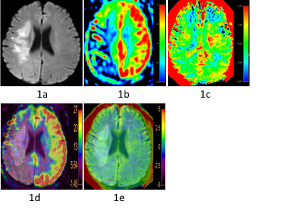 |
Prediction of the Treatment Efficacy of Patients with Subacute Cerebral Infarction using the Heterogeneity of APT-weighted Signals
Yuhan Jiang1, Peipei Chang1, Yingqiu Liuyang1, Bingbing Gao1, Yiwei Che1, Renwang Pu1, Qingwei Song1, Ailian Liu1, Zhiwei Shen2, Jiazheng Wang2, and Yanwei Miao1
1the First Affiliated Hospital of Dalian Medical University, Dalian, China, 2Philips Healthcare, Beijing, China
Amide proton transfer weighted (APTw) MRI has been increasingly applied in the study of stroke based on its ability to detect pH and intracellular proteins content. In subacute infarction, the heterogeneity of APTw signal may reflect the clinical treatment efficacy of patients. In this study, we found that there is negative correlation between the heterogeneity signals (APTwmax-min) in the ischemic regions and the difference of NIHSS scores on the day of hospital admission and leaving (∆NIHSS). Therefore, the heterogeneity of APTw signal is potentially an effective imaging biomarker to predict the efficacy of patients with subacute cerebral infarction.
|
||
0876.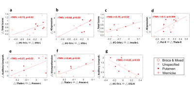 |
Functional Reorganization associated with Language Recovery after Repetitive Transcranial Magnetic Stimulation in Chronic Aphasic Stroke
Bing-Fong Lin1, Po-Yi Tsai2, and Chia-Feng Lu1
1Biomedical Imaging and Radiological Sciences, National Yang Ming University, Taipei, Taiwan, 2Physical Medicine and Rehabilitation, Taipei Veterans General Hospital, Taipei, Taiwan
Low-frequency repetitive transcranial magnetic stimulation (rTMS) provided promising results to facilitate the language recovery in stroke patients with non-fluent aphasia. This study demonstrated that the contralesional inhibitory rTMS treatment can induce the functional reorganization within language networks and recovery of language function in chronic aphasic stroke. The correlation analysis further revealed the association between altered functional connectivity and improved language ability after rTMS treatment. Finally, we reported that the involved functional circuits for the language recovery may depend on lesion location and size of stroke.
|
||
0877.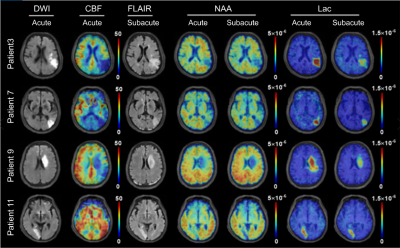 |
Longitudinal Changes in Neurometabolite Levels from Acute to Subacute Stroke: A Pilot Study Using Fast High-resolution 3D 1H-MRSI
Bin Bo1, Tianyao Wang2, Rong Guo3,4, Yudu Li3,4, Yibo Zhao3,4, Tianxiao Zhang1, Zengping Lin1, Ziyu Meng1, Jun Liu2, Xin Yu5, Zhi-Pei Liang3,4, and Yao Li1
1School of Biomedical Engineering, Shanghai Jiao Tong University, Shanghai, China, 2Radiology Department, The Fifth People's Hospital of Shanghai, Fudan University, Shanghai, China, 3Beckman Institute for Advanced Science and Technology, University of Illinois at Urbana-Champaign, Urbana, IL, United States, 4Department of Electrical and Computer Engineering, University of Illinois at Urbana-Champaign, Urbana, IL, United States, 5Department of Biomedical Engineering, Case Western Reserve University, Cleveland, OH, United States
Detection of neurometabolic alterations in stroke patients from acute to subacute stages could provide useful information for brain tissue salvage therapy. Fast high-resolution 3D 1H-MRSI by SPICE has previously provided nearly whole-brain neurometabolic mapping in acute stroke patients. In this pilot study, we investigated the alterations of neurometabolites in a longitudinal cohort of ischemic stroke patients. Our preliminary results showed observable changes of different neurometabolites in different regions within the hypoperfused tissue of stroke patients from acute to subacute stages. Our study may lay a foundation for further investigation of temporal changes of neurometabolic biomarkers during stroke progression.
|
||
0878.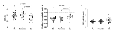 |
Clinical Research of Multi-modal MRI in Ischemic Stroke: Cerebral Oxygen Extraction Fraction, Nonblood Susceptibility and Blood Flow
Di Wu1, Shun Zhang1, and Weiyin Vivian Liu2
1Radiology, Tongji Hospital, Tongji Medical College, Huazhong University of Science and Technology, Wuhan, Hubei, China, 2MR Research, GE Healthcare, Beijing, China, Beijing, China
This study aims to investigate oxygen metabolic, neuroinflammatory and hemodynamic parameters in different parts of the ischemic stroke lesions using multi-modal MRI. Significant difference of cerebral blood flow (CBF) was found between patients with and without penumbra. The oxygen extraction fraction (OEF) and nonblood susceptibility of tissue in the penumbra were significantly different from ischemic core, revealing benign oxygen metabolic compensation and less neuroinflammation in the penumbra. OEF in the diffusion lesion was positively correlated with clinical severity and continuously decreased with time. Multi-modal MRI-based markers can accurately reflect the pathophysiologic discrepancy in different regions of the ischemic stroke lesions.
|
The International Society for Magnetic Resonance in Medicine is accredited by the Accreditation Council for Continuing Medical Education to provide continuing medical education for physicians.