Oral
MRI in Stroke: Vessels, Flow & Tissue Structure
ISMRM & SMRT Annual Meeting • 15-20 May 2021

Oral
MRI in Stroke: Vessels, Flow & Tissue Structure
| Concurrent 6 | 16:00 - 18:00 | Moderators: Thomas Lindner & Lena Vaclavu |
0823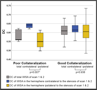 |
Evaluation of primary and secondary collateral pathways in carotid artery stenosis patients before and after revascularization therapy Video Permission Withheld
Lena Schmitzer1, Alexander Seiler2, Nico Sollmann1, Christine Preibisch1,3, Kilian Weiss4, Claus Zimmer1, Fahmeed Hyder5, Jens Göttler1,5, and Stephan Kaczmarz1,5
1School of Medicine, Department of Neuroradiology, Technical University of Munich, Munich, Germany, 2Department of Neurology, Goethe University Frankfurt, Frankfurt, Germany, 3School of Medicine, Clinic of Neurology, Technical University of Munich, Munich, Germany, 4Philips Healthcare, Hamburg, Germany, 5MRRC, Yale University, New Haven, CT, United States
Internal carotid artery stenosis (ICAS) is a well-known risk factor for stroke. While collaterals play a pivotal role in ischemic stroke, their effects in ICAS are not entirely understood. We assessed primary collaterals, i.e., configuration of the Circle of Willis by MR-Angiography, and high Coefficients of Variance in Dynamic Susceptibility Contrast MRI as surrogate of secondary collaterals. Moreover, individual watershed areas (iWSA) were determined in a group of ICAS patients and healthy volunteers. Our results demonstrate post-interventional shifts of iWSA, mainly influenced by primary collateral flow. No secondary collateral flow was found in patients similar to age-matched healthy participants.
|
||
 |
0824.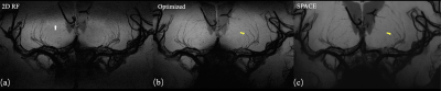 |
Inner volume 3D TSE using optimized spatially selective excitation pulses for vessel wall imaging of intracranial perforating arteries at 7T
Qingle Kong1,2, Yue Wu1, Dehe Weng3, Jing An3, Yan Zhuo1,4, and Zihao Zhang1,4
1Institute of Biophysics, Chinese Academy of Sciences, Beijing, China, 2MR Collaboration, Siemens Healthcare Ltd, Beijing, China, 3Siemens Shenzhen Magnetic Resonance Ltd, Shenzhen, China, 4The Innovation Center of Excellence on Brain Science, Chinese Academy of Sciences, Beijing, China
The impairment of microvessels can lead to neurologic diseases such as stroke and vascular dementia. The imaging of lumen and vessel wall of perforating arteries requires an extremely high resolution due to their small caliber size. In this study, we developed a 3D inner-volume (IV) TSE (SPACE) sequence with optimized spatially selective excitation (SSE) RF pulses. High resolution of isotropic 0.30mm within ten minutes was achieved for the black- blood images of lenticulostriate artery (LSA) for the first time. The IV-SPACE images showed clearer delineation of vessel wall and lumen of LSA than conventional SPACE images.
|
|
 |
0825. |
Evaluation of leptomeningeal collaterals by DSC-based signal variance and hemodynamic features in asymptomatic carotid artery stenosis
Stephan Kaczmarz1,2, Lena Schmitzer1, Jens Göttler1,2, Kilian Weiss3, Christian Sorg1, Claus Zimmer1, Fahmeed Hyder2, Christine Preibisch1, and Alexander Seiler4
1School of Medicine, Department of Neuroradiology, Technical University of Munich (TUM), Munich, Germany, 2MRRC, Yale University, New Haven, CT, United States, 3Philips Healthcare, Hamburg, Germany, 4Department of Neurology, Goethe University Frankfurt, Frankfurt, Germany
Detection of leptomeningeal collateral blood flow has high clinical relevance, but clinically applicable imaging methods are lacking. While a novel approach based on coefficient of variance (CoV) analysis of dynamic susceptibility contrast (DSC) MRI was recently proposed, relations to hemodynamic alterations remained unknown. Moreover, the role of leptomeningeal collaterals in internal carotid artery stenosis (ICAS) is under debate. We present multi-parametric hemodynamic evaluation within high CoV-voxels from 29 asymptomatic ICAS-patients and 30 age-matched healthy controls. Our results suggest no enhanced leptomeningeal collateral recruitment in asymptomatic ICAS. However, hemodynamic characteristics imply detection of voxels that are prone to future leptomeningeal recruitment.
|
|
0826.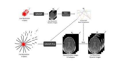 |
GraspMRA: High Temporal Resolution, Non-Contrast Enhanced, Time-Resolved 4D MR Angiography Using Golden-angle Radial Sparse Parallel Imaging
Li Feng1 and Lirong Yan2
1Department of Radiology, Icahn School of Medicine at Mount Sinai, New York, NY, United States, 2USC Stevens Neuroimaging and Informatics Institute, University of Southern California, Los Angeles, CA, United States
Non-contrast enhanced 4D MRA has emerged as a promising approach in charactering flow dynamics. The major challenge of conventional 4D MRA is relatively long scan time. Recently developed 4D MRA combining golden-angle stack-of-stars acquisition and compressed sensing reconstruction can significantly shorten scan time. However, this method only exploits spatial sparsity based on individual frames. The current study aims to test the feasibility of a newly developed low-rank and sparsity-based image reconstruction method to highly accelerate 4D MRA. Our initial results suggest that higher acceleration rates can be achieved using GraspMRA without compromising image quality and temporal fidelity.
|
||
0827.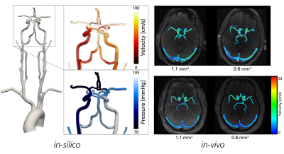 |
Cerebrovascular relative pressure assessment using 4D Flow MRI – accuracy of image-based estimation approaches
David Marlevi1, Jonas Schollenberger2, Maria Aristova3, Edward Ferdian4, Alistair A Young4,5, Elazer R Edelman1, Susanne Schnell3,6, C. Alberto Figueroa2, and David Nordsletten2,5
1Massachusetts Institute of Technology, Cambridge, MA, United States, 2University of Michigan, Ann Arbor, MI, United States, 3Northwestern University, Chicago, IL, United States, 4University of Auckland, Auckland, New Zealand, 5King's College London, London, United Kingdom, 6University of Greifswald, Greifswald, Germany
4D Flow MRI images cerebrovascular blood flow in-vivo, however, estimation of relative pressure is difficult due to the unique flow and anatomies found in the brain. We evaluated the performance of three different techniques (reduced Bernoulli (RB); unsteady Bernoulli (UB); virtual Work-Energy Relative Pressure (vWERP)) for cerebrovascular assessment. Using patient-specific in-silico models, we show that accurate estimations are dependent on sufficient spatial resolution (dx < 0.75 mm3) and used approach (vWERP achieving accurate estimates; RB/UB showing systematic underestimation bias). With similar dependencies indicated in-vivo, these results underline both potentials and challenges of mapping cerebrovascular relative pressure by 4D Flow MRI.
|
||
0828.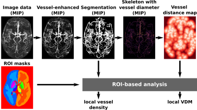 |
Vessel distance mapping for deep gray matter structures
Hendrik Mattern1, Stefanie Schreiber2,3,4, and Oliver Speck1,3,4,5
1Biomedical Magnetic Resonance, Otto-von-Guericke-University Magdeburg, Magdeburg, Germany, 2Department of Neurology, Otto-von-Guericke-University, Magdeburg, Magdeburg, Germany, 3German Center for Neurodegenerative Disease, Magdeburg, Germany, 4Center for Behavioral Brain Sciences, Magdeburg, Germany, 5Leibniz Institute for Neurobiology, Magdeburg, Germany
Vessel distance mapping (VDM) is proposed as a novel tool to enable quantitative and qualitative assessment of vascular patterns in deep gray matter structures. At 7T, ToF angiography and QSM were used to depict the arterial and venous vasculature in six subjects. Based on vessel segmentations, vessel distance maps were generated by computing the Euclidean distance of each non-vessel voxel to its closest vessel. Compared to state-of-the-art methods, VDM interpolates the sparse vessel data to enable new ways to analyze vascular patterns with respect to the surrounding structure.
|
||
0829.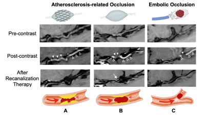 |
Recanalization of Acute Intracranial Large Vessel Occlusions: Novel Findings from High-Resolution Vessel Wall Imaging
Chen Cao1,2, Jing Lei2, Yan Gong3, Song Jin2, Jinxia Zhu4, Ming Wei5, and Shuang Xia6
1Department of Radiology, First Central Clinical College, Tianjin Medical University, Tianjin, China, 2Department of Radiology, Tianjin Huanhu Hospital, Tianjin, China, 3Department of Radiology, Tianjin Medical University Nankai Hospital, Tianjin, China, 4MR Collaboration, Siemens Healthcare Ltd., Beijing, China, 5Department of Neurosurgery, Tianjin Huanhu Hospital, Tianjin, China, 6Department of Radiology, Tianjin First Central Hospital, School of Medicine, Nankai University, Tianjin, China
Pre-operative recanalization assessment for acute intracranial large-vessel occlusion (LVO) can help optimize endovascular therapy and shorten procedural times. We prospectively examined 46 patients with acute intracranial LVO who underwent high-resolution magnetic resonance imaging (HRMRI) before endovascular therapy. HRMRI had good agreement with angiographic assessment of the causes of occlusion (κ=0.89, 95% CI, 0.69–1.00) and length of occlusion (concordance correlation coefficient=0.75, 95% CI, 0.59–0.86). Intraluminal enhancement was associated with procedural complexity (r=0.81, P< .001) and procedural times (r=0.64, P< .001). HRMRI before recanalization can help define the vascular status and assist with endovascular therapy of acute intracranial LVOs.
|
||
0830.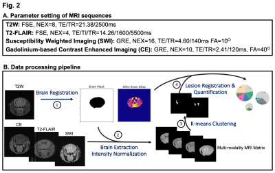 |
Characterization of radiological findings in mouse models of cerebral small vessel diseases using multimodal MRI at 14.1 Tesla
Xiao Gao1, Xiaowei Wang2, Kai Qiao1, Cassandre Labelle-Dumais2, Douglas Gould2, and Myriam M. Chaumeil1
1Department of Radiology, University of California, San Francisco, San Francisco, CA, United States, 2Department of Ophthalmology, University of California, San Francisco, San Francisco, CA, United States
Cerebral small vessel diseases (cSVDs) are a group of conditions that account for up to 30% of strokes. Clinical manifestations associated with cSVD include blood–brain barrier (BBB) leakage, microbleeds, and white matter lesions. Here, we use multimodal MRI at 14.1Tesla to characterize radiological findings observed in innovative mouse models of cSVD caused by mutations in Collagen type IV alpha 1 (Col4a1). By leveraging several image processing toolboxes, we established a workflow that could successfully differentiate between disease subtypes based on the spatial distribution and volume of lesions.
|
||
0831.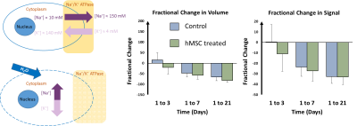 |
Mechanisms of cerebral ischemic stroke recovery from stem cell derived therapies assessed via MRI at 21.1 T
Shannon Helsper1,2, Xuegang Yuan1,2, and Samuel Colles Grant1,2
1National High Magnetic Field Laboratory, Florida State University, Tallahassee, FL, United States, 2Chemical & Biomedical Engineering, FAMU-FSU College of Engineering, Tallahassee, FL, United States
This study evaluates therapeutic efficacy of human mesenchymal stem cell (hMSC) derived treatments applied to an ischemic stroke rat model. The goal is to determine if the presence of hMSC or hMSC derivatives in an ischemic region is required or if delivery of cell secretions alone can improve outcomes, either locally at the lesion or by recruiting regenerative neural progenitor cells. Biochemical markers of tissue recovery measured longitudinally using sodium chemical shift imaging and relaxation-enhanced MR spectroscopy at 21.1 T enables increased sensitivity, enabling insight into ionic and metabolic regulation while enabling to determine therapy efficacy.
|
||
|
0832.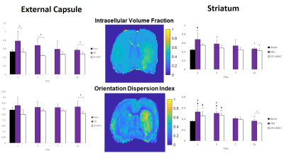 |
Mesenchymal Stem Cell Impacts on Cerebral Microstructural Diffusion Recovery After Ischemic Attack
Frederick A Bagdasarian1,2, Xuegang Yuan1,2, and Samuel Colles Grant1,2
1National High Magnetic Field Laboratory, Florida State University, Tallahassee, FL, United States, 2Chemical & Biomedical Engineering, FAMU-FSU College of Engineering, Tallahassee, FL, United States
DTI, NODDI and DKI techniques were applied to diffusion data acquired at 21.1 T to identify microtissue changes in ischemic brain tissue following adult human mesenchymal stem cell (hMSC) or control treatment. Scanning was conducted 1-21 d post-MCAO and treatment. 2D hMSC significantly reduced cell swelling (ICVF) at nearly every time-point in white matter while preserving orientation integrity (ODI), normal levels of DTI metrics, and early phase kurtosis. In grey matter, 2D hMSC restored DTI metrics to naïve levels quicker than control treatments, as did ICVF and ODI. Kurtosis had high variability, with minor trends evident.
|
The International Society for Magnetic Resonance in Medicine is accredited by the Accreditation Council for Continuing Medical Education to provide continuing medical education for physicians.