Oral
Parkinson & Neurodegeneration
ISMRM & SMRT Annual Meeting • 15-20 May 2021

| Concurrent 6 | 14:00 - 16:00 | Moderators: Emine Can & Khin Tha |
 |
0539. |
Twelve-Year Microstructural Changes in The Deep Gray Nuclei in Parkinson’s Disease: A Serial Diffusion Tensor Imaging Study
Yao-Chia Shih1,2, Qi Rong Leon Ooi3, Septian Hartono2,3, Thomas Welton2,3, Hui-Hua Li2,4, John Carson Allen2, Eng King Tan2,3, and Ling Ling Chan1,2
1Department of Diagnostic Radiology, Singapore General Hospital, Singapore, Singapore, 2Duke-NUS Medical School, Singapore, Singapore, 3Department of Neurology, National Neuroscience Institute (Outram-campus), Singapore, Singapore, 4Health Services Research Unit, Singapore General Hospital, Singapore, Singapore
Diffusion tensor imaging (DTI) characterizes microstructural changes in the basal ganglia in relation to idiopathic Parkinson's disease (PD). However, inconsistent results due to short-interval longitudinal studies with heterogeneous neuropathology across PD stages have been reported. We elucidated microstructural changes in the deep gray nuclei throughout the disease course in a large, prospective, three time-point case-control DTI study in PD over twelve years, with six-year interval gaps. Increased mean striatal diffusivity reflected progressive neurodegeneration, whereas factional anisotropy changes suggested effects of abnormal iron accumulation followed by neuronal loss in the putamen and thalamus as the disease progresses into the late stages.
|
|
0540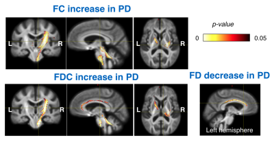 |
Characterizing white matter microstructural alterations in de novo Parkinson’s disease using diffusion MRI Video Permission Withheld
Yiming Xiao1,2, Terry M Peters3,4,5, and Ali R Khan3,4,5,6
1PERFORM Centre, Concordia University, Montreal, QC, Canada, 2Computer Science and Software Engineering, Concordia University, Montreal, QC, Canada, 3Robarts Research Institute, Western University, London, ON, Canada, 4Department of Medical Biophysics, Western University, London, ON, Canada, 5School of Biomedical Engineering, Western University, London, ON, Canada, 6The Brain and Mind Institute, Western University, Lonon, ON, Canada
Understanding microstructural alterations in white matter can be instrumental in revealing pathogenesis and devising effective plans to treat Parkinson’s disease (PD). Diffusion MRI can be used to characterize the status of white matter integrity. With voxel-based analysis using DTI measures and fixel-based analysis (FBA), we demonstrated for the first time strengthened and weakened white matter integrity, which is subject to laterality of motor symptoms in de novo Parkinson’s disease. The findings suggest that the disease gives rise to both functional degeneration and the creation of compensatory networks.
|
||
0541.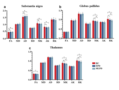 |
Progressive microstructural alterations in subcortical nuclei in Parkinson's disease: a diffusion magnetic resonance imaging study
Xueqin Bai1, Tao Guo1, Xiaojun Guan 1, Cheng Zhou1, Jingjing Wu1, Xiaocao Liu1, Ting Gao1, Luyan Gu1, Xiaojun Xu1, Peiyu Huang1, and Minming Zhang1
1The second Affiliated Hospital, Zhejiang University School of Medicine, Hangzhou, China
In this study, we employed diffusion kurtosis imaging (DKI) and diffusion tensor imaging (DTI) to measure the microstructural alterations in subcortical nuclei across PD patients at different disease stages. Individual diagnostic model was constructed to test the performance of diffusion metrics in identifying PD patients at different stages. We found that PD patients at different stages have progressive microstructural alterations in the main nuclei of widely acknowledged nigral-pallidal and thalamo-cortical pathways. DKI is sensitive to detect microstructural changes in GP and thalamus between early stage PD and moderate-late stage PD patients. The combination of kurtosis and tensor metrics can achieve a good performance in diagnosing PD.
|
||
 |
0542.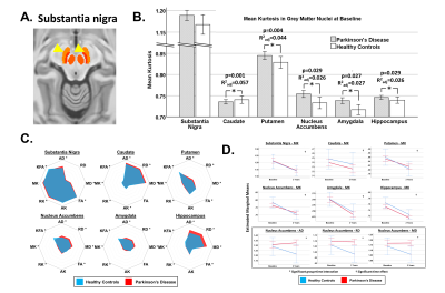 |
Microstructure of grey matter nuclei in early Parkinson’s disease: longitudinal study using diffusion kurtosis imaging
Thomas Welton1,2, Septian Hartono3, Yao-Chia Shih3, Samuel Y-E Ng1, Nicole S Y Chia1, Weiling Lee3, Say Lee Chong3, Eng-King Tan1,2,3, Ling-Ling Chan1,2,3, and Louis CS Tan1,2
1National Neuroscience Institute, Singapore, Singapore, 2Duke-NUS Medical School, Singapore, Singapore, 3Singapore General Hospital, Singapore, Singapore
We show that the diffusion kurtosis characteristics of grey matter nuclei in early Parkinson's disease were abnormal and that this abnormality was maintained over two years. Furthermore, elevated mean kurtosis was associated with worsening motor function. This supports the use of diffusion kurtosis imaging to characterise tissue microstructure and potentially monitor disease progression even in early Parkinson's disease.
|
|
0543.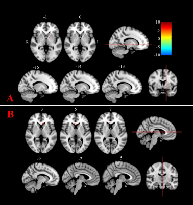 |
Tract Density Imaging in Patients with Parkinson’s Disease Before and After Magnetic Resonance-guided Focused Ultrasound
Yu Shen1, Xianchang Zhang2, Yan Bai1, Rui Zhang1, Rushi Chen1, Wei Wei1, Menghuan Zhang1, and Meiyun Wang1
1Department of Medical Imaging, Henan Provincial People’s Hospital & Zhengzhou University, Zhengzhou, China, 2MR Collaboration, Siemens Healthcare Ltd. Beijing China, Beijing, China
Magnetic resonance-guided focused ultrasound (MRgFUS) is a non-invasive tremor therapy associated with Parkinson disease (PD). Conventional fractional anisotropy analysis might neglect pre- and postoperative microanatomic changes due to low spatial resolution. Finer brain structural depictions could be provided using track density imaging (TDI), a super-resolution reconstruction method. This study conducted TDI on seven patients with PD before and 1-month after MRgFUS and found that tract density values were significantly decreased postoperatively in the genu of the corpus callosum and left globus pallidus. TDI could be a valuable tool for detecting microstructural changes after MRgFUS therapy.
|
||
0544.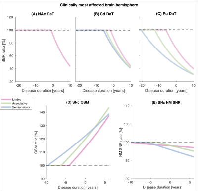 |
Investigating Spatiotemporal Changes in Dopamine, Neuromelanin and Iron in the Nigrostriatal System in Parkinson's Disease
Emma Biondetti1,2,3, Mathieu D. Santin1,2, Romain Valabrègue1,2, Graziella Mangone1,4, Rahul Gaurav1,2,3, Nadya Pyatigorskaya1,3,5, Matthew Hutchison6, Lydia Yahia-Cherif1,2, Nicolas Villain1,7, Marie-Odile Habert8, Isabelle Arnulf1,3,9, Smaranda Leu-Semenescu9, Pauline Dodet9,
Jean-Christophe Corvol1,4,7, Marie Vidailhet1,3,7, and Stéphane Lehéricy1,2,3,5
1Institut du Cerveau – ICM, INSERM U 1127, CNRS UMR 7225, Sorbonne Université, Paris, France, 2ICM, Centre de NeuroImagerie de Recherche – CENIR, Paris, France, 3ICM, Team “Movement Investigations and Therapeutics” (MOV’IT), Paris, France, 4National Institute of Health and Medical Research - INSERM, Clinical Investigation Centre, Pitié-Salpêtrière Hospital, Paris, France, 5Department of Neuroradiology, Pitié-Salpêtrière Hospital, AP-HP, Paris, France, 6Biogen Inc., Cambridge, MA, United States, 7Department of Neurology, Pitié-Salpêtrière Hospital, AP-HP, Paris, France, 8Department of Nuclear Medicine, Pitié-Salpêtrière Hospital, AP-HP, Paris, France, 9National Reference Center for Rare Hypersomnias, Pitié-Salpêtrière Hospital, AP-HP, Paris, France
Parkinson's disease (PD) and idiopathic rapid eye movement sleep behaviour disorder (iRBD, a prodromal condition of Parkinsonism) are characterised by progressive striatal dopaminergic denervation, loss of neuromelanin-containing neurons and increased iron deposition in the substantia nigra. Here, we evaluated neurodegeneration-induced changes in the nigrostriatal system of PD and iRBD patients using longitudinal SPECT and MR imaging compared to healthy control subjects. We showed that dopamine, neuromelanin and iron changes followed similar spatiotemporal gradients of neurodegeneration, and we assessed the temporal onset and ordering of such changes.
|
||
0545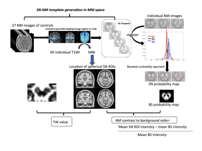 |
Tracking serial Parkinson’s related changes in the substantia nigra using Neuromelanin MRI and free-water diffusion MRI Video Permission Withheld
Yue Xing1,2,3, Saadnah Naidu1,2,3, Halim Abdul-Sapuan1,2,3, Ali-Reza Mohammadi-Nejad2,3, Jonathan Evans4, Ofer Pasternak5, Stamatios Sotiropoulos2,3, Christopher R. Tench1,3, and Dorothee P. Auer1,2,3
1Division of Clinical Neuroscience, Queen’s Medical Centre, University of Nottingham, Nottingham, United Kingdom, 2Sir Peter Mansfield Imaging Centre, University of Nottingham, Nottingham, United Kingdom, 3NIHR Nottingham Biomedical Research Centre, University of Nottingham, Nottingham, United Kingdom, 4Department of Neurology, Nottingham University Hospital Trust, Nottingham, United Kingdom, 5Departments of Psychiatry and Radiology (O.P.), Brigham and Women's Hospital, Harvard Medical School, Boston, MA, United States
Parkinson’s Disease (PD) is characterized by loss of neuromelanin-containing dopaminergic neurons in the substantia nigra (SN), resulting in depigmentation as its pathological hallmark. Neuromelanin (NM)-sensitive-MRI consistently shows PD-related nigral signal-loss with limited evidence for longitudinal change. Nigral free-water (FW) derived from diffusion-MRI can track disease progression. This multimodal-serial study compared subregional annual depigmentation rates and FW in PD and controls. Longitudinal NM signal-loss and FW increase was seen in PD throughout the SN with significant acceleration compared to controls in the ventral-SN. There was no between-metrics correlation, suggesting that these promising serial biomarkers may track different aspects of PD progression.
|
||
0546.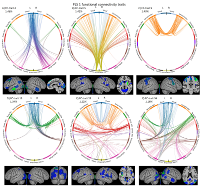 |
Distinct cognitive and anthropometric functional connectivity traits of cognitive decline in Parkinson’s disease using partial least squares.
Vicente Jose Ferrer-Gallardo1, Thomas Bolton2, Manuel Delgado3, Pedro M. Paz-Alonso1, Maricuz Rodriguez-Oroz4, and César Caballero-Gaudes1
1Basque Center on Cognition, Brain and Language, Donostia, Spain, 2Medical image processing, Swiss Federal Institute of Technology Lausanne, Lausanne, Switzerland, 3Department of Neurology, Sierrallana Hospital, Torrelavega, Spain, 4Neurology Department, Clinica Universidad de Navarra, Pamplona, Spain
Cognitive deficits in Parkinson’s Disease (PD) are associated with abnormalities in functional brain networks that can be observed at rest. This study investigates whole-brain independent functional-connectomes in PD patients with mild cognitive impairment (PD-MCI), associating these functional connectomes with cognitive and anthropometric measures using partial least squares (PLS) regression. We found a clear distinction between two sets (PLS-components) with functional connectomes either linked to cognitive scores or anthropometric variables. A PD-specific subcortical-cortical functional connectome was common in the two PLS-components. Furthermore, in PD-MCI a functional connectome defined by attentional and sensorimotor regions linked to attention and memory deficits is critical.
|
||
0547.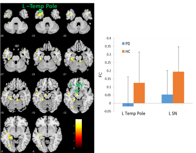 |
Dysfunction of Olfactory Resting-State Functional network in Early-onset Early-stage Parkinson’s Disease
Jianli Wang1, Rachel Stanford1, Lauren Spreen2, Jeffrey Vesek2, Christopher Sica1, Thyagarajan Subramanian3, and Qing X Yang4
1Radiology, Penn State College of Medicine, HERSHEY, PA, United States, 2Molecular Biology, Penn State College of Medicine, HERSHEY, PA, United States, 3Neurology, Penn State College of Medicine, HERSHEY, PA, United States, 4Neurosurgery, Penn State College of Medicine, HERSHEY, PA, United States
Hyposmia is prevalent in Parkinson’s disease (PD) and the central olfactory system is highly affected by PD pathology. Despite the considerable progresses in understanding the pathophysiology of the disease, the mechanism causing hyposmia in PD is still unclear. Given that there is early PD-related neurodegeneration in anterior olfactory nucleus, which is a part of the primary olfactory cortex, we tested the hypothesis that there are PD-related dysfunctions in the central olfactory functional network at the early stage of disease.
|
||
|
0548.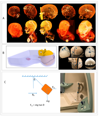 |
Ultra-high field imaging of deep brain stimulation at 7T: The first study of RF safety, displacement force and image artifact
Bhumi Bhusal1, Jason Stockmann2, Azma Mareyam2, John Kirsch2, Lawrence L Wald2, Mark J Nolt1, Joshua Rosenow1, Roberto Lopez-Rosado1, Behzad Elahi1, and Laleh Golestanirad1
1Northwestern University, Chicago, IL, United States, 2Massachusetts General Hospital, Charlestown, MA, United States
We report the results of MRI safety and image artifact assessment of a commercial deep brain stimulation (DBS) system implanted in an anthropomorphic phantom, undergoing MRI at 7T. RF heating was observed to be less than 2°C for all clinically relevant as well as worst-case configurations evaluated in the study. The magnetic force on the pulse generator was found to be within the safe limit. Metal-induced image artifact was comparable to what is observed at lower fields. Our results indicate that 7T MRI could be performed safely in patients with DBS implants under carefully evaluated device model and MRI hardware.
|
The International Society for Magnetic Resonance in Medicine is accredited by the Accreditation Council for Continuing Medical Education to provide continuing medical education for physicians.