Oral
Aging & Dementia
ISMRM & SMRT Annual Meeting • 15-20 May 2021

| Concurrent 6 | 12:00 - 14:00 | Moderators: Frederik Barkhof |
0261.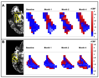 |
Cumulative effects of a statin cocktail on cerebral blood flow and cognitive function in patients with Alzheimer’s Disease
Mohammed Salman Shazeeb1, Elizabeth Degrush2,3, Zeynep Vardar1, Clifford Lindsay1, Matthew Gounis1, and Nils Henninger2,3
1Department of Radiology, University of Massachusetts Medical School, Worcester, MA, United States, 2Department of Neurology, University of Massachusetts Medical School, Worcester, MA, United States, 3Department of Psychiatry, University of Massachusetts Medical School, Worcester, MA, United States
Decreased cerebral metabolism has been implicated in pathogenesis of Alzehimer’s disease. The endothelial nitric oxide synthase (eNOS) pathway plays a major role in cerebral blood flow regulation. This study used DSC-MRI in 10 patients treated with a drug regimen for supporting the eNOS pathway to investigate how perfusion patterns were associated with treatment response based on clinical psychometrics. Regional analysis on rCBF maps showed significant signal changes in the cognitively improved cohort based on ADAS-cog scores while patients that showed cognitive deterioration or no change based on ADAS-cog scores did not show significant rCBF changes.
|
||
0262.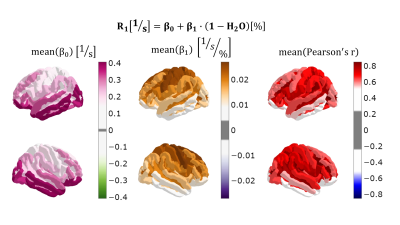 |
The aging quantitative brain: a multiparametric qMRI study
Ana-Maria Oros-Peusquens*1, Jonas Kielmann*1, and N. Jon Shah1,2,3,4
1INM-4, Research Centre Juelich, Juelich, Germany, 2Faculty of Medicine, JARA, RWTH Aachen University, Aachen, Germany, 3INM-11, JARA, Research Centre Juelich, Juelich, Germany, 4Department of Neurology, RWTH Aachen University, Aachen, Germany
Quantitative MRI parameters are determined by the properties of tissue on a microscopic scale and can be expected to reflect microstructural changes created by aging. Here, we investigate a multiparametric qMRI signature of healthy ageing on 26 healthy volunteers, characterised in a high-dimensional parametric space (water content, relaxometry, qMT). Changes with age in the mean values and correlations between parameters are observed and interpreted.
|
||
0263.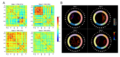 |
Alterations in dynamic functional connectivity in individuals with subjective cognitive decline
Qian Chen1, Jiaming Lu2, Xin Zhang2, Jilei Zhang3, and Bing Zhang1
1Department of Radiology, Drum Tower Hospital, Clinical College of Nanjing Medical University, Nanjing, China, 2Department of Radiology, Drum Tower Hospital, Medical School of Nanjing University, Nanjing, China, 3Philips Healthcare, Shanghai, China
Subjective cognitive decline (SCD) is considered a clinically-based approach for the detection of potential Alzheimer’s disease patients. We observed altered temporal properties of fractional windows, mean dwell time, and the number of transitions by dynamic functional connectivity (DFC) analysis in SCD individuals compared to the control subjects. The altered DFC parameters showed significant associations with cognitive performance. Our findings shed light on the role of DFC in the early detection of subjects with potential Alzheimer’s disease, and the alterations in DFC may suggest the neural basis underlying early cognitive decline in the SCD stage.
|
||
 |
0264. |
Age-related alterations in cortical myelin profile using the Human Connectome Project Aging dataset
Yu Veronica Sui1 and Mariana Lazar1
1Radiology, New York University Grossman School of Medicine, New York, NY, United States
Demyelination is recognized as a major process in both normal aging and neurodegenerative diseases. Using the Human Connectome Project Aging dataset, we investigated intracortical demyelination in normal aging using T1w/T2w maps. To capture the fine changes across cortical layers, we employed a surface-based approach in contrast to the commonly used volumetric approach by constructing a cortical myelin profile for each region, which was then quantified using a nonlinearity index. We showed that the nonlinearity of cortical myelin profile exhibits a steeper decline with aging than cortical thickness and therefore presents the potential of a unique marker of age-related microstructural changes.
|
|
|
0265.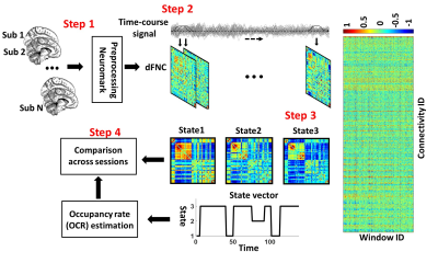 |
Brain instability is a biomarker of Alzheimer’s disease progression
Mohammad S. E. Sendi1, Robyn L Miller2, Elizabeth Mormino3, David H Salat4, and Vince D Calhoun5
1Georgia Institute of Technology/Emory University, ATLANTA, GA, United States, 2Georgia State University, Atlanta, GA, United States, 3Stanford University, Stanford,, GA, United States, 4Harvard University, Cambridge, MA, United States, 5Georgia Institute of Technology, Atlanta, GA, United States
Finding a biomarker predicting the Alzheimer’s disease (AD) progression from a healthy stage to mild dementia is an essential step toward early medical intervention. In recent years, dynamic functional network connectivity (dFNC) from resting state-fMRI, which estimates brain states during the scan, uncovered excellent knowledge about AD progression's underlying mechanism. This study explored whether the AD brain produces similar and stable dFNC states across different scanning sessions and introduced dFNC state (or brain) instability as a potential biomarker of AD progression. Our finding suggests a need for multiple sessions scanning in analyzing rs-fMRI data in this group of patients.
|
|
|
0266.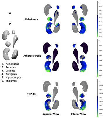 |
Association of age-related neuropathologies with shape of subcortical structures in a large community cohort of older adults
Nazanin Makkinejad1, Ashish A. Tamhane2, David A. Bennett2, Julie A. Schneider2, Boris Gutman1, and Konstantinos Arfanakis1,2
1Department of Biomedical Engineering, Illinois Institute of Technology, Chicago, IL, United States, 2Rush Alzheimer's Disease Center, Rush University Medical Center, Chicago, IL, United States
Age-related neuropathologies have devastating effects on subcortical brain structures. MRI can be used to explore patterns of atrophy associated with various diseases, however, definitive diagnosis of age-related neuropathologies is only possible at autopsy. Therefore, this work combined ex-vivo MRI and detailed pathologic assessment (gold standard) in a large (N=814) community cohort of older adults to investigate the link between age-related neuropathologies and the shape of different subcortical brain structures. The resulting patterns of deformation may contribute towards development or enhancement of in-vivo classifiers of these neuropathologies.
|
|
0267.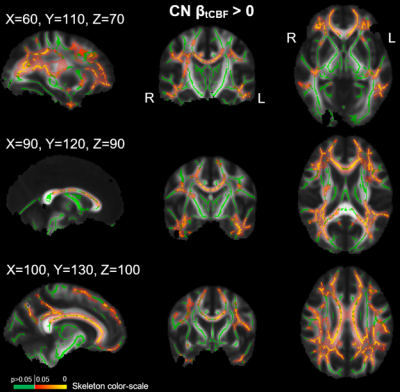 |
Assessment of Cerebrovascular Disease and White Matter Neurite Density in Alzheimer’s Disease
Grant S Roberts1, Leonardo A Rivera-Rivera2, Kevin M Johnson1,3, Sterling C Johnson2, Douglas C Dean III1,4, Andrew L Alexander1,5, Oliver Wieben1, and Laura B Eisenmenger3
1Medical Physics, University of Wisconsin - Madison, Madison, WI, United States, 2Medicine, University of Wisconsin - Madison, Madison, WI, United States, 3Radiology, University of Wisconsin - Madison, Madison, WI, United States, 4Pediatrics, University of Wisconsin - Madison, Madison, WI, United States, 5Psychiatry, University of Wisconsin - Madison, Madison, WI, United States
White matter (WM) microstructural alterations have been shown to occur in Alzheimer’s disease (AD) and may be partially mediated by cerebrovascular disease (CVD). The objective of this study is to use neurite orientation dispersion and density imaging (NODDI) to assess differences in neurite density (NDI) and its relationship to measures of CVD from 4D flow MRI in cognitively normal (CN) and AD subjects. Our results showed differences in NDI between groups in various WM tracts and found correlations between NDI and cerebral blood flow in CN subjects in several WM structures.
|
||
0268.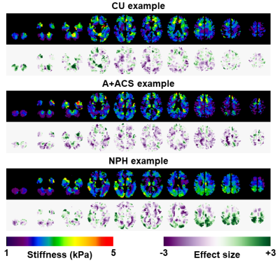 |
Visual interpretation of brain MRE exams using non-parametric statistical mapping to diagnose normal pressure hydrocephalus
Matthew Christopher Murphy1, Petrice M Cogswell1, Joshua D Trzasko1, Armando Manduca2, Matthew L Senjem1, Clifford R Jack, Jr.1, Fredric B Meyer3, Richard L Ehman1, and John Huston, III1
1Radiology, Mayo Clinic, Rochester, MN, United States, 2Physiology and Biomedical Engineering, Mayo Clinic, Rochester, MN, United States, 3Neurosurgery, Mayo Clinic, Rochester, MN, United States
A non-parametric statistical framework is outlined to display the result of brain MRE exams in terms of an effect size. This approach is built upon a neural network-based MRE inversion that estimates the posterior cumulative distribution function at each voxel, and accounts for age, sex, spatial variation in stiffness, and uncertainty in property estimation. Using these effect size maps but no summary statistics, neuoradiologists were able to diagnose normal pressure hydrocephalus with 70% sensitivity and 100% positive predictive value.
|
||
0269.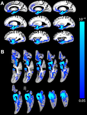 |
Limbic-predominant age-related TDP-43 encephalopathy neuropathological change (LATE-NC) is associated with lower R2 relaxation rate
Mahir Tazwar1, Arnold M Evia Jr.2, Ashish A Tamhane2, David A Bennett2, Julie A Schneider2, and Konstantinos Arfanakis1,2
1Department of Biomedical Engineering, Illinois Institute of Technology, Chicago, IL, United States, 2Rush Alzheimer's Disease Center, Rush University Medical Center, Chicago, IL, United States
Limbic-predominant age-related TDP-43 encephalopathy neuropathological change (LATE-NC) is now recognized as a common age-related neuropathology that has been linked to cognitive decline and dementia. In this work, the spatial pattern of R2 alterations associated with LATE-NC was investigated in a large (N=797) community cohort of older adults. Voxel-wise analysis revealed a pattern of lower R2 for greater LATE-NC burden, controlling for all other neuropathologies and demographics. This pattern involved mainly the temporal, frontal, occipital lobes and basal ganglia. To our knowledge this is the first R2 investigation in LATE-NC.
|
||
0270.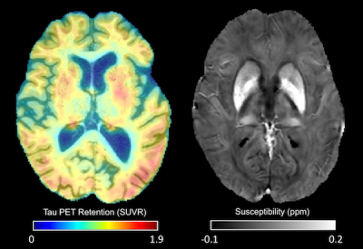 |
Tau correlates with tissue susceptibility and microstructure in APOE-ε4+ mild cognitive impairment
Jason Langley1, Daniel E Huddleston2, Sumanth Dara3, Ilana Bennett4, and Xiaoping P Hu1,3
1Center for Advanced Neuroimaging, University of California Riverside, Riverside, CA, United States, 2Department of Neurology, Emory University, Atlanta, GA, United States, 3Department of Bioengineering, University of California Riverside, Riverside, CA, United States, 4Department of Psychology, University of California Riverside, Riverside, CA, United States
We examine the impact of APOE-ε4 carrier status on cortical iron, gray matter microstructure, and tau-PET signal in mild cognitive impairment. We found significant increases in susceptibility (p=0.01), tau-PET SUVR (p=0.01), and MD (p=0.046) in the temporal lobe of APOE-ε4 positive compared to APOE-ε4 negative participants. Significant correlations were seen between tau-PET SUVR and susceptibility (r=0.717), FA (r=-0.431), and MD (r=0.435) in the temporal lobe of APOE-ε4 positive participants. Taken together, these findings suggest that APOE-ε4 allele increases the risk of developing AD pathology and accumulating iron, which in turn leads to degradation of cortical tissue microstructure.
|
The International Society for Magnetic Resonance in Medicine is accredited by the Accreditation Council for Continuing Medical Education to provide continuing medical education for physicians.