Oral
Cutting-Edge MR & the Brain Tumor Microenvironment
ISMRM & SMRT Annual Meeting • 15-20 May 2021

Oral
Cutting-Edge MR & the Brain Tumor Microenvironment
| Concurrent 6 | 14:00 - 16:00 | Moderators: THANH BINH NGUYEN & Roh Eul Yoo |
 |
0767.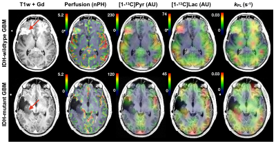 |
Multi-parametric hyperpolarized 13C/1H imaging of human gliomas expressing diverse pathologic mutations
Adam W Autry1, Sana Vaziri1, Marisa LaFontaine1, Jeremy W Gordon1, Hsin-Yu Chen1, Yaewon Kim1, Javier Villanueva-Meyer1, Susan M Chang2, Jennifer Clarke2, Duan Xu1, Janine M Lupo1, Peder EZ Larson1, Daniel B Vigneron1,3, and
Yan Li1
1Department of Radiology and Biomedical Imaging, University of California San Francisco, San Francisco, CA, United States, 2Department of Neurological Surgery, University of California San Francisco, San Francisco, CA, United States, 3Department of Bioengineering and Therapeutic Sciences, University of California San Francisco, San Francisco, CA, United States
This study uses multi-parametric 1H and hyperpolarized carbon-13 (HP-13C) MRI to characterize patients with gliomas that express signature pathologic mutations at the time of recurrence
|
|
0768. |
Glucose oxidation rate as a potential marker for GBM staging: correlation with histopathology and cell proliferation in a mouse model
Rui V Simoes1, Rafael N Henriques1, Beatriz M Cardoso1, Tania Carvalho1, and Noam Shemesh1
1Champalimaud Research, Champalimaud Foundation, Lisbon, Portugal
The dynamic interplay between cancer cells and their microenvironment impacts progression. We used dynamic glucose-enhanced deuterium MRS (DGE 2H-MRS) to investigate the association between functional metabolic heterogeneity and cell proliferation in a syngeneic mouse model of GBM. Taking a stepwise approach, from cell culture studies to in vivo mouse MRI/MRS and post-mortem analysis, our results suggest a potential role for glucose oxidation rate as a marker of cell proliferation and vascular stability. Extending this methodology to other GBM models and/or molecular subtypes could create new opportunities for non-invasive phenotyping of the disease.
|
||
0769.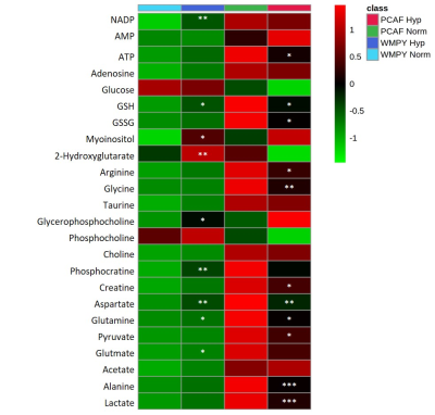 |
Hypoxia alters normal fibroblast metabolism towards a cancer associated fibroblast phenotype
Jesus Pacheco-Torres1, Tariq Shah1, W. Nathaniel Brennen2, Flonne Wildes1, and Zaver M Bhujwalla1,3,4
1Division of Cancer Imaging Research, The Russell H. Morgan Department of Radiology and Radiological Science, The Johns Hopkins University School of Medicine, Baltimore, MD, United States, 2Department of Oncology, The Johns Hopkins University School of Medicine, Baltimore, MD, United States, 3Sidney Kimmel Comprehensive Cancer Center, The Johns Hopkins University School of Medicine, Baltimore, MD, United States, 4Radiation Oncology and Molecular Radiation Sciences, The Johns Hopkins University School of Medicine, Baltimore, MD, United States
Fibroblasts play a pivotal role in cancer progression. In prostate cancer, fibroblasts have been shown to induce growth and increase metastatic potential. To further understand how fibroblasts respond to hypoxic tumor microenvironments that are frequently observed in prostate cancer, we have characterized the effects of hypoxia on normal and cancer associated prostate fibroblast (PCAF) metabolomics and invasion using 1H MRS/I. We found that hypoxia increased matrix degradation by normal fibroblasts. Furthermore, hypoxia metabolically reprogrammed normal prostate fibroblasts to mimic the metabolic pattern of PCAFs, highlighting the potential role of hypoxia in the transition of normal fibroblasts to CAFs.
|
||
0770.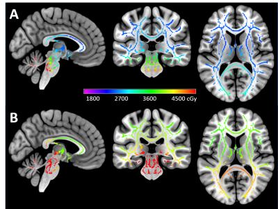 |
Reproducibility study of disrupted white matter integrity and partial recovery in children treated for medulloblastoma
Wilburn E Reddick1, Jared J Sullivan1, John O Glass1, Yian Guo2, Julie H Harreld1, Yimei Li2, Giles W Robinson3, Amar Gajjar3, and Thomas E Merchant4
1Department of Diagnostic Imaging, St. Jude Children's Research Hospital, Memphis, TN, United States, 2Department of Biostatistics, St. Jude Children's Research Hospital, Memphis, TN, United States, 3Department of Oncology, St. Jude Children's Research Hospital, Memphis, TN, United States, 4Department of Radiation Oncology, St. Jude Children's Research Hospital, Memphis, TN, United States
The current reproducibility study evaluates 140 children treated for medulloblastoma and 92 age-similar controls using TBSS analysis of FA and dosimetry accounting for age. Treatment included surgery, post-operative standard or high dose craniospinal irradiation and adjuvant chemotherapy. FA measures at baseline after surgery but prior to therapy demonstrated an immediate decrease due to tumor and surgery, which was then accentuated by irradiation. A partial recovery of FA followed, which was attenuated in patients receiving higher doses. These longitudinal profiles need to be considered when conducting cross-sectional studies of patients at time points during or after therapy.
|
||
0771.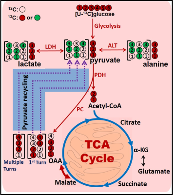 |
Quantification of pyruvate recycling in brain tumor patients
Kumar Pichumani1, Omkar Ijare1, Elizabeth Maher2, Robert M Bachoo2, and David S Baskin1
1Peak Center, Neurosurgery, Houston Methodist Hospital, Houston, TX, United States, 2UT Southwestern Medical Center, Dallas, TX, United States
Pyruvate recycling is the metabolic pathway that generates pyruvate, lactate and alanine from the tricarboxylic acid (TCA) cycle intermediates, oxaloacetate (OAA) and malate. It is active in the liver and the kidney. Although existence and origin of pyruvate recycling mechanism in human brain has been shown to be active, it still remains controversial. Here, we demonstrate that pyruvate recycling mechanism is active in human GBM patients by 13C isotopomer analysis of resected tumor tissues. We have developed a simple method to determine the relative flux of pyruvate recycling with respect to glycolysis using C2 lactate 13C isotopomers.
|
||
0772. |
Pathological validation of MP-MRI intensity-based signatures in brain cancer patients using autopsy tissue samples
Samuel Bobholz1, Allison Lowman2, Michael Brehler2, Savannah Duenweg1, Fitzgerald Kyereme2, Elizabeth Cochran3, Jennifer Connelly4, Wade Mueller5, Mohit Agarwal2, Darren O'Neill2, Anjishnu Banerjee6, and Peter LaViolette2,7
1Biophysics, Medical College of Wisconsin, Milwaukee, WI, United States, 2Radiology, Medical College of Wisconsin, Milwaukee, WI, United States, 3Pathology, Medical College of Wisconsin, Milwaukee, WI, United States, 4Neurology, Medical College of Wisconsin, Milwaukee, WI, United States, 5Neurosurgery, Medical College of Wisconsin, Milwaukee, WI, United States, 6Biostatistics, Medical College of Wisconsin, Milwaukee, WI, United States, 7Biomedical Engineering, Medical College of Wisconsin, Milwaukee, WI, United States
This study investigated the relationship between MRI-intensity values and pathological features of brain cancer using autopsy tissues as ground truth. Mixed effect models were used to examine the association between T1, T1C, FLAIR, and ADC intensity and pathological features (cellularity, cytoplasm density, and extracellular fluid density), as well as to compare the strength of these associations between GBM and non-GBM patients. These analyses confirmed many of the associations seen in prior literature, but with decreased strength than expected. Additionally, this study found that radio-pathomic associations were weaker in GBM patients than non-GBM patients.
|
||
0773.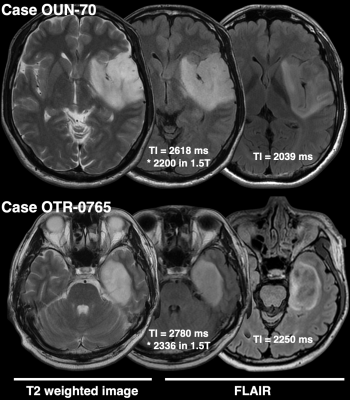 |
Impact of inversion time for FLAIR acquisition on the T2-FLAIR mismatch detectability for IDH-mutant, non-CODEL astrocytomas
Manabu Kinoshita1, Hideyuki Arita1, Masamichi Takahashi2, Takehiro Uda3, Junya Fukai4, Kenichi Ishibashi5, Noriyuki Kijima1, Ryuichi Hirayama1, Mio Sakai6, Atsuko Arisawa7, Hiroto Takahashi7, Katsuyuki Nakanishi6, Naoki Kagawa1,
Kouichi Ichimura8, Yonehiro Kanemura9, Yoshitaka Narita2, and Haruhiko Kishima1
1Neurosurgery, Osaka University Graduate School of Medicine, Suita, Japan, 2Neurosurgery and Neuro-Oncology, National Cancer Center Hospital, Tokyo, Japan, 3Neurosurgery, Osaka City University Graduate School of Medicine, Osaka, Japan, 4Neurological Surgery, Wakayama Medical University, Wakayama, Japan, 5Neurosurgery, Osaka City General Hospital, Osaka, Japan, 6Diagnostic Radiology, Osaka International Cancer Institute, Osaka, Japan, 7Radiology, Osaka University Graduate School of Medicine, Suita, Japan, 8Division of Brain Tumor Translational Research, National Cancer Center Research Institute, Tokyo, Japan, 9Biomedical Research and Innovation, National Hospital Organization Osaka National Hospital, Osaka, Japan
Although the T2-FLAIR mismatch sign is a promising imaging marker specific for IDHmt, non-CODEL astrocytomas, its low sensitivity and NPV hinder its full clinical application. This study discovered that FLAIR acquisition with TI shorter than 2400 ms in 3T could overcome this issue and that the sensitivity and NPV improved to 67% and 74%. Tuning TI for FLAIR acquisition is such a simple technique that clinicians can easily incorporate this procedure into the daily workflow of glioma imaging. Our proposed method would provide a novel yet clinically feasible glioma imaging method in the era of personalized-molecular medicine of cancer.
|
||
0774.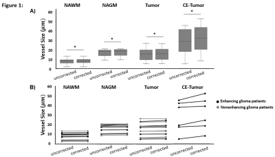 |
Leakage Correction of Dynamic Susceptibility Contrast (DSC-) MRI for vessel size measurements in human glioma
Fatemeh Arzanforoosh1, Paula L. Croal2, Karin Van Garderen1, Marion Smits1, Michael A. Chappell2, and Esther A.H. Warnert1
1Department of Radiology & Nuclear Medicine, ErasmusMC, Rotterdam, Netherlands, 2Radiological Sciences, Division of Clinical Neurosciences, University of Nottingham, Nottingham, United Kingdom
Reliable insight about tumor microvasculature is important for monitoring of disease progression and treatment response. Derived from Dynamic Susceptibility Contrast MRI, transverse relaxation rates are used for vessel size estimation. In high grade glioma, these signals can artificially change by contrast agent extravasation through a disrupted Blood-Brain-Barrier. In this study the effect of applying Boxerman-Schmainda-Weisskoff leakage correction on vessel size estimation has been investigated on a group of 12 glioma patients. The result shows that in Contrast-Enhanced Tumor area applying leakage correction significantly and noticeably changes the vessel size measurements.
|
||
0775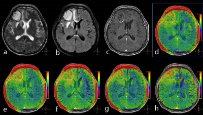 |
3D APTw Brain Tumor Imaging with Compressed SENSE: Comparison of Different Acceleration Factors and with Conventional Parallel Imaging Video Permission Withheld
Nan Zhang1, Qingwei Song2, Ailian Liu2, Haonan Zhang2, Renwang Pu2, Jiazheng Wang3, and Zhiwei Shen3
1The First Affilliated Hospital of Dalian Medical University, Dalian, China, 2The First Affiliated Hospital of Dalian Medical University, Dalian, China, 3Philips Healthcare, Beijing, China, Beijing, China
Amide proton transfer weighted (APTw) imaging is a novel and promising MRI method for brain tumor imaging, but it can be time-consuming. Common parallel imaging methods, like SENSE, can lead to reduced image quality and increased artifact at high acceleration factors. Here, the compressed SENSE (CS) technique with combined strength from both compressed sensing and SENSE was evaluated for the acceleration of APTw imaging in brain. Results showed that it is feasible to apply an CS accelerator factor of 5 to APTw imaging of brain tissue and tumor, which could reduce the scan time to less than 1 min.
|
||
0776.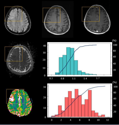 |
MR Imaging Parameters for Noninvasive Prediction of EGFR Amplification in IDH-Wildtype Lower-Grade Gliomas: A Multicenter Study
Yae Won Park1, Ji Eun Park2, Sung Soo Ahn1, Seung Hong Choi3, Ho Sung Kim2, and Seung-Koo Lee1
1Yonsei University College of Medicine, Seoul, Korea, Republic of, 2University of Ulsan College of Medicine, Seoul, Korea, Republic of, 3Seoul National University Hospital, Seoul, Korea, Republic of
Epidermal growth factor receptor (EGFR) amplification status of isocitrate dehydrogenase-wildtype (IDHwt) lower-grade gliomas (LGGs; grade II/III) is one of the key molecular markers for diagnosing molecular glioblastoma. Our study shows that infiltrative or mixed pattern, lower ADC, lower 5th percentile of ADC, and higher 95th percentile of nCBF may be useful imaging biomarkers for the EGFR amplification of IDHwt LGGs. Moreover, quantitative imaging biomarkers may add value to qualitative imaging parameters (with AUCs of 0.71 and 0.88, p = 0.020).
|
The International Society for Magnetic Resonance in Medicine is accredited by the Accreditation Council for Continuing Medical Education to provide continuing medical education for physicians.