Oral
Advances in MR Fingerprinting
ISMRM & SMRT Annual Meeting • 15-20 May 2021

| Concurrent 1 | 18:00 - 20:00 | Moderators: Christian Guenthner |
 |
0165.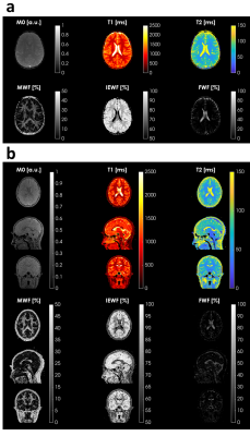 |
Myelin Water Fraction Mapping in developing children using Magnetic Resonance Fingerprinting
Matteo Cencini1,2, Marta Lancione2,3, Laura Biagi1,2, Jan W Kurzawski1,2, Rosa Pasquariello1, Graziella Donatelli2,4, Claudia Dosi1,5, Chiara Ticci1,5, Roberta Battini1,5, Guido Buonincontri1,2, and Michela Tosetti1,2
1IRCCS Stella Maris, Pisa, Italy, 2Imago7 Foundation, Pisa, Italy, 3IMT School for Advanced Studies Lucca, Lucca, Italy, 4Neuroradiology Unit, Azienda Ospedaliero-Universitaria Pisana, Pisa, Italy, 5Department of Clinical and Experimental Medicine, University of Pisa, Pisa, Italy
Magnetic Resonance Fingerprinting (MRF) has been used to obtain Myelin Water Fraction (MWF) estimates in a cohort of developing children by using a three-component signal model. Here, we used a recent approach in which we perform sub-voxel tissue characterization without assumptions on the number and properties of the model components. We then used this signal model to study the myelination process in the developing brain on a 2D MRF dataset. Finally, we measured MWF on a set of subjects acquired with 3D MRF scan and compared the results to the 2D experiment.
|
|
|
0166.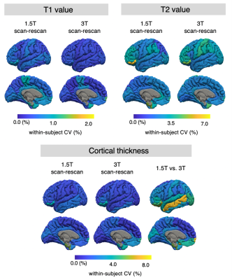 |
Simultaneous morphometry and relaxometry of the human brain using three-dimensional MR fingerprinting at 1.5 and 3T
Shohei Fujita1,2, Matteo Cencini3,4, Guido Buonincontri3,4, Naoyuki Takei5, Rolf F. Schulte6, Wataru Uchida1, Akifumi Hagiwara1, Koji Kamagata1, Osamu Abe2, Michela Tosetti3,4, and Shigeki Aoki1
1Department of Radiology, Juntendo University, Tokyo, Japan, 2Department of Radiology, The University of Tokyo, Tokyo, Japan, 3Imago7 Foundation, Pisa, Italy, 4IRCCS Stella Maris, Pisa, Italy, 5MR Applications and Workflow, GE Healthcare, Tokyo, Japan, 6GE Healthcare, Munich, Germany
Magnetic resonance fingerprinting (MRF) permits simultaneous acquisition of T1 and T2 maps perfectly aligned to the anatomy, allowing morphometry and relaxometry analysis of the brain in a single scan. Here, we examined the reproducibility and repeatability of simultaneous morphology and relaxometry of brain structures in healthy volunteers using three-dimensional MRF at multiple field strengths. Scan-rescan tests of three-dimensional MRF were performed at 1.5 and 3T. The local thickness, volume, T1, and T2 values were calculated for each representative neuroanatomical structure using automatic brain segmentation software. These results can help establish imaging biomarkers using MRF for clinical use.
|
|
|
0167.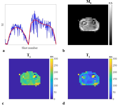 |
3D Magnetic Resonance Fingerprinting at 50 mT
Thomas O`Reilly1, Peter Börnert1,2, Andrew Webb1, and Kirsten Koolstra1
1Radiology, Leiden University Medical Center, Leiden, Netherlands, 2Philips Research Hamburg, Hamburg, Germany
In vivo MR relaxation times, their inter-subject variations, and changes in different diseases have not been widely studied at very low magnetic fields (<100 mT). In this work, we implemented a 3D MRF sequence on a 50 mT Halbach permanent magnet system to efficiently measure relaxation times in vivo. We used a short flip angle train and further accelerate the scans by using random undersampling and matrix completion reconstruction. Initial in vivo and phantom data show good agreement with relaxation time values measured with less efficient conventional techniques.
|
|
 |
0168.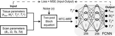 |
Learning-based Optimization of Acquisition Schedule for Magnetization Transfer Contrast MR Fingerprinting
Beomgu Kang1, Byungjai Kim1, Hye-Young Heo2,3, and Hyunwook Park1
1Department of Electrical Engineering, Korea Advanced Institute of Science and Technology, Daejeon, Korea, Republic of, 2Russell H Morgan Department of Radiology and Radiological Science, Johns Hopkins University, Baltimore, MD, United States, 3F.M. Kirby Research Center for Functional Brain Imaging, Kennedy Krieger Institute, Baltimore, MD, United States
Magnetization transfer contrast MR fingerprinting (MTC-MRF) is a novel quantitative imaging method that simultaneously quantifies free bulk water and semisolid macromolecule parameters using pseudo-randomized scan parameters. Here, we propose a framework for learning-based optimization of the acquisition schedule (LOAS), which optimizes RF saturation-encoded MRF acquisitions with a minimum number of acquisitions for tissue parameter estimation. Unlike the optimization methods based on indirect measurements, the proposed approach can optimize scan parameters by directly computing quantitative errors in tissue parameters.
|
|
|
0169.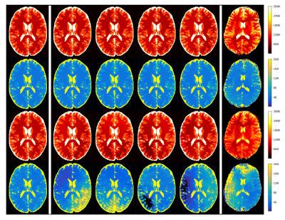 |
Sequence Design for Fast and Robust MR Fingerprinting Scans using Quantum Optimization
Siyuan Hu1, Ignacio Rozada2, Rasim Boyacioglu3, Stephen Jordan4, Sherry Huang3, Matthias Troyer4, Mark Griswold3, Debra McGivney3, and Dan Ma3
1Biomedical Engineering, Case Western Reserve University, Cleveland, OH, United States, 21Qbit, Vancouver, BC, Canada, 3Case Western Reserve University, Cleveland, OH, United States, 4Microsoft, Redmond, WA, United States
MR Fingerprinting is able to quantify multiple tissue properties simultaneously. Here we propose an advanced MR Fingerprinting optimization framework that computes and minimizes the quantitative random errors, undersampling errors and background phase errors in MRF maps simultaneously in the cost function. The optimization is solved by quantum-inspired algorithms. The proposed framework could provide accelerated MRF scans that are robust to undersampling and system imperfections, and outperform the human-designed sequence on the tradeoff between duration and precision.
|
|
|
0170.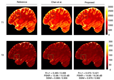 |
Accelerating Submillimeter 3D MR Fingerprinting with Whole-Brain Coverage via Dual-Domain Deep Learning Reconstruction
Feng Cheng1, Yong Chen2, and Pew-Thian Yap3
1Department of Computer Science, University of North Carolina, Chapel Hill, NC, United States, 2Case Western Reserve University, Cleveland, OH, United States, 3Department of Radiology and Biomedical Research Imaging Center, University of North Carolina, Chapel Hill, NC, United States
We accelerate submillimeter 3D MRF using a dual-domain deep learning reconstruction approach that utilizes a graph convolutional network for k-space and a U-Net for image space acceleration. Our preliminary results show that a total of 16x acceleration can be achieved, reducing the acquisition time for whole-brain-coverage at 0.8 mm isotropic resolution to less than 5 mins.
|
|
|
0171.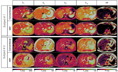 |
Simultaneous comprehensive T1, T2, T2*, T1ρ and Fat Fraction characterization with Magnetic Resonance Fingerprinting
Carlos Velasco1, Gastao Cruz1, René M. Botnar1, and Claudia Prieto1
1School of Biomedical Engineering and Imaging Sciences, King's College London, London, United Kingdom
Quantitative T1, T2, T2* and fat fraction maps are promising imaging biomarkers for the assessment and follow-up of liver disease, while T1⍴ mapping has been reported to be a valuable tool for contrast-free assessment of liver fibrosis. However, these multiple scans are usually performed sequentially during separate breath-holds, leading to long scan times and potentially mis-registered maps. In this study we propose an 8-echo T1, T2 and T1⍴ prepared liver MRF sequence that allows for quantitative T1, T2, T2*,
T1⍴ and FF liver tissue characterization in a single breath-hold scan. The proposed approach has been investigated in healthy subjects.
|
|
0172.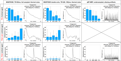 |
Towards optimizing MR vascular fingerprinting
Aurélien Delphin1, Fabien Boux1,2, Clément Brossard1, Jan M Warnking1, Benjamin Lemasson1, Emmanuel Luc Barbier1, and Thomas Christen1
1Univ. Grenoble Alpes, Inserm, U1216, Grenoble Institut Neurosciences, GIN, 38000, Grenoble, France, 2Univ. Grenoble Alpes, Inria, CNRS, G-INP, 38000, Grenoble, France
MR vascular fingerprinting aims at mapping cerebral vascular properties. We propose to improve the method on two levels: (1) by testing new acquisitions patterns using a Monte-Carlo based method that assesses the encoding capacity of MRF sequences; and (2) by testing new geometrical models that represent vascular networks during dictionary simulations. We obtained results suggesting that new MRF-type sequences can be tailored for vascular exploration and should be tested in vivo. We also showed the clear influence of geometry in the simulations and the possibility to include realistic vascular networks.
|
||
|
0173.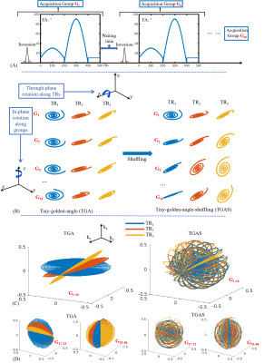 |
Optimized multi-axis spiral projection MRF with subspace reconstruction for rapid 1-mm isotropic whole-brain MRF in 2 minutes
Xiaozhi Cao1,2, Congyu Liao1,2, Siddharth Srinivasan Iyer3,4, Gilad Liberman3, Zijing Dong3,4, Ting Gong5, Zihan Zhou5, Hongjian He5, Jianhui Zhong5,6, and Berkin Bilgic3,7
1Department of Rdiology, Stanford university, Stanford, CA, United States, 2Department of Electrical Engineering, Stanford university, Stanford, CA, United States, 3Athinoula A. Martinos Center for Biomedical Imaging, Massachusetts General Hospital, Charlestown, MA, United States, 4Department of Electrical Engineering and Computer Science, MIT, Cambridge, MA, United States, 5Center for Brain Imaging Science and Technology, Department of Biomedical Engineering, Zhejiang University, Hangzhou, China, 6Department of Imaging Sciences, University of Rochester, Rochester, NY, United States, 7Department of Radiology, Harvard Medical School, Cambridge, MA, United States
To improve the quality and speed of 3D MRF, we applied spatiotemporal subspace reconstruction to 3D MRF and further modified its spiral-projection spatiotemporal encoding scheme. When compared to conventional sliding-window iNUFFT reconstruction, the subspace reconstruction provided markedly improved quantitative maps, with lower artifacts and higher SNR. The optimized spiral-projection encoding scheme, which was designed to increase spatiotemporal incoherency, was also validated to be more robust to artifacts, particularly at high accelerations. The proposed method enables high-quality whole-brain T1, T2, and proton density mapping with 1-mm isotropic resolution in 2 minutes and 0.8-mm isotropic resolution in ~4minutes.
|
|
|
0174. |
Fast acquisition of 31-P creatin kinease chemical exchange rate and relaxation rates of γ-ATP and PCr in vivo human brain at 7T using MRS-FP
Mark Stephan Widmaier1, Song-I Lim2, and Lijing Xin2
1Laboratory for Functional and Metabolic Imaging, CIBM, EPFL, Lausanne, Switzerland, 2Animal Imaging and Technology, CIBM, EPFL, Lausanne, Switzerland
This study shows preliminary results of the feasibility using MRS-FP to measure the relaxation parameters of γ-ATP and PCr as well as the chemical exchange rate kCK. All results were in good agreement with literature values enabling a fast and robust 31 P multi parameter estimation in vivo human brain at 7T .
|
The International Society for Magnetic Resonance in Medicine is accredited by the Accreditation Council for Continuing Medical Education to provide continuing medical education for physicians.