Oral
Quantitative Relaxation Parameter Mapping in the Body
ISMRM & SMRT Annual Meeting • 15-20 May 2021

Oral
Quantitative Relaxation Parameter Mapping in the Body
| Concurrent 1 | 18:00 - 20:00 | Moderators: Martijn Cloos & Mingming Wu |
 |
0609.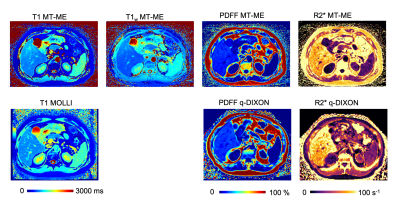 |
Six-Dimensional, Free-Breathing Multitasking Multi-Echo (MT-ME) MRI for Whole-Liver T1, PDFF, and R2* Quantification
Nan Wang1, Tianle Cao1,2, Fei Han3, Yibin Xie1, Xiaodong Zhong3, Sen Ma1, Xinheng Zhang1,2, Xiaoming Bi3, Mazen Noureddin4, Vibhas Deshpande3, Anthony G Christodoulou1, and Debiao Li1
1Biomedical Imaging Research Institute, Cedars-Sinai Medical Center, Los Angeles, CA, United States, 2Bioengineering, University of California, Los Angeles, Los Angeles, CA, United States, 3Siemens Medical Solutions USA, Inc., Los Angeles, CA, United States, 4Department of Medicine, Cedars-Sinai Medical Center, Los Angeles, CA, United States
Chronic liver disease has been a leading cause of mortality worldwide. Multiparametric MRI is a promising tool for non-invasive characterization of liver disease, but has yet to be widely used in clinical practice due to demanding technical challenges. In this work, we proposed a 6D Multitasking multi-echo (MT-ME) technique that allows free-breathing acquisition, whole-liver coverage, and simultaneous T1, PDFF, R2*, and water-specific T1 (T1w) quantification. Phantom study and in vivo studies with 14 volunteers and 1 patient with NAFLD were performed. The quantitative parameters measured from MT-ME were repeatable and showed good agreement with the reference methods.
|
|
|
0610.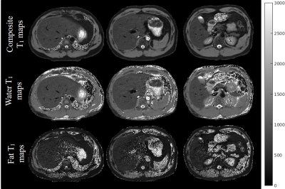 |
Rapid high resolution simultaneous mapping of composite T1, water-only T1 and PDFF in the abdomen with dual-echo IR-radSPGR pulse sequence
Zhitao Li1,2, John M Pauly2, and Shreyas Vasanawala1
1Department of Radiology, Stanford University, Palo Alto, CA, United States, 2Electrical Engineering, Stanford University, Palo Alto, CA, United States
A radial IR-SPGR pulse sequence with bipolar readouts is demonstrated along with a model-based iterative reconstruction to simultaneously map high resolution composite and water-only T1 and proton density fat fraction (PDFF) in the abdomen. The proposed pulse sequence and reconstruction algorithm can yield high-quality T1 and PDFF maps in 2.5 seconds/slice. The resulting maps have an in-plane resolution of 1.25mm and through-plane resolution of 5.00mm. When combined with selective inversion pulse, the technique achieves multi-slice imaging for breath-hold studies.
|
|
|
0611.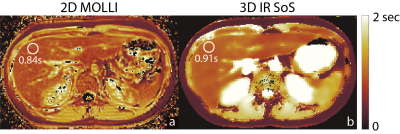 |
Free-Breathing, Confounder Corrected T1 Mapping in the Liver with Stack-of-Stars Inversion Recovery MRI
Yavuz Muslu1,2, Ty A. Cashen3, Sagar Mandava4, and Scott B. Reeder1,2,5,6,7
1Department of Biomedical Engineering, University of Wisconsin-Madison, Madison, WI, United States, 2Department of Radiology, University of Wisconsin-Madison, Madison, WI, United States, 3Global MR Applications and Workflow, GE Healthcare, Madison, WI, United States, 4Global MR Applications and Workflow, GE Healthcare, Atlanta, GA, United States, 5Department of Medical Physics, University of Wisconsin-Madison, Madison, WI, United States, 6Department of Medicine, University of Wisconsin-Madison, Madison, WI, United States, 7Department of Emergency Medicine, University of Wisconsin-Madison, Madison, WI, United States
Quantitative T1 mapping in the liver is an emerging biomarker of hepatic fibrosis and characterization of liver function. The variable flip angle approach with Cartesian sampling is among the most popular T1 mapping methods used in the abdomen. A major drawback of this approach is that T1 estimations are highly sensitive to B1 inhomogeneities. Furthermore, Cartesian sampling suffers from motion related ghosting artifacts and requires breath-holding acquisitions. In this study, we propose to combine stack-of-stars radial sampling with dual-echo inversion recovery (IR-SoS) MRI for confounder corrected T1 mapping in the abdomen.
|
|
 |
0612.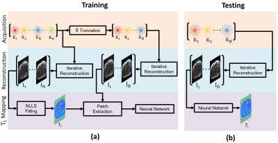 |
Improved Slice Coverage in Inversion Recovery Radial Balanced-SSFP using Deep Learning
Eze Ahanonu1, Zhiyang Fu1,2, Kevin Johnson2, Maria Altbach2,3, and Ali Bilgin1,2,3
1Electrical and Computer Engineering, University of Arizona, Tucson, AZ, United States, 2Medical Imaging, University of Arizona, Tucson, AZ, United States, 3Biomedical Engineering, University of Arizona, Tucson, AZ, United States
Abdominal T1 mapping is important for quantitative evaluation of various pathologies. A recent inversion recovery radial balanced-SSFP (IR-radSSFP) technique allows high resolution T1 mapping of ten slices within a single breath hold period (BHP), but requires multiple BHPs for full abdominal coverage. We propose an accelerated T1 mapping framework which utilizes deep learning to estimate T1 using a fraction of the T1 recovery curve (T1RC). In vivo experiments demonstrate that the proposed framework achieves less than 6% T1 error while using only 25% of the T1RC of the earlier IR-radSSFP technique. This enables full abdominal coverage within a single BHP.
|
|
0613.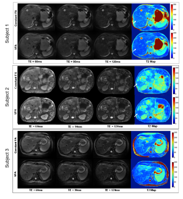 |
Efficient T2 mapping of the Abdomen with low SAR Variable Flip Angle Radial Turbo Spin Echo
Mahesh Bharath Keerthivasan1,2, Lavanya Umapathy2,3, Jean-Philippe Galons2, Diego Martin4, Ali Bilgin2,3,4, and Maria Altbach2
1Siemens Medical Solutions USA Inc, New York, NY, United States, 2Medical Imaging, University of Arizona, Tucson, AZ, United States, 3Electrical and Computer Engineering, University of Arizona, Tucson, AZ, United States, 4Biomedical Engineering, University of Arizona, Tucson, AZ, United States
Radial TSE techniques have been proposed for abdominal T2-weighted (T2w) imaging and T2 mapping. Slice efficiency of breath-held RADTSE is limited by the specific absorption rate (SAR). We present a reduced SAR variable refocusing flip angle RADTSE (RADTSE-VFA) technique designed for efficient slice coverage and improved T2 estimation. The flip angles are designed to (1) minimize T2 estimation error, (2) improve lesion-liver relative contrast, and (3) minimize SAR. RADTSE-VFA generated T2w images with comparable contrast as constant flip angle RADTSE while resulting in a 60% increase in slice coverage at a 1.5x reduction in SAR.
|
||
0614.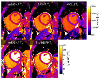 |
Accurate and precise myocardial T1 and T2 mapping in a single breath-hold with multi-parametric SASHA
Kelvin Chow1, Genevieve Hayes2, Jacqueline Flewitt2, Patricia Feuchter2, Carmen Lydell2, Andrew Howarth2, Joseph Pagano3, Richard Thompson4, Peter Kellman5, and James White2
1Cardiovascular MR R&D, Siemens Medical Solutions USA, Inc., Chicago, IL, United States, 2Stephenson Cardiac Imaging Centre, University of Calgary, Calgary, AB, Canada, 3Division of Pediatric Cardiology, University of Alberta, Edmonton, AB, Canada, 4Biomedical Engineering, University of Alberta, Edmonton, AB, Canada, 5National Heart, Lung, and Blood Institute, National Institutes of Health, Bethesda, MD, United States
A novel multi-parametric cardiac mapping sequence, mSASHA, is proposed to calculate T1 and T2 maps in a single breath-hold. mSASHA is validated against spin-echo in phantoms with -0.7±0.4% T1 error and -1.3±1.3% T2 error. In 10 healthy volunteers at 3T, mSASHA had similar T1 values to SASHA in the myocardium (1523±18 ms vs. 1520±18, p>0.05) and blood pool (2054±61 ms vs. 2060±65 ms, p>0.05). mSASHA had similar myocardial T1 coefficient of variation (CoV) to SASHA and MOLLI and similar myocardial T2 CoV to T2p-bSSFP (p>0.05). mSASHA provides accurate and precise T1 and T2 maps in
a single breath-hold.
|
||
|
0615.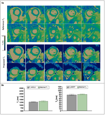 |
3D whole-heart free-breathing isotropic joint T1/T2 quantification: preliminary clinical evaluation
Carlos Velasco1, Giorgia Milotta1, Alina Hua1, Karl Kunze1,2, Radhouene Neji1,2, Tevfik Ismail1, Claudia Prieto1, and René M. Botnar1
1School of Biomedical Engineering and Imaging Sciences, King's College London, London, United Kingdom, 2MR Research Collaborations, Siemens Healthcare Limited, Frimley, United Kingdom
Myocardial tissue characterization, such as quantification of fibrosis and oedema plays an important role in many myocardial diseases. T1 and T2 maps are typically acquired sequentially in 2D under several breath-holds. However, they achieve limited spatial resolution and coverage. To overcome these limitations, a high resolution, motion compensated joint T1/T2 water/fat sequence has been recently proposed and validated in phantom and healthy subjects. In this study, we demonstrate the capability of the proposed approach to obtain whole-heart, motion-compensated, simultaneous and co-registered T1, T2 maps and water and fat images in ~9min in patients with cardiovascular disease.
|
|
|
0616.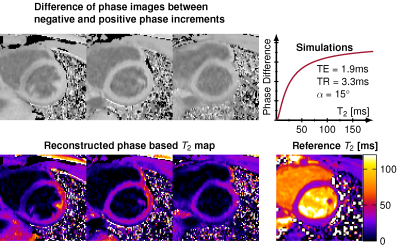 |
Feasibility and insights into transient state phase-based mapping for rapid T2 quantification in the myocardium
Ingo Hermann1,2, Daiki Tamada3, Scott Reeder3, Lothar Schad2, and Sebastian Weingärtner1
1Magnetic Resonance Systems Lab, Department of Imaging Physics, Delft University of Technology, Delft, Netherlands, 2Computer Assisted Clinical Medicine, Medical Faculty Mannheim, University Heidelberg, Mannheim, Germany, 3Department of Radiology, University of Wisconsin, Madison, WI, United States
In this study we explore the use of the signal phase of RF phase modulated gradient echo imaging during steady state for rapid T2 mapping in the myocardium. RF phase modulated GRE images were obtained with quadratic phase increments of ±2° and T2 times were extracted by matching the phase evolution. B1+ sensitivity was incorporated in the reconstruction based on separately acquired Bloch-Siegert maps. Good correlation was found in phantom measurements (R>0.95; p<0.0001) and high visual image quality in vivo.
|
|
0617.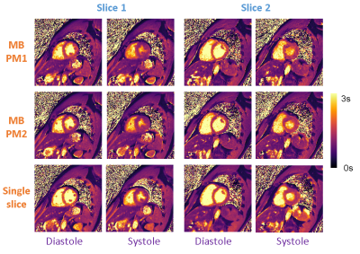 |
Multiband Multitasking for Cardiac T1 Mapping
Qi Liu1, Yuan Zheng1, Jingyuan Lyu1, Zhongqi Zhang2, Yanqun Teng2, Shuheng Zhang2, Jian Xu1, and Weiguo Zhang1
1UIH America, Inc., Houston, TX, United States, 2United Imaging Healthcare, Shanghai, China
Multiband (MB) technique is combined with multitasking for increased spatial coverage of the heart without prolonging scan time. Two different MB multitasking implementations were developed and compared with the conventional multitasking technique on volunteers and phantoms. Both methods demonstrated similar capabilities in solving multiple ‘tasks’ when compared with the reference method and exhibited good agreement in T1 mapping values. MB multitasking is a promising technique for cardiac MR.
|
||
0618. |
Fast T2-mapping in prostate cancer based on echo-time domain compressed sensing
Jochen Keupp1, Petra J. v. Houdt2, Jakob Meineke1, Paul de Bruin3, Johannes M. Peeters3, Leon ter Beek4, and Mariya Doneva1
1Philips Research, Hamburg, Germany, 2Department of Radiation Oncology, The Netherlands Cancer Institute, Amsterdam, Netherlands, 3Philips Healthcare, Best, Netherlands, 4Department of Medical Physics, The Netherlands Cancer Institute, Amsterdam, Netherlands
T2w-MRI plays an important role in prostate cancer providing information on the location and aggressiveness in diagnosis and therapy. T2-mapping may provide objective characterization, but is hampered by long acquisition time, which has been addressed by dedicated acceleration techniques (e.g. k-t T2-mapping). We investigated T2-mapping in a prostate cancer patient based on a 4-minute protocol with Poisson-disk prospective irregular sub-sampling in the ky-TE domain in combination with a low rank and sparsity constraint compressed sensing reconstruction. The regularization parameters were investigated, and compressed sensing results were compared to separately acquired k-t T2-maps with respect to quality and noise.
|
The International Society for Magnetic Resonance in Medicine is accredited by the Accreditation Council for Continuing Medical Education to provide continuing medical education for physicians.