Oral
Machine Learning for Image Reconstruction
ISMRM & SMRT Annual Meeting • 15-20 May 2021

| Concurrent 1 | 14:00 - 16:00 | Moderators: Thomas Küstner & Efrat Shimron |
0271.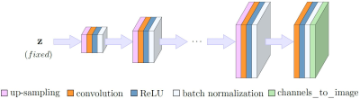 |
Can Un-trained Networks Compete with Trained Ones for Accelerated MRI?
Mohammad Zalbagi Darestani1 and Reinhard Heckel1,2
1Electrical and Computer Engineering, Rice University, Houston, TX, United States, 2Electrical and Computer Engineering, Technical University of Munich, Munich, Germany
Convolutional Neural Networks (CNNs) are highly effective tools for image reconstruction problems. Typically, CNNs are trained on large amounts of images, but, perhaps surprisingly, even without any training data, CNNs such as the Deep Image Prior and Deep Decoder achieve excellent imaging performance. Here, we build on those works by proposing an un-trained CNN for accelerated MRI along with performance-enhancing steps including enforcing data-consistency and combining multiple reconstructions. We show that the resulting method i) achieves reconstruction performance almost on par with baseline as well as state-of-the-art trained CNNs, but without any training, and ii) significantly outperforms competing sparsity-based approaches.
|
||
 |
0272.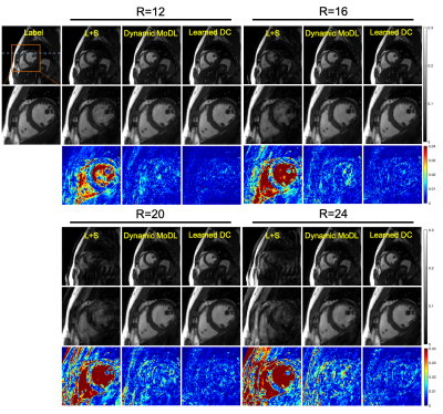 |
Learning data consistency for MR dynamic imaging
Jing Cheng1, Wenqi Huang1, Zhuoxu Cui1, Ziwen Ke1, Leslie Ying2, Haifeng Wang1, Yanjie Zhu1, and Dong Liang1
1Shenzhen Institutes of Advanced Technology, Chinese Academy of Sciences, Shenzhen, China, 2University at Buffalo, The State University of New York, Buffalo, Buffalo, NY, United States
Existing deep learning-based methods for MR reconstruction employ deep networks to exploit the prior information and integrate the prior knowledge into the reconstruction under the explicit constraint of data consistency, without considering the real distribution of the noise. In this work, we propose a new DL-based approach termed Learned DC that implicitly learns the data consistency with deep networks, corresponding to the actual probability distribution of system noise. We evaluated the proposed approach with highly undersampled dynamic cardiac cine data. Experimental results demonstrate the superior performance of the Learned DC.
|
|
0273.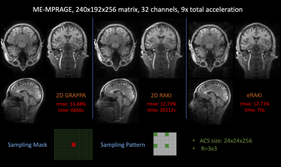 |
eRAKI: Fast Robust Artificial neural networks for K‐space Interpolation (RAKI) with Coil Combination and Joint Reconstruction
Heng Yu1, Zijing Dong2,3, Yamin Arefeen2, Congyu Liao4, Kawin Setsompop4,5, and Berkin Bilgic3,6,7
1Department of Automation, Tsinghua University, Beijing, China, 2Department of Electrical Engineering and Computer Science, Massachusetts Institute of Technology, Cambridge, MA, United States, 3Athinoula A. Martinos Center for Biomedical Imaging, Charlestown, MA, United States, 4Radiological Sciences Laboratory, Stanford University, Stanford, CA, United States, 5Department of Electrical Engineering, Stanford University, Stanford, CA, United States, 6Harvard Medical School, Boston, MA, United States, 7Harvard-MIT Health Sciences and Technology, Massachusetts Institute of Technology, Cambridge, MA, United States
RAKI can perform database-free MRI reconstruction by training models using only auto-calibration signal (ACS) from each specific scan. As it trains a separate model for each individual coil, learning and inference with RAKI can be computationally prohibitive, particularly for large 3D datasets. In this abstract, we accelerate RAKI by more than 200 times by directly learning a coil-combined target and further improve the reconstruction performance using joint reconstruction across multiple echoes together with an elliptical-CAIPI sampling approach. We further deploy these improvements in quantitative imaging and rapidly obtain T2 and T2* parameter maps from a fast EPTI scan.
|
||
 |
0274.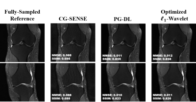 |
Compressed Sensing MRI Revisited: Optimizing $$$\ell_{1}$$$-Wavelet Reconstruction with Modern Data Science Tools
Hongyi Gu1,2, Burhaneddin Yaman2,3, Kamil Ugurbil2, Steen Moeller2, and Mehmet Akcakaya2,3
1Electrical Engineering, University of Minnesota, Minneapolis, MN, United States, 2Center for magnetic resonance research, Minneapolis, MN, United States, 3University of Minnesota, Minneapolis, MN, United States
Deep learning (DL) has shown great promise in improving the reconstruction quality of accelerated MRI. These methods are shown to outperform conventional methods, such as parallel imaging and compressed sensing (CS). However, in most comparisons, CS is implemented with ~2-3 empirically-tuned hyperparameters. On the other hand, DL methods enjoy a plethora of advanced data science tools. In this work, we revisit l1 -regularized CS using these modern tools. Using an unrolled ADMM approach, we show that classical l1-wavelet CS can achieve comparable quality to DL reconstructions, with only 116 parameters compared to hundreds of thousands for the DL approaches.
|
|
0275.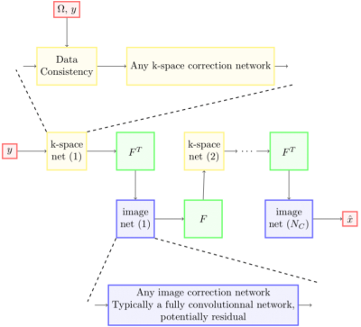 |
XPDNet for MRI Reconstruction: an application to the 2020 fastMRI challenge
Zaccharie Ramzi1,2,3, Jean-Luc Starck2, and Philippe Ciuciu1,3
1Neurospin, Gif-Sur-Yvette, France, 2Cosmostat team, CEA, Gif-Sur-Yvette, France, 3Parietal team, Inria Saclay, Gif-Sur-Yvette, France
We present a new neural network, the XPDNet, for MRI reconstruction from periodically under-sampled multi-coil data. We inform the design of this network by taking best practices from MRI reconstruction and computer vision. We show that this network can achieve state-of-the-art reconstruction results, as shown by its ranking of second in the fastMRI 2020 challenge.
|
||
0276.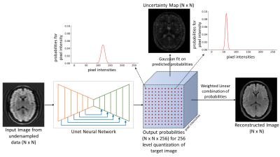 |
Estimating Uncertainty in Deep Learning MRI Reconstruction using a Pixel Classification Image Reconstruction Framework
Kamlesh Pawar1,2, Gary F Egan1,2,3, and Zhaolin Chen1,4
1Monash Biomedical Imaging, Monash University, Melbourne, Australia, 2School of Psychological Sciences, Monash University, Melbourne, Australia, 3ARC Centre of Excellence for Integrative Brain Function, Monash University, Melbourne, Australia, 4Department of Electrical and Computer Systems Engineering, Monash University, Melbourne, Australia
Data-driven deep learning (DL) image reconstruction from undersampled data has become a mainstream research area in MR image reconstruction. The generalization of the model on unseen data and out of sample data distribution is still a concern for the adoption of the DL reconstruction. In this work, we present a method of risk assessment in DL MR image reconstruction by generating an uncertainty map along with the reconstructed image. The proposed method re-casts image reconstruction as a classification problem and the probability of each voxel intensity in the reconstructed image can be used to efficiently estimate its uncertainty.
|
||
0277.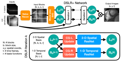 |
DSLR+: Enhancing deep subspace learning reconstruction for high-dimensional MRI
Christopher Michael Sandino1, Frank Ong2, Ke Wang3, Michael Lustig3, and Shreyas Vasanawala2
1Electrical Engineering, Stanford University, Stanford, CA, United States, 2Radiology, Stanford University, Stanford, CA, United States, 3Electrical Engineering and Computer Sciences, UC Berkeley, Berkeley, CA, United States
Unrolled neural networks (UNNs) have enabled state-of-the-art reconstruction of dynamic MRI data, however, they remain limited by GPU memory hindering applications to high-resolution, high-dimensional imaging. Previously, we proposed a deep subspace learning reconstruction (DSLR) method to reconstruct low-rank representations of dynamic imaging data. In this work, we present DSLR+, which improves upon DSLR by leveraging a locally low-rank model and a more accurate data consistency module. We demonstrate improvements over state-of-the-art UNNs with respect to 2D cardiac cine image quality and reconstruction memory footprint, which is greatly reduced by reconstructing compressed representations of the data instead of the data itself.
|
||
0278.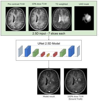 |
Anomaly-aware multi-contrast deep learning model for reduced gadolinium dose in contrast-enhanced brain MRI - a feasibility study
Srivathsa Pasumarthi1, Enhao Gong1, Greg Zaharchuk1, and Tao Zhang1
1Subtle Medical Inc., Menlo Park, CA, United States
Deep learning (DL) has recently been proven to be effective in addressing the safety concerns of Gadolinium-based Contrast Agents (GBCAs). Recent studies have shown that DL-based algorithms are able to reconstruct contrast-enhanced MRI images with only a fraction of the standard dose. This work investigates the feasibility of improving the performance of such DL algorithms using multi-contrast MRI data, combined with an unsupervised anomaly detection based attention mechanism.
|
||
0279.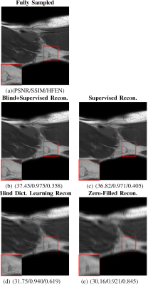 |
Blind Primed Supervised (BLIPS) Learning for MR Image Reconstruction
Anish Lahiri1, Guanhua Wang2, Saiprasad Ravishankar3, and Jeffrey A. Fessler1
1Electrical and Computer Engineering, University of Michigan, Ann Arbor, MI, United States, 2Biomedical Engineering, University of Michigan, Ann Arbor, MI, United States, 3Computational Mathematics, Science and Engineering, and Biomedical Engineering, Michigan State University, East Lansing, MI, United States
This work examines a combined supervised-unsupervised framework involving dictionary-based blind learning and deep supervised learning for MR image reconstruction from under-sampled k-space data. A major focus of the work is to investigate the possible synergy of learned features in traditional shallow reconstruction using sparsity-based priors and deep prior-based reconstruction. Specifically, we propose a framework that uses an unrolled network to refine a blind dictionary learning based reconstruction. We compare the proposed method with strictly supervised deep learning-based reconstruction approaches on several datasets of varying sizes and anatomies.
|
||
0280. |
Joint estimation of coil sensitivities and image content using a deep image prior
Guanxiong Luo1, Xiaoqing Wang1, Volkert Roeloffs1, Zhengguo Tan1, and Martin Uecker1,2
1Institute for Diagnostic and Interventional Radiology, University Medical Center Göttingen, Germany, Göttingen, Germany, 2Campus Institute Data Science (CIDAS), University of Göttingen, Germany, Göttingen, Germany
Parallel imaging for reduction of scanning time is now routinely used in clinical practice. The spatial information from the coils’ profiles are exploited for encoding. The nonlinear inversion reconstruction is a calibrationless parallel imaging technique, which jointly estimate coil sensitivities and image content. In this work, we demonstrate how to combine such a calibrationless parallel imaging technique with an advanced neural network based image prior for efficient MR imaging.
|
The International Society for Magnetic Resonance in Medicine is accredited by the Accreditation Council for Continuing Medical Education to provide continuing medical education for physicians.