Oral
Optimized Signal Representation for Acquisition & Reconstruction
ISMRM & SMRT Annual Meeting • 15-20 May 2021

Oral
Optimized Signal Representation for Acquisition & Reconstruction
| Concurrent 1 | 12:00 - 14:00 | Moderators: Dana Peters & Martin Uecker |
 |
0437.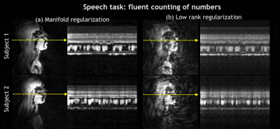 |
Rapid dynamic speech imaging at 3Tesla using combination of a custom airway coil, variable density spirals and manifold regularization
Rushdi Zahid Rusho1, Wahidul Alam1, Abdul Haseeb Ahmed2, Stanley J. Kruger3, Mathews Jacob2, and Sajan Goud Lingala1,3
1Roy J. Carver Department of Biomedical Engineering, The University of Iowa, Iowa City, IA, United States, 2Department of Electrical and Computer Engineering, The University of Iowa, Iowa City, IA, United States, 3Department of Radiology, The University of Iowa, Iowa City, IA, United States
We propose a novel rapid dynamic speech MRI scheme that leverages multi-coil acquisitions from a dedicated 16 channel airway coil, variable density spirals, and manifold regularization. The variable density spirals enables self-navigation to extract the Laplacian manifold matrix from low spatial but high temporal resolution data. Our scheme allows for efficient exploitation of similarities between image frames that are distant in time without the need of explicit binning. We demonstrate robust reconstructions on free running speech data containing complex spatio-temporal dynamics at a temporal resolution of 15 ms/frame.
|
|
 |
0438. |
An open dataset for speech production real-time MRI: raw data, synchronized audio, and images
Yongwan Lim1, Asterios Toutios1, Yannick Bliesener1, Ye Tian1, Krishna S. Nayak1, and Shrikanth Narayanan1
1University of Southern California, Los Angeles, CA, United States
We introduce the first-ever public domain real-time MRI raw dataset for the study of human speech production. The dataset consists of raw, multi-receiver-coil MRI data with non-Cartesian, spiral sampling trajectory and reconstructed images derived using a reference reconstruction method along with synchronized audio for 72 subjects performing 32 linguistically motivated speech tasks. This dataset can be used to develop traditional and machine learning / artificial intelligence approaches for dynamic image reconstruction in the context of fast aperiodic motion, which is currently an unsolved problem, as well as for artifact correction, feature extraction, and direct extraction of linguistically relevant biomarkers.
|
|
|
0439.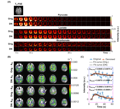 |
Denoising of Hyperpolarized 13C MR Images of the Human Brain Using Patch-based Higher-order Singular Value Decomposition
Yaewon Kim1, Hsin-Yu Chen1, Adam W. Autry1, Javier Villanueva-Meyer1, Susan M. Chang2, Yan Li1, Peder E. Z. Larson1, Jeffrey R. Brender3, Murali C. Krishna3, Duan Xu1, Daniel B. Vigneron1,2, and Jeremy W. Gordon1
1Department of Radiology and Biomedical Imaging, University of California, San Francisco, CA, United States, 2Department of Neurological Surgery, University of California, San Francisco, CA, United States, 3Center for Cancer Research, National Cancer Institute, National Institutes of Health, Bethesda, MD, United States
Quantifying metabolism in hyperpolarized (HP) 13C MRI can be challenging because of low signal-to-noise ratio for downstream metabolites. To overcome this limitation, we investigated a new patch-based singular value decomposition method to denoise dynamic imaging data and tested it in numerical simulations and on 6 HP [1-13C]pyruvate EPI human brain datasets. The sensitivity enhancement provided by denoising significantly improved quantification of metabolite dynamics. With denoising, [1-13C]pyruvate and its metabolites [1-13C]lactate and [13C]bicarbonate had ≥5-fold sensitivity gain, improving the number of quantifiable voxels for mapping pyruvate-to-bicarbonate conversion rates (kPB) by 2-fold, and providing whole-brain coverage for mapping pyruvate-to-lactate conversion rates (kPL).
|
|
|
0440.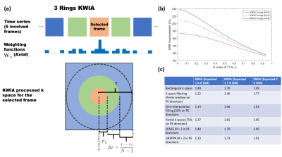 |
k-Space Weighted Image Average (KWIA) for ASL-based Dynamic MRA and Perfusion Imaging
Chenyang Zhao1, Xingfeng Shao1, Lirong Yan1, and Danny JJ Wang1
1Laboratory of Functional MRI Technology (LOFT), Mark & Mary Stevens Neuroimaging and Informatics Institute, University of Southern California, Los Angeles, CA, United States
The intrinsically low SNR of ASL techniques is a main limitation that hinders their clinical translations. This work presented a novel denoising algorithm for dynamic MRI termed KWIA and evaluated its use in multi-delay ASL and ASL-based 4D dMRA. KWIA improves SNR without compromising spatial and temporal resolution by progressively increasing the neighboring time frames for view-shared averaging for more distal k-space regions. Experimental results showed that KWIA can provide significant SNR improvement that enables better visualization and quantification for both multi-delay ASL and ASL-based 4D dMRA as well as other dynamic MRI techniques.
|
|
0441.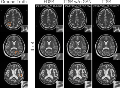 |
MR image super-resolution using attention mechanism: transfer textures from external database
Mengye Lyu1, Guoxiong Deng1, Yali Zheng1, Yilong Liu2,3, and Ed X. Wu2,3
1College of Health Science and Environmental Engineering, Shenzhen Technology University, Shenzhen, China, 2Laboratory of Biomedical Imaging and Signal Processing, The University of Hong Kong, Hong Kong, China, 3Department of Electrical and Electronic Engineering, The University of Hong Kong, Hong Kong, China
Super-resolution (SR) is useful to reduce scan time and/or enhance MR images for better visual perception. High-resolution reference images may improve super-resolution quality, but most previous studies focused on using references from the same subject. Here, we use an image search module to find similar images from other subjects and use transformer based neural networks to learn and transfer the relevant textures to the output. We demonstrate that this approach can outperform single-image super-resolution, and is feasible to achieve high-quality super-resolution at large factors. As the reference images are not limited within a subject, it potentially has wide applications.
|
||
0442.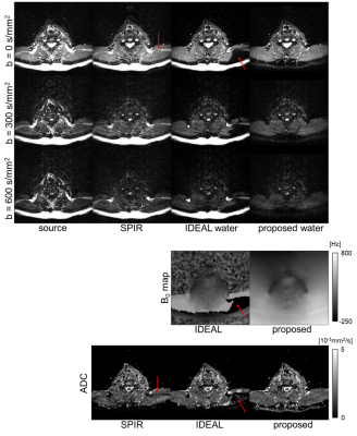 |
A regularized joint water/fat separation and B0 map estimation for 2D-navigated interleaved EPI based diffusion MRI
Yiming Dong1, Kirsten Koolstra2, Malte Riedel3, Matthias J.P. van Osch1, and Peter Börnert1,4
1C.J. Gorter Center for High Field MRI, Department of Radiology, LUMC, Leiden, Netherlands, 2Division of Image Processing, Department of Radiology, LUMC, Leiden, Netherlands, 3University of Lübeck, Lübeck, Germany, 4Philips Research Hamburg, Hamburg, Germany
Multi-shot EPI enables high-resolution diffusion-weighted imaging with reduced geometric distortions. However, fat is often a confounding factor in EPI, especially in regions with severe B0 inhomogeneities. For the proposed method, data is acquired using TE-shifted interleaved EPI and 2D-navigators to sense the motion-induced shot phases. The reconstruction includes a multi-peak fat model and corrects for the fat frequency-specific chemical shift displacements in phase-encoding direction by a time-efficient image-space formulation. In-vivo results show that the proposed algorithm provides improved water/fat separation compared with the conventional technique and fat-suppressed acquisition.
|
||
|
0443.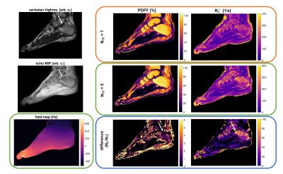 |
On quantification errors of R2* and PDFF mapping in trabecularized bone marrow induced by the static dephasing regime
Sophia Kronthaler1, Christof Boehm1, Kilian Weiss2, Marcus R. Makowski1, and Dimitrios C. Karampinos1
1Department of Diagnostic and Interventional Radiology, School of Medicine, Technical University of Munich, Munich, Germany, 2Philips Healthcare, Hamburg, Germany
Chemical shift encoding-based (CSE) water-fat separation techniques are becoming more common in the study of bone marrow changes as they allow the simultaneous assessment of tissue fat-fraction and R2*. A typical acquisition strategy in CSE-MRI aims to minimize the first TE to increase SNR. However, the R2* decay in the presence of trabecular bone microstructure is known to be nontrivial due to the occurrence of the static dephasing regime. The present work investigates, with the help of UTE acquisitions, the quantification errors of R2* and PDFF maps in trabecularized bone marrow due to the presence of the static dephasing regime.
|
|
|
0444.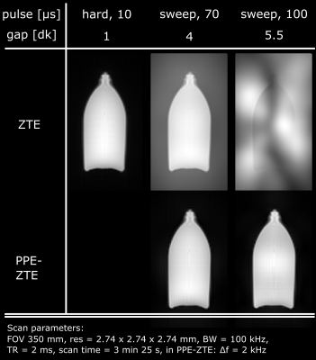 |
Algebraic reconstruction of missing data in zero echo time MRI with pulse profile encoding (PPE-ZTE)
Romain Froidevaux1, Markus Weiger1, and Klaas Paul Pruessmann1
1Institute for Biomedical Engineering, ETH Zurich and University of Zurich, Zurich, Switzerland
Short-T2 imaging needs immediate and rapid encoding, as provided by zero echo time (ZTE) MRI. However, in ZTE, excitation and early encoding occur simultaneously and preclude data acquisition in the k-space center, leading to local undersampling or gap. One way of retrieving the missing data involves algebraic reconstruction, but it is limited to small gaps and thus requires short RF pulses that restrain achievable SNR and contrast options. Here, we demonstrate a method for algebraic reconstruction of large gaps, based on the knowledge of excitation pulses. It enables the use of longer pulses and overcomes ZTE flip angle limitations.
|
|
|
0445.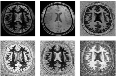 |
Simultaneous optimisation of MP2RAGE UNI and FLAWS brain images at 7T using Extended Phase Graph (EPG) Simulations
Ayse Sila Dokumaci1, Fraser R. Aitken1, Jan Sedlacik1, Philippa Bridgen1, Raphael Tomi-Tricot1,2, Tom Wilkinson1, Ronald Mooiweer1, Sharon Giles1, Joseph V. Hajnal1, Shaihan Malik1, Jonathan O'Muircheartaigh1, and David W. Carmichael1
1Division of Imaging Sciences and Biomedical Engineering, King's College London, London, United Kingdom, 2MR Research Collaborations, Siemens Healthcare Limited, Frimley, United Kingdom
The MP2RAGE sequence is typically used at 7T to produce UNI image with maximised contrast between WM-GM and GM-CSF while mitigating B1- field variability. It can also be optimised to obtain Fluid and White Matter Suppression (FLAWS) images but this is typically done separately. Here, the Extended Phase Graph formalism was used to optimise both FLAWS and UNI images at 7T within one acquisition while minimising B1+ sensitivity. Different combinations were tested in healthy subjects with 0.65mm isotropic resolution demonstrating that UNI and FLAWS images could be obtained together while largely maintaining image quality.
|
|
0446.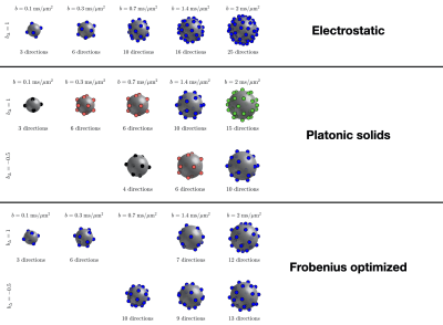 |
Frobenius optimization of tensor-valued diffusion sampling schemes
Alexis Reymbaut1
1Random Walk Imaging AB, Lund, Sweden
Tensor-valued diffusion encoding has allowed for increased specificity in diffusion MRI, probing the diffusion patterns of water molecules in vivo along new dimensions. From an intuitive standpoint, a versatile sampling scheme should be sensitive to a diverse set of diffusion profiles in any given voxel. However, while optimization strategies based on electrostatic repulsion achieve this for conventional diffusion sampling scheme, no equivalent optimization exists for tensor-valued diffusion data. In this work, we derive an optimization strategy based on maximizing the Frobenius distance between b-tensors. Its evaluation in silico demonstrates that it increases the accuracy of diffusion tensor distribution imaging.
|
The International Society for Magnetic Resonance in Medicine is accredited by the Accreditation Council for Continuing Medical Education to provide continuing medical education for physicians.