Oral
Motion Correction Strategies
ISMRM & SMRT Annual Meeting • 15-20 May 2021

| Concurrent 1 | 16:00 - 18:00 | Moderators: Tess Wallace & Xucheng Zhu |
 |
0119.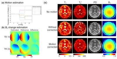 |
Motion-corrected 3D-EPTI with 4D navigator for fast and robust whole-brain quantitative imaging
Zijing Dong1,2, Fuyixue Wang1,3, Jie Xiang4, and Kawin Setsompop5,6
1Athinoula A. Martinos Center for Biomedical Imaging, Massachusetts General Hospital, Charlestown, MA, United States, 2Department of Electrical Engineering and Computer Science, MIT, Cambridge, MA, United States, 3Harvard-MIT Health Sciences and Technology, MIT, Cambridge, MA, United States, 4Tsinghua University, Beijing, China, 5Department of Radiology, Stanford University, Stanford, CA, United States, 6Department of Electrical Engineering, Stanford University, Stanford, CA, United States
A motion-correction method is developed for the recently proposed 3D-EPTI to achieve fast and motion-robust quantitative imaging of the human brain. A 4D-navigator (x-y-z-echoes) is inserted into the relaxation-recovery dead-time of the sequence to provide accurate estimations of 3D-motion and B0-inhomogeneity changes at every TR (~2-3s), which are incorporated into a motion-and-phase corrected subspace reconstruction. The navigator utilizes an optimized spatiotemporal encoding to acquire central 3D k-space for accurate motion-estimation using just 4 small-flip-angle excitations, resulting in negligible signal-recovery reduction (<1%) to the 3D-EPTI acquisition. Simulation and in-vivo experiments preliminarily validate the accuracy of estimation and effectiveness of the reconstruction.
|
|
|
0120.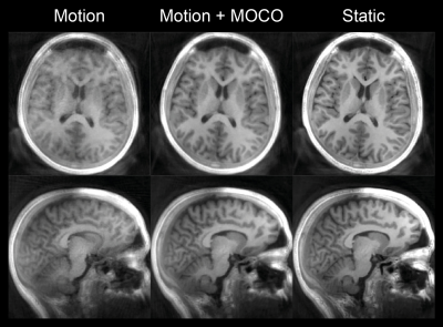 |
MERLIN: Motion Insensitive Silent Neuroimaging
Emil Ljungberg1, Tobias Wood1, Ana Beatriz Solana2, Steven C.R. Williams1, Gareth J. Barker1, and Florian Wiesinger1,2
1Neuroimaging, Institute of Psychiatry, Psychology, and Neuroscience, King's College London, London, United Kingdom, 2ASL Europe, GE Healthcare, Munich, Germany
In this work we present MERLIN (Motion Elimination in Radial acquisition Leveraging Interleaved Navigators): a new method for silent, motion insensitive, MRI using self-navigated zero echo time (ZTE) imaging. Using T1w ZTE neuroimaging as an example, we demonstrate that MERLIN can correct for rigid body motion and markedly improve image quality. Such a silent and motion insensitive neuroimaging protocol can save time and money in both clinical and research settings.
|
|
0121.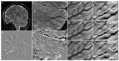 |
Visualizing the cerebellar cortical layers with prospective motion correction
Nikos Priovoulos1, Mads Andersen2, Vincent O Boer3, and Wietske van der Zwaag1
1Spinoza Center, Amsterdam, Netherlands, 2Philips Healthcare, Copenhagen, Denmark, 3Danish Research Centre for Magnetic Resonance, Centre for Functional and Diagnostic Imaging and Research, Copenhagen University Hospital, Hvidovre, Denmark
The cerebellar cortical microstructure is important for several functions, like motor control, but remain largely underexplored in vivo due to its small thickness. Here, we combined a susceptibility-weighted acquisition with in-plane resolution of just 0.2x0.2mm2 at 7Tesla along with a fat-navigator-based prospective motion correction. Using this setup, the cerebellar layers could be seen in the phase images of all 4 MRI-naive participants. Our results show that the cerebellar layers can be consistently visualized opening new neuroscientific and clinical dimensions.
|
||
0122.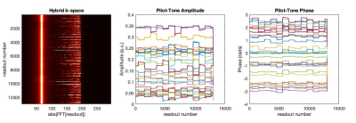 |
Motion Estimation for Brain Imaging at Ultra-High Field Using Pilot-Tone: Comparison with DISORDER Motion Compensation
Tom Wilkinson1,2, Felipe Godinez1,2, Yannick Brackenier1,2, Raphael Tomi-Tricot1,2,3, Lucilio Cordero-Grande1,2,4, Philippa Bridgen1,2, Sharon Giles1,2, Joseph V Hajnal1,2, and Shaihan J Malik1,2
1Biomedical Engineering Department, School of Biomedical Engineering and Imaging Sciences, Kings College London, London, United Kingdom, 2Centre for the Developing Brain, School of Biomedical Engineering and Imaging Sciences, Kings College London, London, United Kingdom, 3MR Research Collaborations, Siemens Healthcare Limited, Frimley, United Kingdom, 4Biomedical Image Technologies, ETSI Telecomunicación, Universidad Politécnica de Madrid and CIBER-BNN, Madrid, Spain
A ‘pilot-tone’ implementation for 7T head MRI was constructed by broadcasting RF at the appropriate frequency into the scanner room during data acquisition. This signal was demonstrated to enable motion estimation, when calibrated first by correlating measurements with motion estimates from image registration. Subsequently these estimates were compared with others obtained from the iterative DISORDER joint motion estimation & reconstruction method. These independent methods of motion estimation can potentially improve or replace other methods of motion correction at ultra-high field where motion-correction is particularly relevant.
|
||
0123.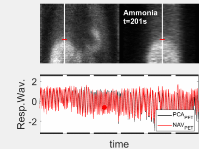 |
PET/MR respiratory motion gating for free
Florian Wiesinger1, Timothy Deller2, Floris Jansen2, Jose de Arcos Rodriguez1, Ronny R Buechel3, Philipp A Kaufmann3, and Edwin EGW ter Voert3
1GE Healthcare, Munich, Germany, 2GE Healthcare, Waukesha, WI, United States, 3Department of Nuclear Medicine, University Hospital Zurich, Zurich, Switzerland Respiratory motion correction is a long-standing problem in hybrid PET/MR imaging with many partial solutions. Here we present a novel method based on extracting respiratory motion information from the PET data using the ultra-fast listmode reconstruction framework. Doing so a highly accurate respiratory waveform derived from the inside of the body (i.e. lung liver interface) is obtained for free without requiring an extra motion sensor or complicating the PET/MR imaging workflow. |
||
0124.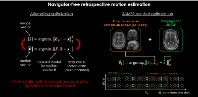 |
Separable motion estimation and correction for 2D TSE imaging using a rapid 3D volumetric scout acquisition
Daniel Polak1,2, Daniel Nicolas Splitthoff1, Berkin Bilgic2,3,4, Lawrence L. Wald2,3,4, Kawin Setsompop5, and Stephen F. Cauley2,3,4
1Siemens Healthcare GmbH, Erlangen, Germany, 2Department of Radiology, A. A. Martinos Center for Biomedical Imaging, Charlestown, MA, United States, 3Department of Radiology, Harvard Medical School, Boston, MA, United States, 4Harvard-MIT Health Sciences and Technology, Massachusetts Institute of Technology, Cambridge, MA, United States, 5Department of Radiology, Stanford, Stanford, CA, United States
SAMER is a navigation-free retrospective motion-correction technique which achieves rapid motion estimation using an ultra-fast, low-resolution scout scan as an image prior. In this work, the SAMER framework is extended to 3D volumetric reconstructions of 2D TSE imaging data. The optimized 3D volumetric scout scan is combined with a distributed 2D TSE slice ordering for fully separable motion estimation with negligible added scan time. The motion correction performance was evaluated in-vivo for representative motion trajectories and compatibility to highly accelerated Simultaneous Multi-Slice acquisitions is demonstrated.
|
||
0125.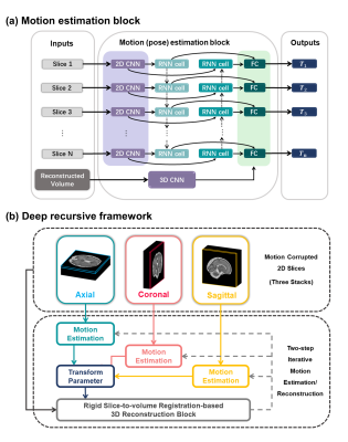 |
Automated motion correction of multi-slice fetal brain MRI using a deep recursive framework
Wen Shi1,2,3, Jiwei Sun1, Yamin Li3, Cong Sun4, Tianshu Zheng1, Yi Zhang1, Guangbin Wang4, and Dan Wu1
1Key Laboratory for Biomedical Engineering of Ministry of Education, Department of Biomedical Engineering, College of Biomedical Engineering & Instrument Science, Zhejiang University, Hangzhou, China, 22. Department of Biomedical Engineering, Johns Hopkins University School of Medicine, Baltimore, MD, United States, 3School of Biomedical Engineering, Shanghai Jiao Tong University, Shanghai, China, 4Department of Radiology, Shandong Medical Imaging Research Institute, Cheeloo College of Medicine, Shandong University, Jinan, China
Prenatal MRI of fetal brain is vulnerable to unpredictable fetal motion and maternal movement. The conventional registration-based motion correction methods sometimes fail in excessive motion. In this work, we proposed a learning-based scheme to estimate fetal brain motion using a deep recursive framework, which replicated the iterative slice-to-volume registration and 3D volumetric reconstruction process. The network outperformed the previous learning-based methods and with good computational efficiency compared to traditional method. It also achieved high super-resolution reconstruction accuracy on simulated motion-corrupted slices, and therefore, is promising for fetal brain MRI analysis.
|
||
|
0126.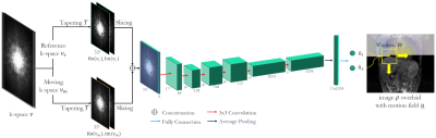 |
LAPNet: Deep-learning based non-rigid motion estimation in k-space from highly undersampled respiratory and cardiac resolved acquisitions
Thomas Küstner1,2, Jiazhen Pan3, Haikun Qi2, Gastao Cruz2, Kerstin Hammernik3,4, Christopher Gilliam5, Thierry Blu6, Sergios Gatidis1, Daniel Rueckert3,4, René Botnar2, and Claudia Prieto2
1Department of Radiology, Medical Image and Data Analysis (MIDAS), University Hospital of Tübingen, Tübingen, Germany, 2School of Biomedical Engineering and Imaging Sciences, King's College London, London, United Kingdom, 3AI in Medicine and Healthcare, Klinikum rechts der Isar, Technical University of Munich, München, Germany, 4Department of Computing, Imperial College London, London, United Kingdom, 5RMIT, University of Melbourne, Melbourne, Australia, 6Chinese University of Hong Kong, Hong Kong, Hong Kong
Estimation of non-rigid motion is an important task in respiratory and cardiac motion correction. Usually, this problem is formulated in image space via diffusion, parametric-spline or optical flow methods. However, image-based registration can be impaired by aliasing artefacts or by estimating in low image resolution in cases of highly accelerated acquisitions. In this work, we propose a novel deep learning-based non-rigid motion estimation directly in k-space, named LAPNet. The proposed method, inspired by optical flow, is compared against registration in image space and tested for respiratory and cardiac motion as well as different acquisition trajectories providing a generalizable diffeomorphic registration.
|
|
0127.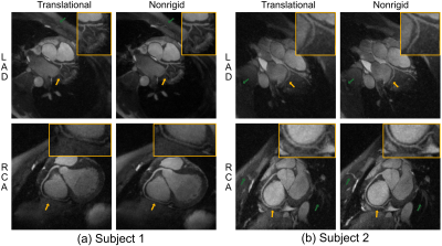 |
Nonrigid Motion-corrected Reconstruction Using Image-space Gridding for Free-breathing Cardiac MRI
Kwang Eun Jang1,2, Mario O. Malavé1, Dwight G. Nishimura1, and Shreyas S. Vasanawala3
1Magnetic Resonance Systems Research Lab (MRSRL), Department of Electrical Engineering, Stanford University, Stanford, CA, United States, 2Department of Bioengineering, Stanford University, Stanford, CA, United States, 3Department of Radiology, Stanford University, Stanford, CA, United States
Motion remains a major challenge in MRI. Many motion-corrected reconstruction methods are available, yet models are often simplified. We propose image-space gridding that resamples images onto arbitrary grids, which provides a pair of operators that represents the forward and adjoint of a nonrigid transform. This allows existing nonrigid image registration techniques to be incorporated into model-based reconstructions. We apply this method to correct for respiratory motion in free-breathing cardiac MRI. Data from individual heartbeats are binned to reconstruct image-based self-navigators. Nonrigid motion is estimated using a diffeomorphic demons algorithm, and corrected by solving an optimization problem with image-space gridding operators.
|
||
0128.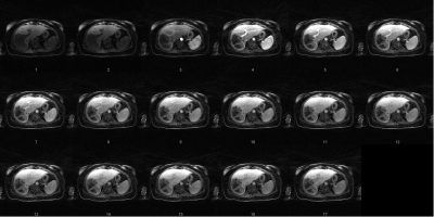 |
Forward-Fourier Motion-Corrected Reconstruction for Free-Breathing Liver DCE-MRI
Sihao Chen1, Cihat Eldeniz1, Weijie Gan1, Ulugbek Kamilov1, Tyler Fraum1, and Hongyu An1
1Washington University in St. Louis, Saint Louis, MO, United States
Dynamic contrast-enhanced MRI (DCE-MRI) of the liver offers structural and functional information for assessing the contrast uptake visually. However, respiratory motion and the requirement of high temporal resolution make it difficult to generate high-quality DCE-MRI. In this study, we proposed a novel forward-Fourier motion-corrected reconstruction utilizing deep learning based 3D motion information on severely undersampled DCE-MRI. With no need to use view-sharing or DCE contrast smoothness constraint, this approach avoids enhancement spillover from adjacent DCE contrasts and reconstructs high-quality motion-free DCE images with reduced artifacts and enhanced sharpness.
|
The International Society for Magnetic Resonance in Medicine is accredited by the Accreditation Council for Continuing Medical Education to provide continuing medical education for physicians.