Oral
Novel Acquisitions
ISMRM & SMRT Annual Meeting • 15-20 May 2021

| Concurrent 1 | 12:00 - 14:00 | Moderators: Gareth Barker & Craig Meyer |
0001.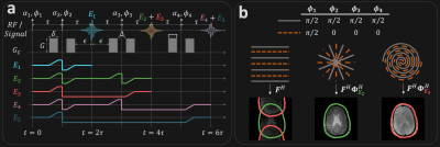 |
Magnetic Resonance Coherence Pathway Unraveling
Nikolai Mickevicius1 and Eric Paulson2
1Medical College of Wisconsin, Milwaukee, WI, United States, 2Department of Radiation Oncology, Medical College of Wisconsin, Milwaukee, WI, United States The magnetic resonance coherence pathway unraveling (MR-CPU) method acquires primary and stimulated echoes simultaneously, and encodes them using CAIPIRINHA RF phase cycling such that they can be separated during image reconstruction. This initial study demonstrates the feasibility of unaliasing overlapped coherence pathway images, and future studies will investigate its use for quantitative T1, T2, and diffusion coefficient mapping. |
||
0002.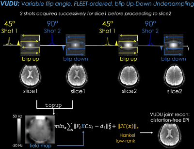 |
VUDU: motion-robust, distortion-free multi-shot EPI
Jaejin Cho1,2, Avery JL Berman1,2, Borjan Gagoski2,3, Congyu Liao4, Jason Stockmann1,2, Jonathan R Polimeni1,2, and Berkin Bilgic1,2
1Martinos Center for Biomedical Imaging, Department of Radiology, Massachusetts General Hospital, Charlestown, MA, United States, 2Harvard Medical School, Boston, MA, United States, 3Fetal-Neonatal Neuroimaging and Developmental Science Center, Boston Children's Hospital, Boston, MA, United States, 4Radiological Sciences Laboratory, Stanford University, Palo Alto, CA, United States
We introduce VUDU (Variable flip, blip-Up and -Down Undersampling) for motion-robust, distortion-free multi-shot EPI (msEPI) acquisition. VUDU uses FLEET-ordering to acquire all shots of a given slice successively before proceeding to the next slice, and employs variable flip angle (vfa) excitation to maximize the signal. Phase encoding polarities are reversed between shots to estimate and eliminate distortions, and low-rank constraint mitigates shot-to-shot inconsistencies. VUDU thus utilizes vfa-FLEET excitation and blip-up and -down acquisition (BUDA) to encode each slice in 250ms. We demonstrate VUDU with GRE, SE/diffusion contrasts in the brain, and expect that this will enable msEPI in the abdomen.
|
||
0003.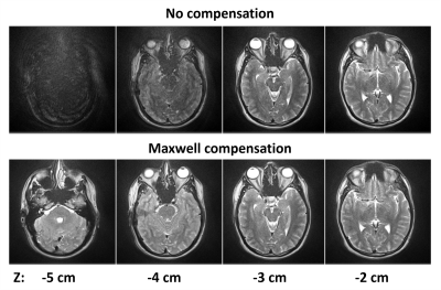 |
Maxwell Compensation for Spiral Turbo-Spin-Echo Imaging
John P. Mugler1, Adrienne E. Campbell-Washburn2, Rajiv Ramasawmy2, Josef Pfeuffer3, and Craig H. Meyer1
1University of Virginia, Charlottesville, VA, United States, 2Cardiovascular Branch, Division of Intramural Research, National Heart, Lung, and Blood Institute, National Institutes of Health, Bethesda, MD, United States, 3Siemens Healthcare GmbH, Erlangen, Germany
Spiral TSE imaging presents challenges for compensating concomitant (Maxwell) gradient effects because spiral waveforms vary along the echo train, as opposed to Cartesian imaging for which the same readout waveform is used for every echo. Since Maxwell terms are proportional to 1/Bo, compensation is particularly important at low field strength. An interleaved-spiral T2-weighted 2D-TSE pulse sequence was developed that incorporates gradient waveform modifications to achieve compensation of the self-squared Maxwell terms at both the echoes and over echo spacings. This approach provided substantial improvement in image quality at 0.55T for degradation associated with self-squared concomitant-gradient effects.
|
||
0004.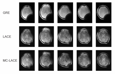 |
Low-Angle Combined-Echo (LACE) Imaging in Highly Inhomogeneous B0 Magnetic Fields
Sebastian Theilenberg1, Chathura Kumaragamage2, Scott McIntyre2, Terry W. Nixon2, Christoph Juchem1,3, and Robin A. de Graaf2
1Biomedical Engineering, Columbia University, New York, NY, United States, 2Department of Radiology and Biomedical Imaging, Magnetic Resonance Research Center, Yale University School of Medicine, New Haven, CT, United States, 3Radiology, Columbia University Medical Center, New York, NY, United States
In line with recent developments in scanner design for more cost effective and more accessible scanners, we propose a low-angle combined-echo sequence similar to a conventional spin echo sequence capable of producing high quality images in the presence of strong B0 inhomogeneity. We present simulation results investigating the signal dependence on the sequence’s timings and flip angles as well as image contrast for typical relaxation times of normal brain white and gray matter. The simulations were validated in vivo at 4 T. Lastly, we show the feasibility to utilize multi-coil generated image encoding fields for this sequence.
|
||
 |
0005.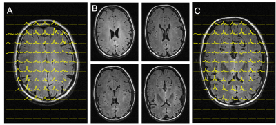 |
Interleaved MRI and DMI on human brain in vivo
Yanning Liu1, Henk M. De Feyter1, Scott McIntyre1, Terence W. Nixon1, and Robin A. de Graaf1
1MRRC Yale University, New Haven, CT, United States
Deuterium metabolic imaging (DMI) is a powerful method to map metabolism in vivo. To integrate DMI with clinical MRI, we propose and demonstrate an interleaved MRI and DMI routine, including the necessary hardware and sequence modifications. Using interleaved FLAIR MRI+DMI as an example, we demonstrate that MR image quality and DMI sensitivity as well as information content are preserved, both in phantoms and in the human brain in vivo. The interleaved MRI+DMI technology provides full flexibility to extend any MRI protocol with DMI, thereby offering a metabolic component to the range of MR imaging contrasts.
|
|
|
0006. |
Accelerated diffusion and relaxation-diffusion magnetic resonance imaging using time-division multiplexing echo-planar imaging (TDM-EPI)
Yang Ji1,2, Borjan Gagoski 2,3, W. Scott Hoge 1,2, Yogesh Rathi 1,2, and Lipeng Ning 1,2
1Brigham and Women’s Hospital, Boston, MA, United States, 2Harvard Medical School, Boston, MA, United States, 3Boston Children’s Hospital, Boston, MA, United States
Recently, several studies have shown that brain tissue consists of microscopically heterogeneous components that are characterized by different T2 values and diffusivity. The joint relaxation-diffusion MRI technique has been developed to probe the intrinsic tissue microstructure that cannot be probed using standard dMRI. However, a major limitation of the relaxation-diffusion MRI technique is the long scan time for acquiring dMRI with multiple TEs. In order to significantly reduce the scan time, we propose a time-division multiplexing based echo-planar imaging (TDM-EPI) sequence, which can accelerate relaxation-diffusion MRI and standard dMRI by 2 or 3 folds.
|
|
0007.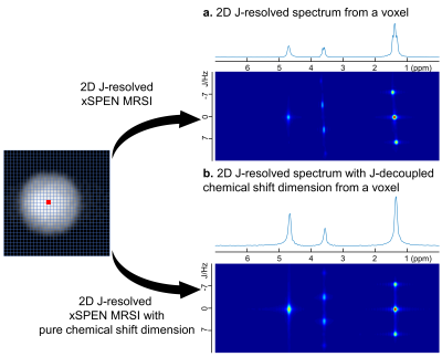 |
Fast 2D J-resolved MRSI combining echo planar imaging acquisition and turbo spin echo train evolution
Ke Dai1, Qingjia Bao2,3, Hao Chen1, Yiling Liu1, and Zhiyong Zhang1
1School of Biomedical Engineering, Shanghai, China, 2Wuhan United Imaging Life Science Instruments Co., Ltd, Wuhan, China, Wuhan, China, 3Department of Chemical and Biological Physics, Weizmann Institute of Science, Rehovot, Israel
J-resolved MRSI is a powerful tool for separating overlapping resonances and detecting coupled species such as GABA and glutamate, which are of great interest to brain studies. However, a major practical limitation of J-resolved MRSI lies in its long data acquisition time. In this work, we present a novel fast fully sampled 2D J-resolved MRSI, termed as J-resolved xSPEN spectroscopy, combining echo planar imaging acquisition and turbo spin echo train evolution. Our preliminary phantom results demonstrate the proposed method can achieve highly efficient fully sampled 2D J-resolved MRSI with increasing chemical shift separation and detection of coupled species.
|
||
0008.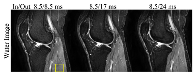 |
Dual Spin-Echo Proton Density-Weighted and T2-Weighted Knee Imaging with Asymmetric Spiral In-out Trajectories
Dinghui Wang1, Francis I. Baffour1, Daniel D. Borup2, Tzu-Cheng Chao1, and James G. Pipe1
1Radiology, Mayo Clinic, Rochester, MN, United States, 2Royal Philips, MR R&D, Rochester, MN, United States
This work proposes a spiral dual echo spin-echo sequence with asymmetric in-out trajectories to increase the scan efficiency of the second echo. The sequence has been applied for simultaneous sagittal proton density-weighted and T2-weighted Dixon knee imaging. Volunteer scans have demonstrated the feasibility of using the proposed method to achieve up to 36% SNR improvement for T2-weighted images. High quality water and fat images can be obtained with comparable total scan time as the conventional non-Dixon Cartesian fast (turbo) spin-echo sequences with SENSE factors from 1.5 to 2. T2 maps can also be estimated from proton density-weighted and T2-weighted images.
|
||
0009.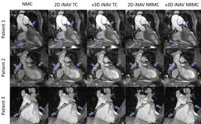 |
Whole-heart CMRA non-rigid motion compensation with autofocus virtual 3D iNAV
Alina Psenicny1, Gastao Cruz1, Camila Munoz1, Reza Hajhosseiny1, Thomas Kuestner1, Karl P Kunze2, Radhouene Neji1,2, René M Botnar1, and Claudia Prieto1
1School of Biomedical Engineering and Imaging Sciences, King's College London, London, United Kingdom, 2MR Research Collaborations, Siemens Healthcare Limited, Frimley, United Kingdom
3D whole-heart coronary MR angiography (CMRA) acquisition remains lengthy and can suffer from residual motion and/or undersampling related artifacts. 2D image-navigator based non-rigid respiratory motion compensation has been recently proposed to accelerate the CMRA scan. This framework combines 2D beat-to-beat translational and 3D bin-to-bin non-rigid motion correction. However, beat-to-beat anterior-posterior motion is not corrected for with this approach, which can result in significant residual motion. Here we propose a virtual 3D iNAV approach that exploits autofocus motion correction to further enable beat-to-beat anterior-posterior translational motion correction, assuming a linear relationship between the translational foot-head and anterior-posterior movement of the heart.
|
||
0010.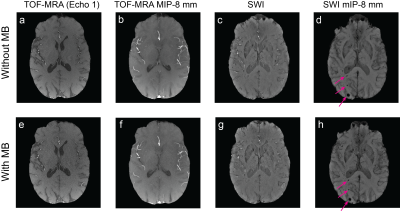 |
Multi-Band Multi-Slab 3D Multi-Echo Acquisition for Simultaneous Time-of-Flight MR Angiography and Susceptibility-Weighted Imaging at 3T
Misung Han1, Brian L Burns2, Suchandrima Banerjee2, and Janine M Lupo1,3
1Radiology and Biomedical Imaging, University of California, San Francisco, San Francisco, CA, United States, 2Applications and Workflow, GE Healthcare, Menlo Park, CA, United States, 3UCSF-UC Berkeley Graduate Program in Bioengineering, University of California, San Francisco and University of California, Berkeley, San Francisco, CA, United States
A single scan of multi-slab, multi-echo acquisition can simultaneously provide 3D time-of-flight (TOF) MR angiography and susceptibility-weighted imaging (SWI) MR venography, which allows for the assessment of vascular injury in the form of cerebral microbleeds in association with arteries and veins. However, the acquisition for high-resolution multi-slab 3D TOF-MRA/SWI with whole brain coverage takes over 10 minutes to acquire at 3T. In this work, we developed a 3D multi-slab, multi-echo acquisition for TOF-MRA/SWI with multi-band acceleration to reduce acquisition time and compared the resulting TOF-MRA and SWI images in patients with radiation-induced microbleeds.
|
The International Society for Magnetic Resonance in Medicine is accredited by the Accreditation Council for Continuing Medical Education to provide continuing medical education for physicians.