Oral
Optimized Sampling & Sequence Design
ISMRM & SMRT Annual Meeting • 15-20 May 2021

| Concurrent 1 | 18:00 - 20:00 | Moderators: Maria Engel & Rebecca Feldman |
0833.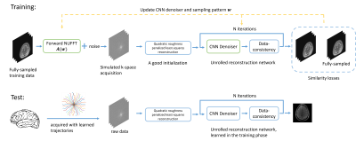 |
B-spline Parameterized Joint Optimization of Reconstruction and K-space Sampling Patterns (BJORK) for Accelerated 2D Acquisition
Guanhua Wang1, Tianrui Luo1, Jon-Fredrik Nielsen1, Jeffrey A. Fessler2, and Douglas C. Noll1
1Biomedical Engineering, University of Michigan, Ann Arbor, MI, United States, 2Electrical Engineering and Computer Science, University of Michigan, Ann Arbor, MI, United States
The proposed approach, BJORK, provides a robust and generalizable workflow to jointly optimize non-Cartesian sampling patterns and a physics-informed reconstruction. Several approaches, including re-parameterization of trajectories, multi-level optimization, and non-Cartesian unrolled neural networks, are introduced to improve training effect and avoid sub-optimal local minima. The in-vivo experiments show that the networks and trajectories learned on simulation dataset are transferable to the real acquisition even with different parameter-weighted MRI contrasts and noise-levels, and demonstrate improved image quality compared with previous learning-based and model-based trajectory optimization methods.
|
||
0834. |
Accelerated Ultrahigh Temporal-Resolution MRI with Random k-Space Undersampling
Qingfei Luo1, Zheng Zhong1,2, Kaibao Sun1, and Xiaohong Joe Zhou1,2,3
1Center for MR Research, University of Illinois at Chicago, Chicago, IL, United States, 2Department of Bioengineering, University of Illinois at Chicago, Chicago, IL, United States, 3Departments of Radiology and Neurosurgery, University of Illinois at Chicago, Chicago, IL, United States
It has been reported that a new sequence, epi-SPEEDI, can offer sub-millisecond temporal resolution, but its scan time is relatively long due to phase-encoding. In this study we propose an accelerated version of epi-SPEEDI (named epi-SPEEDI-kt) that improves the acquisition efficiency of epi-SPEEDI by randomly undersampling the k-space, followed by image reconstruction using joint spatiotemporal partial separability and sparsity constraints. Through a human imaging example for visualizing the dynamics of aortic valve, we demonstrated that epi-SPEEDI-kt can provide comparable image quality to epi-SPEEDI and reduce the scan time by ~50% concurrently. This acceleration is expected to enhance SPEEDI applications.
|
||
0835.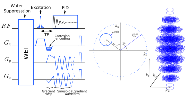 |
ECcentric Circle ENcoding TRajectorIes for Compressed-sensing (ECCENTRIC): A fully random non-Cartesian sparse k-space sampled MRSI at 7 Tesla
Antoine Klauser1,2,3, Bernhard Strasser2,4, Wolfgang Bogner4, Lukas Hingerl4, Claudiu Schirda5, Bijaya Thapa2, Daniel Cahill6, Tracy Batchelor7, François Lazeyras1,3, and Ovidiu Andronesi2
1Radiology and Medical Informatics, University of Geneva, Geneva, Switzerland, 2Athinoula A. Martinos Center for Biomedical Imaging, Department of Radiology, Massachusetts General Hospital, Harvard Medical School, Boston, MA, United States, 3CIBM Center for Biomedical Imaging, Geneva, Switzerland, 4High‐Field MR Center, Department of Biomedical Imaging and Image‐guided Therapy, Medical University of Vienna, Vienna, Austria, 5Department of Radiology, University of Pittsburgh, Pittsburgh, PA, United States, 6Department of Neurosurgery, Massachusetts General Hospital, Harvard Medical School, Boston, MA, United States, 7Department of Neurology, Brigham and Women, Harvard Medical School, Boston, MA, United States
A new encoding trajectory for magnetic resonance spectroscopic imaging was developed and implemented on a 7T human scanner. ECcentric Circle ENcoding TRajectorIes for Compressed-sensing (ECCENTRIC) is a spatial-spectral encoding strategy optimized for random non-Cartesian sparse Fourier domain sampling. Acceleration by undersampling ECCENTRIC prevents coherent aliasing artefacts in the spatial response function. ECCENTRIC allows smaller circles to avoid temporal interleaving for large matrix size, which is beneficial for spectral quality. Circle trajectories need limited gradient slewrate without rewinding deadtime, and are robust to timing imperfection and eddy-current delays.
|
||
 |
0836.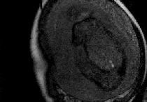 |
Faster fetal whole-heart anatomical and blood flow 4D cine MRI with k-t SWEEP
Thomas A Roberts1, Laurence H Jackson1, Joshua FP van Amerom2, Alena Uus1, Anthony N Price1, Johannes K Steinweg1, David FA Lloyd1,3, Milou PM van Poppel1, Kuberan Pushparajah3, Mary A Rutherford1, Reza Razavi1,3, Maria Deprez1, and Joseph V Hajnal1
1Biomedical Engineering Department, School of Biomedical Engineering and Imaging Sciences, King's College London, London, United Kingdom, 2Division of Pediatric Cardiology, The Hospital for Sick Children, Toronto, ON, Canada, 3Department of Congenital Heart Disease, Evelina Children's Hospital, London, United Kingdom
MRI of the fetal heart is challenging due to fetal motion and its small size. Previously, we have demonstrated a method for simultaneous 4D anatomical and blood flow imaging of the whole fetal heart. However, the acquisition takes at least 14 minutes, which is unfavourable for routine clinical usage. Here, we combine k-t SENSE with SWEEP excitation to acquire a continuous sequence of images through the heart. The removal of dummy pulses due to SWEEP excitation reduces our acquisition time by 17%. We also acquire one-third fewer frames compared to our conventional method, resulting in a 45% faster acquisition time.
|
|
|
0837.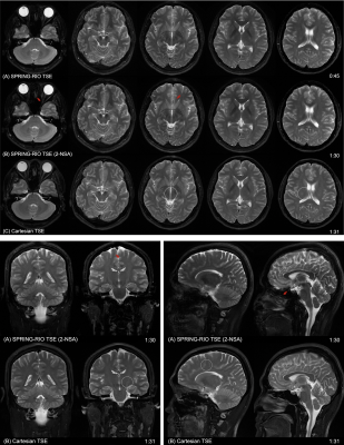 |
SPRING-RIO TSE: 2D T2-Weighted Turbo Spin-Echo Brain Imaging using SPiral RINGs with Retraced In/Out Trajectories
Zhixing Wang1, Steven Allen1, Xue Feng1, John P. Mugler2, and Craig H. Meyer1
1Biomedical Engineering, University of Virginia, Charlottesville, VA, United States, 2Radiology & Medical Imaging, University of Virginia, Charlottesville, VA, United States
This study presents a new approach to 2D TSE imaging using annular spiral rings with a retraced in/out trajectory, dubbed “SPRING-RIO TSE”, for fast T2-weighted brain imaging. Annular rings with retraced in/out segments offer benefits for reducing T2 decay induced artifacts and self-correction of moderate off-resonance effects. Phantom and human results show that high-quality T2-weighted images can be acquired with higher scan efficiency and reduced SAR, when compared with Cartesian TSE imaging.
|
|
0838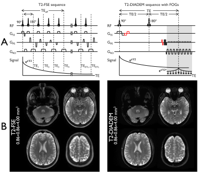 |
Rapid T2-DIADEM Echo-Planar Imaging as an Alternative to T2-FSE: A Clinical Feasibility Study Video Permission Withheld
Myung-Ho In1, Norbert G Campeau1, John III Huston1, Zijing Dong2,3, Kawin Setsompop4,5, Daehun Kang1, Uten Yarach6, Yunhong Shu1, Joshua D Trzasko1, and Matt A Bernstein1
1Department of Radiology, Mayo Clinic, Rochester, MN, United States, 2A. A. Martinos Center for Biomedical Imaging, Department of Radiology, Massachusetts General Hospital, Charlestown, MA, United States, 3Department of Electrical Engineering and Computer Science, MIT, Cambridge, MA, United States, 4Department of Radiology, Stanford University, Stanford, CA, United States, 5Stanford University, Stanford, CA, United States, 6Department of Radiologic Technology, Faculty of Associated Medical Sciences, Chiang Mai University, Chiang Mai, Thailand
Rapid high-resolution echo-planar-imaging (EPI) with improved image quality could conceivably be utilized as a high-speed alternative to conventional T2-weighted fast-spin-echo imaging (T2-FSE). A variant of multi-shot EPI, DIADEM (distortion-free imaging: a double encoding method), was recently described (5) and shown to yield high quality, high resolution images free of spatial distortion. In this work, a T2-weighted DIADEM pulse sequence was optimized with self-calibrated, tilted-CAIPI reconstruction scheme and then comparatively evaluated to T2-FSE images of the brain in 24 human subjects by two neuroradiologists.
|
||
0839. |
Spiral Cardiac bSSFP with Phase-Modulation to Enable Long Repetition Times
Michael Schär1
1Radiology, Johns Hopkins University School of Medicine, Baltimore, MD, United States
Recently, it was shown that phase-modulated balanced steady-state free precession (bSSFP) offers banding-free radial cine bSSFP images. Because this novel approach for bSSFP overcomes the need for short repetition times (TR), this work proposes to use more efficient spiral readouts with long TR of 15 ms. Banding free bSSFP cine images are demonstrated for either high temporal or high spatial resolution. The long TR furthermore facilitates fat suppression with spatial-spectral water-only excitation pulses which could improve depiction of the coronary arteries. Out of slice signal pileup at dark band frequencies is shown to be reduced with pre-saturation of those frequencies.
|
||
|
0840. |
Echo-train bSSFP for Rapid Golden-angle Radial Sampling
Kaibao Sun1, Zheng Zhong1,2, Kezhou Wang1, and Xiaohong Joe Zhou1,2,3
1Center for MR Research, University of Illinois at Chicago, Chicago, IL, United States, 2Department of Bioengineering, University of Illinois at Chicago, Chicago, IL, United States, 3Departments of Radiology and Neurosurgery, University of Illinois at Chicago, Chicago, IL, United States
Golden-angle radial sampling based on single-echo bSSFP has been increasingly used in MRI. In a method known as ETGAR (echo-train golden-angle radial), a series of spokes separated by a golden angle are acquired within a TR to accelerate data sampling. A main disadvantage is that the large inter-echo steering gradients can lead to a longer TR and trigger eddy currents. We herein demonstrate an alternative golden-angle radial sequence – ETGAR-II – to overcome the afore-mentioned issues. This novel sequence, together with an integrated phase-correction algorithm has been demonstrated on phantoms and human to obtain high quality images with 36% scan time reduction.
|
|
0841.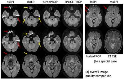 |
A whole-blade turboPROP technique with motion correction for rapid and robust DWI
Zhiqiang Li1, Melvyn B Ooi2, and John P Karis1
1Neuroradiology, Barrow Neurological Institute, Phoenix, AZ, United States, 2Philips Healthcare, Gainesville, FL, United States
DW-MRI is an invaluable technique for the diagnosis of neurological disorders. ssEPI is time efficient but suffers from geometric distortions. DW-PROPELLER and its variants, including turboPROP, have been proposed for generating distortion-free images. This project proposes a new whole-blade acquisition mode with phase correction in reconstruction to increase the blade width and consequently reduce the scan time by a factor of ~2, relative to the original turboPROP. Furthermore, the increased blade width allows for robust motion correction, which was typically not performed in PROPELLER-based DWI. In vivo results demonstrate advantages over ssEPI, and recently released product msEPI and SPLICE-PROPELLER.
|
||
 |
0842.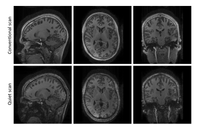 |
Fast and quiet 3D MPRAGE using a silent gradient axis - sequence development
Edwin Versteeg1, Sarah M. Jacobs1, Ícaro A.F. Oliveira2, Dennis W.J. Klomp1, and Jeroen C.W. Siero1,2
1Radiology, University Medical Center Utrecht, Utrecht, Netherlands, 2Spinoza centre for neuroimaging Amsterdam, Amsterdam, Netherlands
Sound levels in MRI can be reduced by switching a silent gradient axis beyond the hearing threshold. In this work, we implemented a silent readout module that applies such a silent gradient axis (at 7T) to a 3D MPRAGE sequence. This resulted in a sequence that featured a much lower peak sound level (26 dB reduction), similar image contrast and imaging time compared to a conventional MPRAGE-scan. This shows that a silent gradient axis provides a pathway to fast and quiet brain imaging with the potential to translate to other field strengths.
|
The International Society for Magnetic Resonance in Medicine is accredited by the Accreditation Council for Continuing Medical Education to provide continuing medical education for physicians.