Oral
Molecular Imaging & X-Nuclei
ISMRM & SMRT Annual Meeting • 15-20 May 2021

| Concurrent 2 | 12:00 - 14:00 | Moderators: Myriam Chaumeil & James Grist |
 |
0231.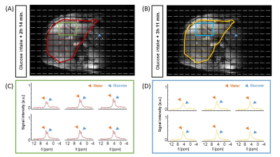 |
Deuterium Echo-Planar Spectroscopic Imaging (DEPSI) to Dynamically Monitor Deuterated Glucose in the Liver at 7T
Kyung Min Nam1, Ayhan Gursan1, Alex Bhogal1, Jannie Wijnen1, Dennis Klomp1, Jeanine Prompers1, and Arjan D. Hendriks1
1University of Medical Center Utrecht, Utrecht, Netherlands
Deuterium Echo-Planar Spectroscopic Imaging (DEPSI) is proposed as a way to increase the spatial and temporal resolution of deuterium metabolic imaging (DMI) at 7T. Typically, DMI uses traditional, slow MRSI sequences, which cannot capture rapid dynamic metabolic processes in large organs with sufficient spatial and/or temporal resolution. With DEPSI, in vivo glucose metabolism of the liver could be monitored after intake of [6,6′-2H2]-glucose with 20 mm nominal voxel size, full liver coverage, and a scan time of less than 10 minutes. DEPSI was combined with Hamming weighted acquisition in the phase encoding directions to maximize SNR.
|
|
0232.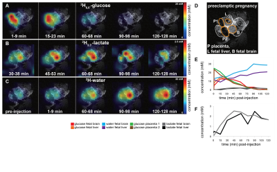 |
Deuterium metabolic imaging (DMI) of glucose metabolism in pregnant preeclamptic mice at 15.2 tesla.
Stefan Markovic1, Tangi Roussel2, Michal Neeman3, and Lucio Frydman1
1Department of Chemical and Biological Physics, Weizmann Institute of Science, Rehovot, Israel, 2Center for Magnetic Resonance in Biology and Medicine, Marseille, France, 3Department of Biological Regulation, Weizmann Institute of Science, Rehovot, Israel
Deuterium Metabolic Imaging (DMI) was used to follow metabolism in wildtype and l-NAME induced preeclamptic pregnant mice, after intravenous administration of 2H6,6’-glucose. Maps for 2H6,6’-glucose and its metabolic products 2H3,3’-lactate and 2H-water were measured over 2 hours by 2H CSI at 15.2 tesla. 2H-water was generated as main metabolic product in fetoplacental units; placentas and fetal organs also generated 2H3,3’-lactate, but this was not detected in other
maternal organs. Lactate levels were more elevated and its clearance was slower in preeclamptic fetuses than in healthy controls. DMI thus may aid in development and monitoring of future intervention for early preeclampsia.
|
||
 |
0233.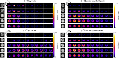 |
Hyperpolarized [2-13C]Pyruvate Molecular Imaging with Whole Brain Coverage
Yaewon Kim1, Brian T. Chung1, Jeremy W. Gordon1, Adam W. Autry1, Chou T. Tan2, Chris Suszczynski2, Susan M. Chang3, Yan Li1, Duan Xu1, and Daniel B. Vigneron1,3
1Department of Radiology and Biomedical Imaging, University of California, San Francisco, CA, United States, 2ISOTEC Stable Isotope Division, MilliporeSigma, Merck KGaA, Miamisburg, OH, United States, 3Department of Neurological Surgery, University of California, San Francisco, CA, United States
Hyperpolarized (HP) [2-13C]pyruvate MRI has great clinical potential for monitoring pyruvate-to-glutamate conversions through the TCA cycle in addition to pyruvate-to-lactate glycolytic metabolism. This new method for human molecular imaging has enabled whole brain acquisitions with sufficient signal-to-noise. Using a specialized multi-slice EPI sequence, volumetric, dynamic images of HP [2-13C]pyruvate and its downstream metabolites [5-13C]glutamate and [2-13C]lactate were acquired from healthy brain volunteers. The downfield and upfield lactate signals were acquired separately with no artifacts arising from J-coupling were observed. This work demonstrated the capability of volumetric and dynamic [2-13C]pyruvate EPI to interrogate both glycolytic and oxidative cerebral energy metabolism simultaneously.
|
|
|
0234.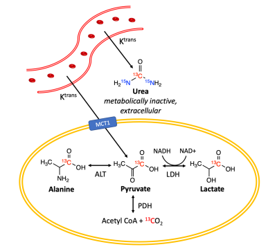 |
Clinical translation of simultaneous metabolic and perfusion imaging with hyperpolarized [1-13C]pyruvate and [13C, 15N2]urea
Hecong Qin1,2, Shuyu Tang1, Andrew Riselli1, Robert A. Bok1, Romelyn Delos Santos1, Mark Van Criekinge1, Jeremy W. Gordon1, Rahul Aggarwal3, Evelyn Escobar1, Rui Chen4, Chunxin Tracy Zhang5, Gregory Goddard5, Albert Chen4,
Galen Reed4, Ruscitto M. Daniel5, Renuka Sriram1, James Slater1, Peder E.Z. Larson1,2, Daniel B. Vigneron1,2, and John Kurhanewicz1,2
1Radiology and Biomedical Imaging, University of California, San Francisco, San Francisco, CA, United States, 2Graduate Program in Bioengineering, UC Berkeley – UCSF, San Francisco, CA, United States, 3Medicine, University of California, San Francisco, San Francisco, CA, United States, 4GE Healthcare, Waukesha, WI, United States, 5GE Research, Niskayuna, NY, United States
Altered metabolism and perfusion are implicated in cancer’s underlying pathophysiology. Prior preclinical and clinical studies have shown that metabolic and perfusion imaging could provide a sensitive and specific evaluation of tumor grade and therapeutic response. We aim to develop a dual-agent hyperpolarized 13C MR technique for simultaneous metabolic and perfusion imaging in humans. Here, we report the technical developments towards its clinical translation: 1) formulation and co-polarization system of 13C pyruvate and urea, 2) imaging probe characterization, 3) safety-related studies, and 4) multi-probe imaging sequence. Upon FDA approval, this work would lead to the first-in-human dual-agent hyperpolarized MR study.
|
|
|
0235. |
High-resolution 3D Phosphorus Metabolic Imaging of the Human Brain at 7T using SPICE
Hannes Michel Wiesner1, Rong Guo2,3, Yudu Li2,3, Yibo Zhao2,3, Zhi-Pei Liang2,3, Xiao-Hong Zhu1, and Wei Chen1
1CMRR, Department of Radiology, University of Minnesota, Minneapolis, MN, United States, 2Beckman Institute for Advanced Science and Technology, University of Illinois at Urbana-Champaign, Urbana, IL, United States, 3Departments of Electrical and Computer Engineering, University of Illinois at Urbana-Champaign, Urbana, IL, United States
In vivo 31P MRS imaging (MRSI) is unique to study brain energy metabolism including ATPase and creatine kinase metabolic rates, and the NAD redox ratio especially at ultrahigh field (UHF) with significant improvements in detection sensitivity and spectral resolution. Nevertheless it is still challenging to achieve high spatial resolution even at UHF, owing to extremely low concentration of phosphorus metabolites. In this study, we employed the subspace‐based image reconstruction method called SPICE to largely reduce spectral noise and increase the signal-to-noise ratio (SNR) for achieving high-resolution 3D 31P MRSI covering the entire human brain at 7T.
|
|
|
0236.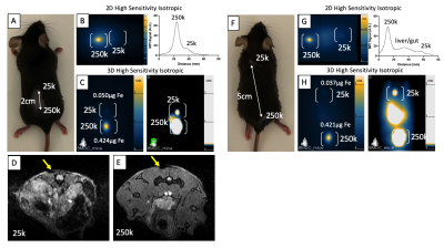 |
Assessment of sensitivity, resolution, and quantification of Synomag-D labeled dendritic cells with magnetic particle imaging
Julia J Gevaert1,2, Corby Fink3,4, Jimmy D Dikeakos3, Gregory A Dekaban3,4, and Paula J Foster1,2
1Department of Medical Biophysics, University of Western Ontario, London, ON, Canada, 2Cellular and Molecular Imaging Group, Robarts Research Institute, London, ON, Canada, 3Department of Microbiology and Immunology, University of Western Ontario, London, ON, Canada, 4Biotherapeutics Research Laboratory, Robarts Research Institute, London, ON, Canada
The purpose of this study was to explore magnetic particle imaging (MPI), which directly detects superparamagnetic iron oxide nanoparticles (SPIOs) as a novel technique for tracking dendritic cells (DCs). DCs were labeled using Vivotrax, a commonly used MPI-SPIO, and Synomag-D, a tailored MPI-SPIO, with and without transfection agents. Vivotrax with TAs showed high levels of extracellular iron, overestimating iron content and cellular sensitivities. The use of TAs improved cellular sensitivity of Synomag-D to as few as 6k cells with 1min 2D images. Signal from 250k and 25k cells could be resolved at a distance of 2cm with 3D imaging.
|
|
|
0237.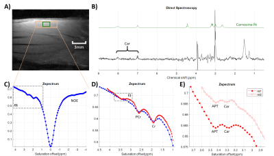 |
First in vivo detection of carnosine using CEST
Solène Bardin1,2, Michele Lecis1,3, Davide Boido1,2, Fawzi Boumezbeur1,2, and Luisa Ciobanu1,2
1NeuroSpin, CEA, Gif-sur-Yvette, France, 2Paris-Saclay University, Saclay, France, 3Technical University of Munich, Munich, Germany
Accelerated CEST acquisitions using a linescan sequence coupled with an ultra-high magnetic field allows, for the first time, the detection of carnosine in vivo.
|
|
|
0238.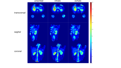 |
Quantitative Sodium MRI of the Human Kidney at 7T Applying Respiratory Sorting
Anna K. Scheipers1, Johanna Lott1, Armin M. Nagel1,2, Peter Bachert1, Mark E. Ladd1, and Tanja Platt1
1Medical Physics in Radiology, German Cancer Research Center (DKFZ), Heidelberg, Germany, 2University Hospital Erlangen, Institute of Radiology, Friedrich‐Alexander‐Universität Erlangen‐Nürnberg (FAU), Erlangen, Germany Sodium (23Na) plays an important role in many cellular processes, making it an interesting nuclei to investigate using MRI. Low in-vivo signals and short relaxation times require high magnetic field strengths, dedicated hardware and pulse sequences as well as correction methods to obtain reliable tissue sodium concentrations. Therefore we compare the influence of different correction methods on 23Na concentration investigations in the human kidney and validate our methods with phantom measurements. We employ retrospective respiratory self‐gating for the quantitative 23Na images as well as for the acquired B1+ maps to reduce the influence of image blurring due to motion artifacts. |
|
|
0239.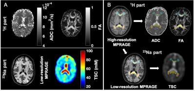 |
Quantitative sodium and diffusion imaging of mild traumatic brain injury: regional analysis findings
Anna M Chen1, Teresa Gerhalter1, Seena Dehkharghani1,2, Rosemary Peralta1, Fatemeh Adlparvar1, James S Babb1, Tamara Bushnik3, Jonathan M Silver4, Brian S Im3, Stephen P Wall5, Ryan Brown1,6, Steven Baete1,6, Ivan I Kirov1,2,6,
and Guillaume Madelin1
1Center for Biomedical Imaging, Department of Radiology, New York University Grossman School of Medicine, New York, NY, United States, 2Department of Neurology, New York University Grossman School of Medicine, New York, NY, United States, 3Department of Rehabilitation Medicine, New York University Grossman School of Medicine, New York, NY, United States, 4Department of Psychiatry, New York University Grossman School of Medicine, New York, NY, United States, 5Ronald O. Perelman Department of Emergency Medicine, New York University Grossman School of Medicine, New York, NY, United States, 6Center for Advanced Imaging Innovation and Research, Department of Radiology, New York University Grossman School of Medicine, New York, NY, United States
27 mTBI patients and 19 controls were scanned at 3T. Total sodium concentration (TSC), fractional anisotropy (FA) and apparent diffusion coefficient (ADC) were measured with voxel averaging in 12 grey and white matter regions-of-interest (ROIs). Patients had lower mean TSC than controls across all ROIs, however, statistical significance was only reached in the caudate. Statistically significant FA differences also occurred in only one region, frontal white matter (WM), while none were observed for ADC. TSC changes existed in mTBI and occurred with similar frequency as FA, but the FA finding had a higher effect size, and correlated with symptoms.
|
|
|
0240.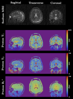 |
Optimized simultaneous 3D proton MRF and sodium MRI
Zidan Yu1,2, Olga Dergahyova1, Daniel K. Sodickson1,2, Guillaume Madelin1,2, and Martijn A. Cloos3,4
1Center for Biomedical Imaging, Department of Radiology, New York University School of Medicine, New York, NY, United States, 2Vilcek Institute of Graduate Biomedical Sciences, NYU Langone Health, New York, NY, United States, 3Centre for Advanced Imaging, The University of Queensland, Brisbane, Australia, 4ARC Training Centre for Innovation in Biomedical Imaging Technology, The University of Queensland, Brisbane, Australia
In this work, we present an optimized 3D technique that can simultaneously acquire quantitative 1H density, T1, T2, B1+ maps and a 23Na image of the whole head in a reasonable scan time (~20 min).
|
The International Society for Magnetic Resonance in Medicine is accredited by the Accreditation Council for Continuing Medical Education to provide continuing medical education for physicians.