Oral
Spectroscopy: Acq/Recon/Analysis
ISMRM & SMRT Annual Meeting • 15-20 May 2021

| Concurrent 2 | 14:00 - 16:00 | Moderators: Yao Li & John Port |
0281.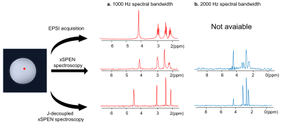 |
xSPEN spectroscopy: a self-navigated fast chemical shift encoded echo planar imaging acquisition
Ke Dai1, Hao Chen1, Hongda Shao2, Jianjun Liu2, and Zhiyong Zhang1
1School of Biomedical Engineering, Shanghai Jiao Tong University, Shanghai, China, 2Departments of Nuclear Medicine, Renji Hospital, School of Medicine, Shanghai Jiao Tong University, Shanghai, China
xSPEN is a single-shot echo-planar imaging-based MRI approach with exceptional resilience to chemical shifts and field inhomogeneities. We introduce a time increasing (t1) evolution as chemical shift encoding to fast obtain multiple-voxel spectroscopy. The new method endows a 2D self-navigated motion correction and a unique J-decoupled spectrum by splitting the t1 evolution into τ-t1/2 and t1/2 on two sides of π pulse, which results a constant-τ J coupling evolution. We present in vitro results, demonstrating an alternative fast MRSI and increasing chemical shift separation and detection with the robustness to the in-plane motion and the unique J-decoupled spectrum capability.
|
||
0282.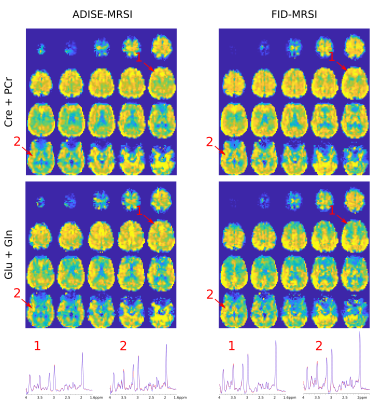 |
Fast Adiabatic Spin-Echo MRSI Sequence for Whole-Brain 5mm-isotropic metabolic imaging
Antoine Klauser1,2, Sebastien Courvoisier1,2, Michel Kocher1,2, and François Lazeyras1,2
1Radiology and Medical Informatics, University of Geneva, Geneva, Switzerland, 2CIBM Center for Biomedical Imaging, Geneva, Switzerland
A fast 1H 3D Adiabatic Spin-Echo (ADISE) MRSI sequence was implemented to measure metabolite distributions over the whole brain with 5mm isotropic resolution. MRSI data were measured on volunteers and compared with FID-MRSI sequence. ADISE-MRSI and FID-MRSI acquisitions were accelerated with compressed-sensing and reconstructed with a Low-Rank TGV-constrained model.
|
||
0283.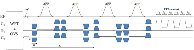 |
Diffusion-weighted Echo Planar Spectroscopic Imaging Using semi‐LASER Localization at 3T: A Pilot Study
Manoj Kumar Sarma1,2, Andres Saucedo3, and M. Albert Albert Thomas3
1Advanced Imaging Research Center, UT Southwestern Medical Center, Dallas, TX, United States, 2Radiology, UT Southwestern Medical Center, Dallas, TX, United States, 3Radiology, UCLA School of Medicine, Los Angeles, CA, United States
Diffusion-weighted spectroscopy (DW-MRS) is an excellent tool to explore the compartment specific assessment of tissue microstructure. Although there has been growing interest in DW-MRS for clinical applications, most of the studies involving human brain metabolites so far have used single-voxel methods, which limit its application to specific white matter tracts or arbitrarily selected regions of interest. There has been only a few attempts to evaluate the diffusion properties of brain metabolites with DW-MRSI. Here, we propose an echo planar-based diffusion-weighted spectroscopic imaging using semi-LASER localization and bipolar diffusion gradients. Initial results show good spectral quality and spatial localization.
|
||
 |
0284.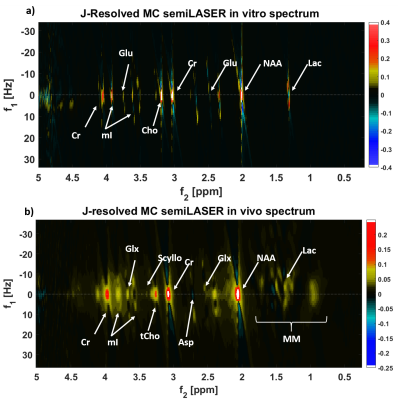 |
Quantification of Human Brain Metabolites using Two-Dimensional J-Resolved Metabolite-Cycled semiLASER at 9.4 T
Saipavitra Murali-Manohar1,2, Tamas Borbath1,2, Andrew Martin Wright1,3, and Anke Henning1,4
1High Field Magentic Resonance, Max Planck Institute for Biological Cybernetics, Tuebingen, Germany, 2Faculty of Science, University of Tuebingen, Tuebingen, Germany, 3IMPRS for Cognitive Neuroscience, Tuebingen, Germany, 4Advanced Imaging Research Center, UT Southwestern Medical Center, Dallas, TX, United States
Crowded proton spectra with severely overlapped J-coupled resonances pose a challenge in the reliable quantification of metabolites in the human brain. Several advanced techniques such as editing methods, multi-dimensional spectroscopy methods, sophisticated processing or quantification pipelines were proposed in the past. In this work, we present a two-dimensional metabolite-cycled semiLASER technique at 9.4 T with maximum echo sampling scheme. This method helps well resolve the J-coupled peaks and clearly distinguish them. 2D spectral fitting is performed using ProFit2.0 and the metabolites are quantified using internal water referencing after correcting the fitted concentration for tissue content and relaxation effects.
|
|
 |
0285.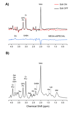 |
GABA measurement at 7T: short-TE or MEGA editing?
Song-I Lim1,2 and Lijing Xin1,2
1CIBM Center for Biomedical Imaging, Lausanne, Switzerland, 2Animal Imaging and Technology, EPFL, Lausanne, Switzerland
The purpose of the study is to compare short-TE and MEGA editing methods and verify reproducibility of GABA measurement in the motor cortex at 7T. The measured average GABA/Cr and GABA/NAA ratios were 0.121 ± 0.034 and 0.057 ± 0.019 respectively for short-TE sSPECIAL measurement and 0.083 ± 0.014 and 0.052 ± 0.008 respectively for MEGA-sSPECIAL sequence. 6 healthy volunteers were scanned two times and average coefficient of variances were 21.2 ± 11.8 % for short-TE measurement and 4.5 ± 3.9 for MEGA-sSPECIAL sequence.
|
|
|
0286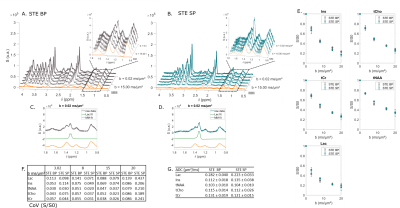 |
Using selective RF pulses in diffusion-weighted MRS for lactate diffusion measurements with minimal J-modulation Video Permission Withheld
Eloïse Mougel1, Sophie Malaquin1, Melissa Vincent1, and Julien Valette1
1Université Paris-Saclay, Commissariat à l'Energie Atomique et aux Energies Alternatives (CEA), Centre National de la Recherche Scientifique (CNRS), Molecular Imaging Research Center (MIRCen), Laboratoire des Maladies Neurodégénératives, Fontenay aux Roses, France
Brain lactate compartmentation is an important but debated neuroscience question. By assessing local microstructure where lactate is diffusing in, diffusion-weighted MRS has unique potential to non-invasively assess lactate compartmentation. We propose to increase lactate signal using selective pulses (SP) to cancel J-modulation. We compare lactate signal behavior in diffusion-weighted experiments performed in vivo, using either spin echo or stimulated echo sequence relying on selective pulses. We verify here that the signal increases in both cases, compared to conventional cases using broad pulses. Spin echo using SP appears the most valuable option to measure lactate diffusion at high b-values.
|
|
|
0287.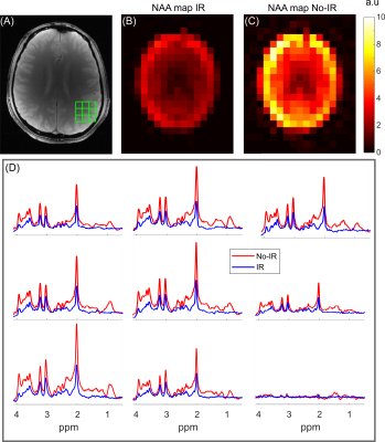 |
Short-TE ECLIPSE for Macromolecular-Nulled MRSI in the Human Brain
Chathura Kumaragamage1, Anastasia Coppoli1, Peter B Brown1, Scott McIntyre1, Terence W Nixon1, Henk M De Feyter1, Graeme Mason1, and Robin A de Graaf1
1Department of Radiology and Biomedical Imaging, Magnetic Resonance Research Center, Yale University, New Haven, CT, United States
An ECLIPSE-IVS based MRSI method was developed utilizing 3 ms GOIA-WURST RF pulses (BW = 15 kHz), operating at an RF amplitude B1(95%) = 0.87 kHz. The ECLIPSE-IVS method was preceded with a water suppression module incorporating an optional inversion recovery (IR) component, to achieve macromolecule-nulled acquisitions. MRSI in vivo demonstrate robust extracranial lipid suppression with reliable, artifact-free metabolic maps generated with peak integration and LCModel fitting.
|
|
0288.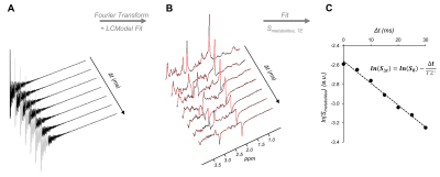 |
T2* of human brain metabolites estimated from a single proton MRS acquisition
Chloé Najac1, Marjolein Bulk1, Hermien E. Kan1, Andrew G. Webb1, and Itamar Ronen1
1C.J. Gorter Center for High Field MRI, Department of Radiology, Leiden University Medical Center, Leiden, Netherlands
We propose a method to calculate T2* values of brain metabolites from a series of time-shifted datasets obtained from a single 1H magnetic resonance spectroscopy acquisition. T2* values from five brain metabolites were measured in the posterior cingulate cortex. Robust T2* values were obtained for all five metabolites, including J-coupled metabolites such as glutamate and myo-inositol, for which T2* estimation is otherwise not possible. We show in a subsequent reproducibility study that the water linewidth within the same volume can be used to account for variability in local B0 inhomogeneity and reduce the associated variability across measurements.
|
||
0289.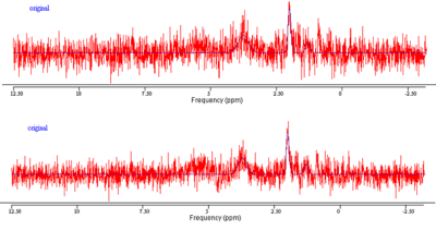 |
A method for high quality magnetic resonance spectroscopy of discs during normal breathing
Frida Johansson1,2, Helena Brisby2,3, Hanna Hebelka2,4, Maria Ljungberg1,2, and Kerstin Lagerstrand1,2
1Medical Physics and Biomedical Engineering, Sahlgrenska University Hospital, Gothenburg, Sweden, 2Institute of Clinical Sciences, Gothenburg University, Gothenburg, Sweden, 3Department of Orthopaedics, Sahlgrenska University Hospital, Gothenburg, Sweden, 4Department of Radiology, Sahlgrenska University Hospital, Gothenburg, Sweden This study aimed to evaluate the effect of respiratory motion on disc MRS and propose an MRS-method that improves the signal-to-noise-ratio. Findings showed that the phase signal of the disc changes substantially between expiration and inspiration. With the proposed postprocessing method, all spectra gave a higher signal-to-noise-ratio (largest gain=30%). Present study shows that respiratory motion affects the disc phase signal and should be taken into consideration when evaluating the disc using MRS. The proposed method improved the quality of the MRS-spectrum and, thus, showed feasibility in measuring the molecular disc content non-invasively during normal breathing. |
||
|
0290.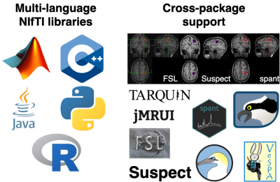 |
NIfTI MRS: A standard format for spectroscopic data
William T Clarke1, Tiffany Bell2,3,4, Uzay Emir5,6, Mark Mikkelsen7,8, Georg Oeltzschner7,8, Benjamin C Rowland9, Amirmohammad Shamaei10,11, Brian J Soher12, Sofie Tapper7,8, and Martin Wilson13
1Wellcome Centre for Integrative Neuroimaging, NDCN, University of Oxford, Oxford, United Kingdom, 2Department of radiology, University of Calgary, Calgary, AB, Canada, 3Hotchkiss brain institute, University of Calgary, Calgary, AB, Canada, 4Alberta Children’s Hospital Research Institute, University of Calgary, Calgary, AB, Canada, 5School of Health Sciences, Purdue University, West Lafayette, IN, United States, 6Weldon School of Biomedical Engineering, Purdue University, West Lafayette, IN, United States, 7Russell H. Morgan Department of Radiology and Radiological Science, The Johns Hopkins University School of Medicine, Baltimore, MD, United States, 8F. M. Kirby Research Center for Functional Brain Imaging, Kennedy Krieger Institute, Baltimore, MD, United States, 9Division of Cancer Science, School of Medical Sciences, Faculty of Biology, Medicine and Health, University of Manchester, Manchester, United Kingdom, 10Institute of Scientific Instruments of the CAS, Brno, Czech Republic, 11Department of Biomedical Engineering, Faculty of Electrical Engineering and Communication, Brno University of Technology, Brno, Czech Republic, 12Center for Advanced MR Development, Department of Radiology, Duke University Medical Center, Durham, NC, United States, 13Centre for Human Brain Health and School of Psychology, University of Birmingham, Birmingham, United Kingdom
We propose a flexible data format for the storage of multidimensional MRS data. The lack of a single widely used data format in MRS hinders widespread use, consistent analyses, and open dissemination of data. Here, we adapt the widely adopted NIfTI format to store MRS data, extending NIfTI to include the additional meta-data required for MRS data interpretation. We present a pipeline for end-to-end analysis of MRS data and an open-source conversion tool. The community is encouraged to contribute to the format.
|
The International Society for Magnetic Resonance in Medicine is accredited by the Accreditation Council for Continuing Medical Education to provide continuing medical education for physicians.