Oral
Contrast Agents & Preclinical Studies
ISMRM & SMRT Annual Meeting • 15-20 May 2021

| Concurrent 2 | 14:00 - 16:00 | Moderators: Giovanna Diletta Ielacqua & Zheng-Rong Lu |
0073.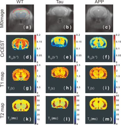 |
Altered pH in early Alzheimer’s disease detected by creatine chemical exchange saturation transfer magnetic resonance imaging
Lin Chen1,2, Peter C.M. van Zijl1,2, Zhiliang Wei1,2, Hanzhang Lu1,2, Wenzhen Duan3, Philip C. Wang4,5, Tong Li4,5, and Jiadi Xu1,2
1Department of Radiology and Radiological Science, Johns Hopkins University, Baltimore, MD, United States, 2F.M. Kirby Research Center for Functional Brain Imaging, Kennedy Krieger Research Institute, Baltimore, MD, United States, 3Department of Psychiatry and Behavioral Sciences, Johns Hopkins University, Baltimore, MD, United States, 4Department of Pathology, Johns Hopkins University, Baltimore, MD, United States, 5Department of Neuroscience, Johns Hopkins University, Baltimore, MD, United States
We demonstrate the feasibility of creatine chemical exchange saturation transfer (CrCEST) MRI in detecting altered pH in Alzheimer’s disease (AD) mouse brain. In two early-stage AD models, namely Tau and APP mice, CrCEST contrast in the brain was significantly reduced compared to that of WT mice at 6-7 months (P<0.007). From MRS experiments, the brain creatine concentration between WT and AD mice was the same within error, which indicates that the reduced CrCEST contrast in the AD brain can be contributed mainly to pH reduction. Immunohistochemical analysis showed neuroinflammation in the APP mice, a potential factor for causing pH reduction.
|
||
 |
0074.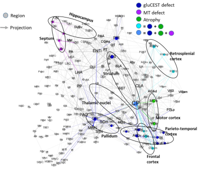 |
DTI and gluCEST imaging reveal the key role of white matter alteration in the pathogenesis in a mouse model of Huntington’s Disease
Jean-Baptiste Perot1, Marina Célestine1, Marc Dhenain1, Sandrine Humbert2, Emmanuel Brouillet1, and Julien Flament1
1Université Paris-Saclay, Commissariat à l’Energie Atomique et aux Energies Alternatives (CEA), Centre National de la Recherche Scientifique (CNRS), Molecular Imaging Research Center (MIRCen), Laboratoire des Maladies Neurodégénératives, Fontenay-aux-Roses, France, 2Université Grenoble-Alpes, Grenoble Institute of Neurosciences (GIN), INSERM U1216, Grenoble, France
Huntington’s Disease (HD) is a neurodegenerative disorder caused by the expansion of CAG repeats on the exon 1 of the HTT gene. Although genetic origin of HD is well-established, early and predictive biomarkers of disease onset and progression are still lacking. In the present study, we performed a multiparametric longitudinal MRI study on a mouse model of HD. Our results in gluCEST, Magnetization Transfer, morphometry and Diffusion Tensor Imaging revealed early modifications of white matter followed by progressive functional and anatomical changes in HD mice. Such network seems to point out the central role of white matter in HD pathogenesis.
|
|
|
0075.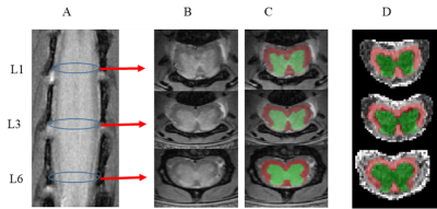 |
Multimodal MRI study of multiple sclerosis: the therapeutic role of complement system
Abdullah Althobity1,2, Nemat Khan3, Trent Woodruff3, Gary Cowin1,4, Ian Brereton1,4, and Nyoman Kurniawan1
1Centre for Advanced imaging, University of Queensland, Brisbane, Australia, 2Ministry of Education, Riyadh, Saudi Arabia, 3Faculty of Medicine, School of Biomedical Sciences, University of Queensland, Brisbane, Australia, 4National Imaging Facility, Brisbane, Australia
Multiple sclerosis (MS) is an autoimmune disease with uncertain aetiology. In this work, we used two genetically modified mice, each derived by gene ablation to complement protein C5aR1 and C5L2 receptors, designed to investigate their roles in mediating the disease model experimental autoimmune encephalomyelitis (EAE). MR spectroscopy and DTI were measured using a 9.4T MRI with cryoprobe at the level of lumbar spinal cord. Changes in metabolites and DTI parameters indicate that the ablation of C5L2, to a greater extent than the ablation of C5aR1, made these mice less susceptible to EAE induced neuronal damage compared to wild-type mice.
|
|
0076.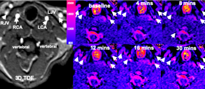 |
In vivo methemoglobin modulation as an intravascular contrast agent for magnetic resonance imaging: Rabbit Model with T1 measurement
Seong-Eun Kim1, J Scott McNally1, Matthew Alexander1, Dennis L Parker1, Matthew S Zabriskie 1, and Ronald Day2
1UCAIR, Department of Radiology and Imaging Sciences, University of Utah, Salt Lake City, UT, United States, 2Department of Pediatrics, University of Utah, Salt Lake City, UT, United States
After intravenous injection, gadolinium(GBCA), commonly used for MR contrast, distributes from intravascular to the extravascular space and rapidly cleaned by renal excretion. This pharmacologic behavior makes GBCA unfavorable as in blood pool agent. One potential alternative agent is endogenous intracellular methemoglobin (MetHb), a paramagnetic molecule in our blood cells. Intracellular levels of MetHb can be increased by exposing blood to sodium nitrite. We evaluated change of T1 of blood according to a transient increase in intracellular MetHb in in vivo animal model. Our results demonstrated that MetHb modulation resulted T1 shortening of blood and soft tissue enhancement.
|
||
 |
0077.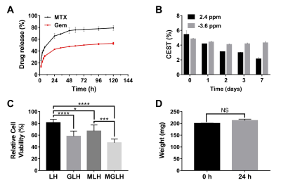 |
CEST-MRI guided sequential drug delivery using injectable hydrogel for local treatment in the brain
Xiongqi Han1, Jianpan Huang1, and Kannie Wai Yan Chan1,2,3
1Biomedical Engineering, City University of Hong Kong, Hong Kong, Hong Kong, 2Biomedical Engineering, Shenzhen Research Institute, City University of Hong Kong, Shenzhen, China, 3Russell H. Morgan Department of Radiology and Radiological Science, The Johns Hopkins University School of Medicine, Baltimore, MD, United States
Glioblastoma (GBM) is the most malignant brain tumor with notorious heterogeneous, infiltrative and chemoresistance. Here, we developed a hydrogel-based drug delivery system for sequential and sustainable local drug delivery with rheological properties that favor applications to the brain. We designed methotrexate (MTX) to release prior to gemcitabine (Gem). Using CEST, the multicomponent of hydrogel matrix could be detected at 3 T, including the contrast of loaded drugs (5.4%) at 2.2-2.4 ppm and liposomal hydrogel matrix at -3.6 ppm in vitro. Furthermore, it showed combined cytotoxicity on U87 cells, demonstrating its potential on CEST MRI guided local treatment.
|
|
0078. |
Fluorine Magnetic Resonance Imaging for Natural Killer Cell Tracking with a Dual Tuned 1H/19F Torso Coil at 3T
Paul Begovatz1, Lawrence Lechuga1, Monica Cho2, Mallery Olsen2, Rachel McMahon3, David Vail3,4, and Sean Fain1,5,6
1Medical Physics, Carbone Cancer Center, University of Wisconsin School of Medicine and Public Health, Madison, WI, United States, 2Pediatrics, Carbone Cancer Center, University of Wisconsin School of Medicine and Public Health, Madison, WI, United States, 3Medical Sciences, School of Veterinary Medicine, University of Wisconsin-Madison, Madison, WI, United States, 4Carbone Cancer Center, University of Wisconsin-Madison, Madison, WI, United States, 5Radiology, Carbone Cancer Center, University of Wisconsin School of Medicine and Public Health, Madison, WI, United States, 6Biomedical Engineering, Carbone Cancer Center, University of Wisconsin School of Medicine and Public Health, Madison, WI, United States
Fluorine magnetic resonance imaging (19F-MRI) has been demonstrated as a non-invasive method to track and quantify immune cells in vivo. However due to the low 19F spin density of immune cell labeling, these studies have been mostly conducted on ultra-high field MRI systems, or with small sensitive surface coils at clinical field strengths. This feasibility study found that concentrations of perfluoropolyether (PFPE), and phantoms consisting of fewer than one million PFPE labeled NK cells were reliably detected through 19F-MRI with the combination of a cartesian 3D fast spin echo imaging sequence, and a dual tuned 1H/19F torso coil at 3T.
|
||
0079 |
A Mn-based probe targeted towards organic-anion transporting polypeptides Video Permission Withheld
Nivin N Nyström1, Hanlin Liu2, Francisco Martínez-Santiesteban1, Xiao-an Zhang2, Timothy J Scholl1, and John A Ronald1
1Robarts Research Institute, London, ON, Canada, 2University of Toronto, Toronto, ON, Canada
We have developed a manganese(III) porphyrin probe for targeted imaging of cells expressing organic-anion transporting polypeptides. OATP1 targeting and relaxation characteristics were evaluated in vitro in engineered cells, and in vivo and ex vivo in different organs in mice.
|
||
0080. |
MR Measurements of Placental Perfusion in Normal Sheep Pregnancies
Dimitra Flouri1,2, Jack RT Darby3, Stacey L Holman3, Sunthara R Perumal4, Anna L David5,6, Janna L Morrison3, and Andrew Melbourne2,7
1School of Biomedical Engineering & Imaging Sciences, King's College London, London, United Kingdom, 2Department of Medical Physics & Biomedical Engineering, University College London, London, United Kingdom, 3Early Origins of Adult Health Research Group, University of South Australia, Adelaide, Australia, 4Preclinical Imaging and Research Laboratories, South Australian Health and Medical Research Institute, Adelaide, Australia, 5Elizabeth Garrett Anderson Institute for Women’s Health, University College London, London, United Kingdom, 6NIHR Biomedical Research Centre, University College London Hospitals, London, United Kingdom, 7School of Biomedical Engineering and Imaging Sciences, King's College London, London, United Kingdom
MRI techniques are considered to give additional placental information in vivo to support clinical decision-making. Preclinical models such as in pregnant sheep provide an invasive method to validate MRI measurements, as they allow for controlled experiments and analysis during pregnancy. Here we characterised diffusion and perfusion properties of normal sheep placenta such as apparent diffusion coefficient, T2 measurements and fractional anisotropy analysis. We also presented the first application of multi-compartment MRI model to normal sheep placenta.
|
||
0081. |
Detectable velocity range in single-cell tracking by time-lapse MRI
Enrica Wilken1, Felix Freppon1, Max Masthoff1, and Cornelius Faber1
1Translational Research Imaging Center, Clinic of Radiology, University Hospital Muenster, Muenster, Germany
Repetitive T2*-weighted image acquisition in vivo and contrast simulations were used to show that single iron oxide nanoparticle labeled cells can be resolved and tracked non-invasively by time-lapse MRI. Calculation of the velocity of intravascular moving immune cells in mice brain and velocity-dependent blurring of time-lapse contrast in simulations indicate that cell dynamics slower than 1 µm/s are detectable. Therefore, time-lapse MRI is able to reveal patrolling immune cells as hypointense spots and can be a suitable tool to study inflammatory diseases and the progression of cancer metastasis.
|
||
0082.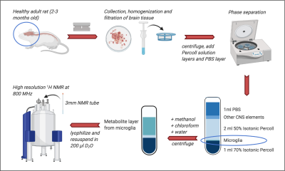 |
Investigating the microglial metabolome with high resolution 1H NMR
Lydia M. Le Page1, Jayson Ball2, Linda Watkins2, and Myriam Chaumeil1
1Physical Therapy and Rehabilitation Science, Radiology and Biomedical Imaging, UCSF, San Francisco, CA, United States, 2Department of Psychology & Neuroscience & the Center for Neuroscience, University of Colorado, Boulder, CO, United States
Microglia are an essential part of the brain’s immune system. In 2006, Frank et al. described a rapid isolation technique to obtain high-purity quiescent microglia from the rat brain, enabling study of their role in health and disease. Metabolomics via NMR is a robust, data-rich strategy for understanding cell metabolism. Given the central role of microglial metabolic reprogramming in numerous diseases, we investigated whether ultra-high-field 1H NMR could detect key metabolites in freshly isolated adult microglia. We optimized the isolation and extraction protocols and showed that several key microglia metabolites can be reliably quantified using high-resolution 1H NMR at 18.8Tesla.
|
The International Society for Magnetic Resonance in Medicine is accredited by the Accreditation Council for Continuing Medical Education to provide continuing medical education for physicians.