Oral
Microstructure: Modelling Gray & White Matter Diffusion
ISMRM & SMRT Annual Meeting • 15-20 May 2021

Oral
Microstructure: Modelling Gray & White Matter Diffusion
| Concurrent 4 | 18:00 - 20:00 | Moderators: Francesco Grussu & Ken Sakaie |
 |
0639.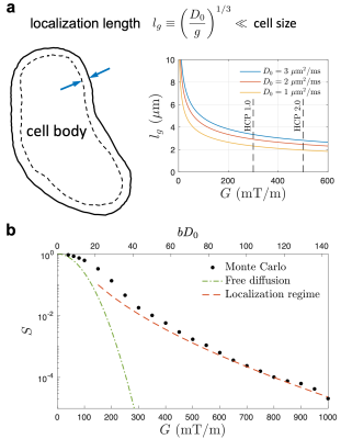 |
Localization regime of diffusion in human gray matter on a high-gradient MR system: Sensitivity to soma size
Hong-Hsi Lee1, Els Fieremans1, Susie Y Huang2, Qiyuan Tian2, and Dmitry S Novikov1
1New York University School of Medicine, New York, NY, United States, 2Department of Radiology, A. A. Martinos Center for Biomedical Imaging, Massachusetts General Hospital, Charlestown, MA, United States
In vivo estimation of cell dimension using diffusion MRI usually requires biophysical modeling of multiple compartments in biological tissues. However, the signal decomposition of multiple compartments is non-trivial due to limited amount of data and scan time. Instead, by applying strong diffusion gradients, a universal localization regime emerges: Magnetization far away from the cell boundaries vanishes, and only that near the boundaries within a thickness of localization length contributes to signal. Here, using Connectome gradients, we for the first time achieve the localization regime in vivo, and estimate the soma size in cortical brain gray matter of two healthy subjects.
|
|
 |
0640. |
Parameter estimation for the GRAMMI (GRAy Matter Microstructure Imaging) model of two exchanging compartments in the rat cortex in vivo
Alexandre de Skowronski1, Marco Palombo2, Dmitry S. Novikov3, and Ileana O. Jelescu4
1Dept. of Physics, Ecole Polytechnique Fédérale de Lausanne, Lausanne, Switzerland, 2Centre for Medical Image Computing and Dept. of Computer Science, University College London, London, United Kingdom, 3Center for Biomedical Imaging, Dept. of Radiology, New York University, New York, NY, United States, 4CIBM Center for Biomedical Imaging, Ecole Polytechnique Fédérale de Lausanne, Lausanne, Switzerland
Developing a relevant model for brain gray matter is a complex task. As opposed to white matter, features such as inter-compartment water exchange or soma should likely be modeled. In this work we examine the performance of a variant of the Kärger Model, called GRAMMI, that accounts for exchange, both on synthetic and experimental data. We show q-t coverage is necessary for reliable model parameter estimation at the individual voxel level and compare two regression approaches. Future work includes protocol optimization and the extension of the GRAMMI model to account for soma.
|
|
|
0641.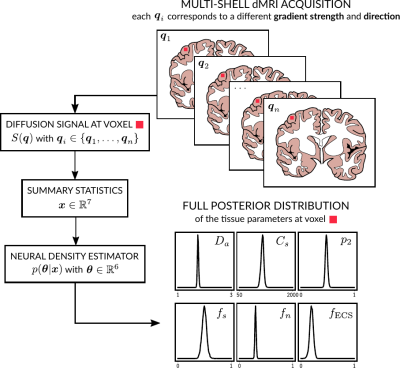 |
Diffusion MRI-Based Cytoarchitecture Measurements in Brain Gray Matter using Likelihood-Free Inference
Maëliss Jallais1, Pedro L. C. Rodrigues1, Alexandre Gramfort1, and Demian Wassermann1
1Université Paris-Saclay, Inria, CEA, Palaiseau, France
We propose a new method to solve the inverse problem of relating the diffusion MRI signal with cytoarchitectural characteristics in brain gray matter. Specifically, our method has quantitative sensitivity to soma density and volume. Our solution is twofold. First, we propose a new forward model that relates summary statistics of the dMRI signal with tissue parameters, relying on six b-shells only. We then apply a likelihood-free inference based algorithm to invert the proposed model, which not only estimates the tissue parameters that best describe the acquired diffusion signal, but also a full posterior distribution over the parameter space.
|
|
0642.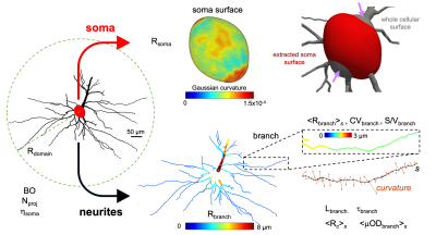 |
Large-scale analysis of brain cell morphometry informs microstructure modelling of gray matter
Marco Palombo1, Daniel C. Alexander1, and Hui Zhang1
1Centre for Medical Image Computing, University College London, London, United Kingdom
Diffusion-weighted MRI (dMRI) is a formidable technique for non-invasively characterizing brain microstructure. Biophysical modelling is often necessary to gain specificity to cellular structure. However, designing sensible biophysical models and appropriate dMRI acquisitions is challenging, especially for gray matter (GM), as little is known about typical values of relevant features of brain-cell morphology contributing to dMRI signal. This study addressed this unmet need: we analysed ~3,500 cells from mouse, rat, monkey and human brains to determine statistical distributions of 13 morphological features relevant to GM microstructure modelling. Illustrative examples demonstrate how this study can inform biophysical modelling.
|
||
|
0643.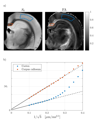 |
Power-law scaling of the diffusion signal in gray matter and the influence of exchange
Jonas L. Olesen1,2, Noam Shemesh3, and Sune N. Jespersen1,2
1Center of Functionally Integrative Neuroscience (CFIN) and MINDLab, Aarhus University, Aarhus, Denmark, 2Department of Physics and Astronomy, Aarhus University, Aarhus, Denmark, 3Champalimaud Research, Champalimaud Centre for the Unknown, Lisbon, Portugal
Diffusion models can be validated by observing their functional dependencies, exemplified by the b-1/2 power-law scaling recently used to validate the “stick” compartment in white matter. In contrast, such behavior has not been observed in grey matter (GM), potentially due to a) water exchange between dendrites and extra-neurite space and/or b) a distinct signal contribution from somas. We report the first observation of the stick power-law in GM at very large b-values consistent with b). Nevertheless, the dependence on diffusion time indicates significant water exchange affecting the scaling range. The combined observations thus offer a window into more complicated microstructure.
|
|
0644.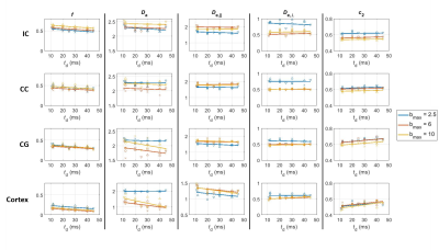 |
What can a rat tell about physics beyond Standard Model: Exchange or structural disorder?
Ileana O. Jelescu1,2 and Dmitry S. Novikov3
1CIBM Center for Biomedical Imaging, Lausanne, Switzerland, 2Animal Imaging and Technology, Ecole Polytechnique Federale de Lausanne, Lausanne, Switzerland, 3Dept. of Radiology, New York University School of Medicine, New York, NY, United States
One fundamental challenge in brain microstructure is to establish the biophysical origin of effects beyond the “Standard Model” (SM) picture of non-exchanging Gaussian compartments. The intra-compartmental structural disorder competes with inter-compartmental water exchange. Here we show that in rats, the exchange dominates, and offer the picture of diffusion time effectively filtering out the contribution of unmyelinated axons with stronger dispersion. At long times, only the myelinated (non-exchanging) axons contribute to the intra-axonal SM compartment, and the rest is attributed to extra-axonal space.
|
||
|
0645.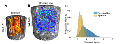 |
Feasibility of axon diameter estimation in complex fiber architectures by powder averaging of the diffusion MRI signal
Mariam Andersson1,2, Marco Pizzolato1,2,3, Hans Martin Kjer1,2, Henrik Lundell1, and Tim B. Dyrby1,2
1Danish Research Centre for Magnetic Resonance, Hvidovre, Denmark, 2Technical University of Denmark, Kgs. Lyngby, Denmark, 3Signal Processing Laboratory (LTS5), École Polytechnique Fédérale de Lausanne, Lausanne, Switzerland
Orientation dispersion bias in axon diameter measurements can be removed by powder averaging of the diffusion MRI signal in isotropically distributed directions, but has not been validated in complex fiber architectures. Here, we demonstrate the success of the spherical mean technique (SMT) and a recent power law (PL) implementation in removing orientation-related bias in diameter estimates of real axons from the splenium corpus callosum and a complex crossing fiber region of the vervet monkey brain. In the crossing fiber region, we find a significant population of very large axons, indicating a need for sensitivity to a wide range of diameters.
|
|
|
0646.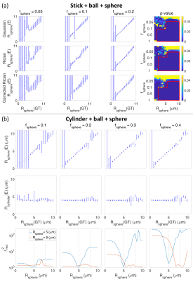 |
SPHERIOUSLY? The challenges of estimating spherical pore size non-invasively in the human brain from diffusion MRI
Maryam Afzali1, Markus Nilsson2, Marco Palombo3, and Derek K Jones1
1Cardiff University Brain Research Imaging Centre (CUBRIC), School of Psychology, Cardiff University, Cardiff, United Kingdom, 2Clinical Sciences Lund, Radiology, Lund University, Lund, Sweden, 3Centre for Medical Image Computing, Department of Computer Science, University College London, London, United Kingdom
Soma and Neurite Density Imaging (SANDI) was recently proposed to disentangle cylindrical and spherical geometries, attributed to neurite and soma compartments. In this work, using: (i) ultra-strong gradients; (ii) a combination of linear, planar, and spherical b-tensor encodings; and (iii) analysing the signal in the frequency domain, three main challenges were identified; First, the Rician noise floor biases estimation of soma properties. Second there is an empirical lower bound on the spherical signal fraction and pore-size. Third, if there is sensitivity to the transverse intra-cellular diffusivity in cylindrical structures, estimation of spherical pore-size is challenging.
|
|
0647.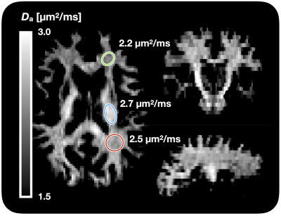 |
Estimation of intra-axonal axial diffusivity by tensor-valued dMRI and powder-averaging
Markus Nilsson1, Samuel St-Jean1,2, Christian Beaulieu2, and Filip Szczepankiewicz1
1Clinical Sciences Lund, Lund University, Lund, Sweden, 2Department of Biomedical Engineering, University of Alberta, Edmonton, AB, Canada
The intra-axonal axial diffusivity (Da) has a prominent role in describing and modeling the white matter microstructure, but cannot be obtained from regular diffusion tensor imaging due to the influence of orientation dispersion and extracellular water. It may be estimated using higher b-values and modeling, however, its estimation is still an ill-posed problem or requires knowledge of the orientation distribution function. Here, we show that using b-tensor encoding and powder averaging turns the problem into a well-posed one and allows rapid mapping across the whole brain yielding Da values of 2.2-2.7 µm2/ms.
|
||
0648.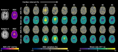 |
Dynamic Changes in Brain Tissue Strain and ADC over the Cardiac Cycle quantified at 7T MRI
Jacob-Jan Sloots1, Martijn Froeling1, Geert Jan Biessels2, and Jaco Zwanenburg1
1Radiology, University Medical Center Utrecht, Utrecht, Netherlands, 2Neurology, University Medical Center Utrecht, Utrecht, Netherlands
The apparent diffusion coefficient (ADC) in brain tissue slightly varies over the cardiac cycle. In this study, we investigate to what extent ADC variations can be explained by brain tissue strain, which affects the measured MRI signals. To this end, we developed a high-field MRI sequence that simultaneously measures both ADC and tissue strain. Preliminary results in 2 volunteers show that ADC fluctuations over the cardiac cycle are an order of magnitude larger than could be explained from measurement errors induced by tissue strains. Consequently, ADC fluctuations in the brain probably reflect physiology.
|
The International Society for Magnetic Resonance in Medicine is accredited by the Accreditation Council for Continuing Medical Education to provide continuing medical education for physicians.