Oral
Brain Microstructure: Application & Validation Across Species
ISMRM & SMRT Annual Meeting • 15-20 May 2021

Oral
Brain Microstructure: Application & Validation Across Species
| Concurrent 3 | 14:00 - 16:00 | Moderators: Manisha Aggarwal |
0291.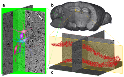 |
Multi-modal, multi-resolution imaging of a single mouse brain
Sean Foxley1, Vandana Sampathkumar1, Vincent De Andrade2, Scott Trinkle1, Anstasia Sorokina1, Katrina Norwood1, Patrick La Riviere1, and Narayanan Kasthuri1
1University of Chicago, Chicago, IL, United States, 2Advanced Photon Source, Argonne National Laboratory, Lamont, IL, United States
Mammalian neurons operate at length scales spanning many orders of magnitude; micron-scale-diameter myelinated axons project millimeters across brain regions, forming nanometer scale synapses. Capturing these disparately sized anatomical features requires imaging samples with multiple independent imaging modalities. Directly correlating features across modalities requires that all imaging is performed in the same brain. Here, we imaged the same postmortem mouse brain over five orders of spatial resolution using MRI, synchrotron x-ray tomography (μCT), and electron microscopy. This pipeline provides an unprecedented look across a single brain's multi-scaled organization and a vehicle for studying the brain’s multi-scaled pathologies.
|
||
 |
0292. |
Pre- and post-neonatal in vivo DTI on mice: Targeting brain microstructures at 15.2T
Odélia Jacqueline Chitrit1, Qingjia Bao1, Maxime Yon1, and Lucio Frydman1
1Department of Chemical and Biological Physics, Weizmann institute of Science, Rehovot, Israel
DTI is a well-established technique for mapping brain microstructure. Brain’s microstructural features derive from white matter and change over the course of maturation; hence methods that can acquire DTI in utero and immediately post-partum, are of interest. The present study explores the use of a customized 3D phase-encoded Spatiotemporal Encoding (SPEN) MRI approach that can be used to overcome the motional and susceptibility challenges arising in such instances, delivering quality DTI volumetric data at 15.2T. Maps of ADC, MDD and FA could thus be collected for mice fetal brains in utero, as well as within the first week post-partum.
|
|
0293. |
Measuring apparent water exchange using Filter Exchange Imaging and diffusion time dependent kurtosis imaging in post-mortem mouse brains
Chenyang Li1,2, Els Fieremans1, Dmitry S. Novikov1, Yulin Ge1, and Jiangyang Zhang1
1Department of Radiology, Center for Biomedical Imaging, NYU Grossman School of Medicine, New York, NY, United States, 2Vilcek Institute of Graduate Biomedical Sciences, NYU Grossman School of Medicine, New York, NY, United States
Filter exchange imaging (FEXI) and diffusion time dependent diffusion kurtosis imaging (DKI(t)) are two techniques that are sensitive to water exchange between tissue compartments. However, built on different theoretical frameworks and models, many questions remain on the interpretations of FEXI and (DKI(t)) results in the brain. In this study, we measured water exchange effects in post-mortem mouse brains using FEXI and (DKI(t)) and observed a correlation between them, suggesting that they are sensitive to similar exchange processes.
|
||
 |
0294. |
Exploring the epileptic rat hippocampus using oscillating gradients, 3D electron microscopy and Monte Carlo simulations
Jonathan Scharff Nielsen1, Alejandra Sierra2, Ilya Belevich3, Eija Jokitalo3, and Manisha Aggarwal1
1Department of Radiology and Radiological Science, Johns Hopkins University School of Medicine, Baltimore, MD, United States, 2A.I. Virtanen Institute of Molecular Sciences, University of Eastern Finland, Kuopio, Finland, 3Institute of Biotechnology, University of Helsinki, Helsinki, Finland
Oscillating gradient spin-echo (OGSE) diffusion MRI (dMRI) is sensitive to small-scale restrictions and may provide a sensitive probe of gray matter microstructural changes in brain disorders such as temporal lobe epilepsy. However, relating the OGSE spectral changes to specific microstructural features is a difficult challenge. Here, we combined OGSE-dMRI with serial block-face electron microscopy volumes of healthy and status epilepticus exhibiting rat hippocampi. From tissue parameters extracted from these volumes, we generated 3D digital substrates for Monte-Carlo random-walk simulations, which allowed us to elucidate the relative contributions of underlying gray matter microstructural features to the OGSE measurements.
|
|
0295.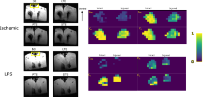 |
Towards differentiation of white matter pathologies through B-tensor encoding.
Ricardo Rios-Carrillo1, Ricardo Coronado-Leija2, Hiram Luna-Munguía1, Alonso Ramírez-Manzanares3, and Luis Concha1
1Instituto de Neurobiología, Universidad Nacional Autónoma de México, Querétaro, Mexico, 2Radiology, New York University School of Medicine, New York, NY, United States, 3Centro de Investigación en Matemáticas, Guanajuato, Mexico
This work explores the ability of B-tensor encoding methods to disentangle axonal degeneration and inflammation through DW-MRI. In particular, we tested Q-space trajectory encoding and diffusion tensor distribution imaging. Both methods clearly differentiated between damaged and intact nerves, and showed moderately different diffusion characteristics between the two experimental conditions.
|
||
|
0296.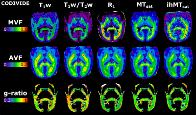 |
g-Ratio in the common marmoset: a comparison across different myelin-sensitive MRI metrics with b-tensor encoded diffusion
Christopher D Rowley1,2, Ilana R Leppert2, Jennifer SW Campbell2, Filip Szczepankiewicz3,4, Stephen Nuara5, Markus Nilsson3, G Bruce Pike6, and Christine L Tardif1,2,7
1Neurology and Neurosurgery, McGill University, Montreal, QC, Canada, 2McConnell Brain Imaging Centre, Montreal Neurological Institute and Hospital, Montreal, QC, Canada, 3Diagnostic Radiology, Clinical Sciences Lund, Lund University, Lund, Sweden, 4Radiology, Brigham and Women's Hospital, Harvard Medical School, Boston, MA, United States, 5Comparative Medicine and Animal Resources Center, McGill University, Montreal, QC, Canada, 6Hotchkiss Brain Institute and Departments of Radiology and Clinical Neuroscience, University of Calgary, Calgary, AB, Canada, 7Department of Biomedical Engineering, McGill University, Montreal, QC, Canada
The g-ratio quantifies the relative thickness of the myelin sheath and can be estimated from myelin volume fraction (MVF) and axonal volume fraction (AVF) maps. The best MRI methods for deriving these metrics are still under investigation. This study examines the use of inhomogeneous magnetization transfer (ihMTsat) along with other myelin-sensitive metrics, and diffusion MRI with b-tensor encoding for calculating the g-ratio in a marmoset brain. We find that while the different myelin-sensitive metrics and diffusion microstructural models produce different MVF and AVF maps, the g-ratio values follow similar trends across the white matter.
|
|
0297.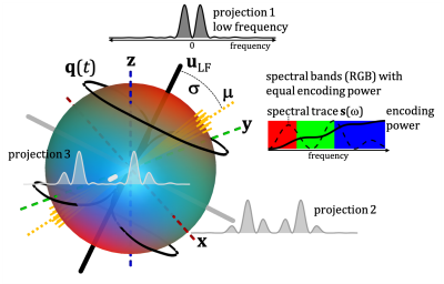 |
The spectral tilt plot (STP) – new microstructure signatures from spectrally anisotropic b-tensor encoding
Samo Lasic1,2, Filip Szczepankiewicz3, Markus Nilsson3, Tim B. Dyrby1,4, and Henrik Lundell1
1Danish Research Centre for Magnetic Resonance, Centre for Functional and Diagnostic Imaging and Research, Copenhagen University Hospital Hvidovre, Copenhagen, Denmark, 2Random Walk Imaging, Lund, Sweden, 3Clinical Sciences, Lund University, Lund, Sweden, 4Department of Applied Mathematics and Computer Science, Technical University of Denmark, Copenhagen, Denmark
Tensor-valued diffusion encoding can probe diffusion tensor distribution unconfounded by orientation and heterogeneity. Since different cell morphologies may yield similar apparent diffusion tensors, inferring specific microstructural features remains challenging. Further information can be accessed by considering time-dependent diffusion. We show that rotational dependence of spherical tensor encoding, caused by spectral anisotropy, can be prominent on a preclinical scanner. The presented analysis may reveal different microstructural signatures depending on cell shape, which could be relevant for tissue modelling. Our results on fixed monkey brain suggest that brain cells exhibit anisotropic restricted diffusion along all directions, unlike in the cylindrical diffusion model.
|
||
|
0298.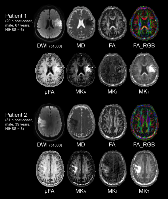 |
Tensor-valued Diffusion MRI Shows Elevated Microscopic Anisotropy and Tissue Heterogeneity in White and Grey Matter of Acute Ischemic Stroke
Mi Zhou1, Robert Stobbe1, Filip Szczepankiewicz2,3, Mar Lloret4, Brian Buck4, Paige Fairall4, Ken Butcher4, Ashfaq Shuaib4, Derek Emery5, Markus Nilsson2, Carl-Fredrik Westin3, and Christian Beaulieu1
1Biomedical Engineering, University of Alberta, Edmonton, AB, Canada, 2Clinical Sciences Lund, Lund University, Lund, Sweden, 3Radiology, Brigham and Women’s Hospital, Harvard Medical School, Boston, MA, United States, 4Neurology, University of Alberta, Edmonton, AB, Canada, 5Radiology and Diagnostic Imaging, University of Alberta, Edmonton, AB, Canada
Novel diffusion encoding modalities, such as tensor-valued encoding, can disentangle the effects of intra-voxel orientation dispersion and diffusion anisotropy, thereby resolving the fiber density from tissue heterogeneity. A rapid 2.5-minute protocol for tensor-valued diffusion MRI was applied for the first time to acute stroke. Microscopic anisotropy (µFA and MKA) and tissue heterogeneity (MKI) were higher in lesions of white and grey matter, in contrast to reduced DTI-derived fractional anisotropy at the voxel level. Elevated microscopic anisotropy in acute stroke may reflect increased trapped water in swollen axons, a measure independent of tract orientation dispersion.
|
|
|
0299.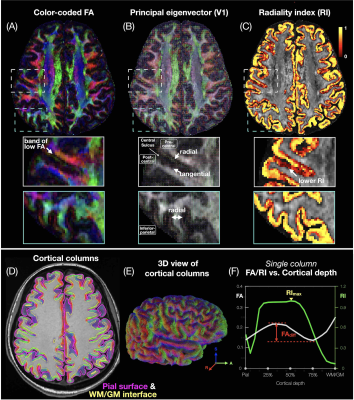 |
Column-based cortical depth analysis of the diffusion anisotropy in submillimeter whole-brain DTI of the human gray matter
Yixin Ma1,2, Trong-Kha Truong1,2, Iain P. Bruce1, Chun-Hung Yeh3, Jeffrey R. Petrella1,2, and Allen W. Song1,2
1Brain Imaging and Analysis Center, Duke University, Durham, NC, United States, 2Medical Physics Graduate Program, Duke University, Durham, NC, United States, 3Institute for Radiological Research, Chang Gung University and Chang Gung Memorial Hospital, Taoyuan, Taiwan
High-resolution diffusion tensor imaging (DTI) can noninvasively probe the microstructural integrity of cortical gray matter in vivo. We propose a column-based method that samples submillimeter isotropic whole-brain DTI data along radially oriented cortical columns to achieve a quantitative analysis of the fractional anisotropy and radiality index dependence on the cortical depth, curvature, and brain regions across the whole brain. This method is robust across repeated scans and healthy subjects, and can capture characteristic diffusion anisotropy and radiality patterns in the cortical gray matter, potentially providing quantitative biomarkers for various neurological disorders.
|
|
0300. |
Ex-vivo whole human brain high b-value diffusion MRI at 550 micron with a 3T Connectom scanner
Gabriel Ramos-Llordén1, Chiara Maffei1, Qiyuan Tian1, Berkin Bilgic1,2, Thomas Witzel3, Boris Keil4, Anatasia Yendiki1, and Susie Huang1,2
1Athinoula A. Martinos Center for Biomedical Imaging, Department of Radiology, Masachusetts General Hospital, Harvard Medical School, Charlestown, MA, United States, 2Harvard-MIT Division of Health Sciences and Technology, Massachusetts Institute of Technology, Cambridge, MA, United States, 3Q Bio Inc, San Carlos, CA, United States, 4Institute of Medical Physics and Radiation Protection, Mittelhessen University of Applied Sciences, Giessen, Germany
We demonstrate high quality, high b-value diffusion MRI (up to 10 000 s / mm2) in an ex-vivo human brain at ultra-high spatial resolution (550 micrometer). The high signal quality allows us to obtain high diffusion contrast and delineate fine structures that cannot be resolved within in vivo acquisitions. We present DTI and DKI results of the whole brain, show diffusivity on the primary somatosensory, auditory, and visual cortex areas as well as illustrate tractography of the hippocampus and thalamus structures, revealing internal connectivity at a high level of detail.
|
The International Society for Magnetic Resonance in Medicine is accredited by the Accreditation Council for Continuing Medical Education to provide continuing medical education for physicians.