Oral
Diffusion in Cancer: Clinical Studies & Validation
ISMRM & SMRT Annual Meeting • 15-20 May 2021

Oral
Diffusion in Cancer: Clinical Studies & Validation
| Concurrent 4 | 12:00 - 14:00 | Moderators: Eleftheria Panagiotaki & Rebecca Rakow-Penner |
0695.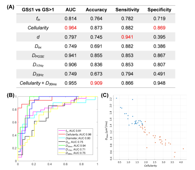 |
Microstructural mapping with diffusion-time dependent diffusion MRI improves diagnosis of prostate cancer at 3T
Dan Wu1, Kewen Jiang2, Yi-Cheng Hsu3, Yi Sun3, Yi Zhang1, and Yudong Zhang2
1Biomedical Engineering, Zhejiang University, Hangzhou, China, 2Radiology, the First Affiliated Hospital with Nanjing Medical University, Nanjing, China, 3MR Collaboration, Siemens Healthcare Ltd., Shanghai, China
Diffusion-time-dependent diffusion MRI (dMRI) has shown potentials in characterizing tumor microstructure. This study investigated the diagnostic value of time-dependent dMRI to differentiate prostate cancer pathological grades at 3T. Oscillating and pulsed gradient dMRI was performed in 55 patients, and the data were fitted with the IMPULSED model to estimate cell diameter, intracellular fraction, cellularity, and diffusivities. We found fin and cellularity increased as Gleason score increased, while diameter and Dex decreased. Cellularity achieved the highest diagnostic accuracy with an accuracy of 0.87 and area-under-the curve of 0.96, and the combination of cellularity and ADC further improved the accuracy to 0.91.
|
||
 |
0696.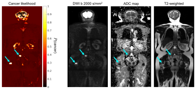 |
Leveraging a multicompartmental signal model for improved classification of prostate-cancer bone metastases in whole-body DWI
Christopher C Conlin1, Christine H Feng2, Leonardino A Digma2, Ana E Rodriguez-Soto1, Joshua M Kuperman1, Dominic Holland3, Rebecca Rakow-Penner1, Tyler M Seibert1,2,4, Anders M Dale1,3,5, and Michael E Hahn1
1Department of Radiology, UC San Diego School of Medicine, La Jolla, CA, United States, 2Department of Radiation Medicine and Applied Sciences, UC San Diego School of Medicine, La Jolla, CA, United States, 3Department of Neurosciences, UC San Diego School of Medicine, La Jolla, CA, United States, 4Department of Bioengineering, UC San Diego Jacobs School of Engineering, La Jolla, CA, United States, 5Halıcıoğlu Data Science Institute, UC San Diego, La Jolla, CA, United States
Multicompartmental diffusion modeling shows promise for overcoming the limitations of conventional DWI methods and may help to improve the clinical evaluation of prostate-cancer bone involvement. In this study, we applied multicompartmental modeling to develop an empirical tissue classifier for identifying bone lesions in whole-body DWI. The proposed classifier relates signal contributions from model compartments with lower diffusion coefficients to the likelihood that such contributions are from cancerous tissue. This approach proved effective for detecting metastatic lesions in whole-body DWI data, considerably outperforming a classifier based on conventional ADC values.
|
|
0697.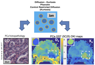 |
Biomimetic phantoms of impeded diffusion in prostate cancer using lipid nanoparticles
Scott D. Swanson1, Thomas L. Chenevert1, Prasad R. Shankar1, Ted Lynch2, and Dariya I. Malyarenko1
1Department of Radiology, University of Michigan, Ann Arbor, MI, United States, 2CIRS, Norfolk, VA, United States
PI-RADS guidelines use quantitative mono-exponential apparent diffusion coefficient (ADC) for risk stratification of small potentially aggressive prostate cancer (PCa). Inherently, multi-exponential diffusion in complex tumor microenvironment, limits accuracy of ADC-based thresholds and clinical utility of DWI acquisition protocols. Biomimetic phantoms that provide ground-truth diffusion parameters help establish accuracy of advanced multi-b DWI protocols and parametric diffusion models in prostate tumors. This work demonstrates development of stable phantoms based on lipid nanoparticles that provide diffusion parameter ranges typical of PCa.
|
||
0698.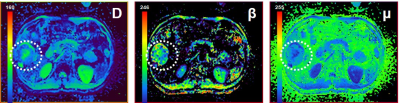 |
Evaluate the micro-vascular invasion in HCC with a Fractional Order Calculus DWI Model
Xiuzhong Yao1, Yunfei Zhang2, Mengsu Zeng1, and Yongming Dai2
1Zhongshan Hospital affiliated to Fudan University, Shanghai, China, 2Central Research Institute, United Imaging Healthcare, Shanghai, China
The determination of micro-vascular invasion (MVI) in patients with hepatocellular carcinoma (HCC) is significant for prognostic prediction. However, histologic assessment is the gold standard for evaluating the MVI, which is accompanied with many disadvantages including invasiveness, potential sampling bias and so forth. DWI MRI has shown clinical potential for assessing the MVI. However, some recently-developed non-gaussian DWI techniques have hardly been applied for predicting the MVI. This research, hence, sought to evaluate the MVI with a Fractional Order Calculus DWI Model (FROC-DWI). The results indicated that FROC-derived parameters were valuable for accurately identifying the MVI in patients with HCC.
|
||
|
0699.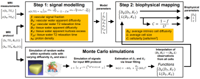 |
DR-HIGADOS: a new diffusion-relaxation framework for clinically feasible microstructural imaging of the liver
Francesco Grussu1, Ignasi Barba2, Kinga Bernatowicz1, Irene Casanova-Salas3, Alba Escriche Villarroya4, Natalia Castro3, Emanuela Greco4, Juan Francisco Corral5,6, Marta Vidorreta7, Manuel Escobar Amores5,6, Núria Roson5,6, Xavier Merino5,6, Richard Mast5,6,
Nahúm Calvo‐Malvar5,8, Joaquin Mateo3, Paolo Nuciforo9, María Abad4, Josep R. Garcia-Bennett8, and Raquel Perez-Lopez1,6
1Radiomics Group, Vall d'Hebron Institute of Oncology, Vall d'Hebron Barcelona Hospital Campus, Barcelona, Spain, 2NMR Lab, Vall d'Hebron Institute of Oncology, Vall d'Hebron Barcelona Hospital Campus, Barcelona, Spain, 3Prostate Cancer Translational Research Group, Vall d'Hebron Institute of Oncology, Vall d'Hebron Barcelona Hospital Campus, Barcelona, Spain, 4Cellular Plasticity and Cancer Group, Vall d'Hebron Institute of Oncology, Vall d'Hebron Barcelona Hospital Campus, Barcelona, Spain, 5IDI (Institut de Diagnòstic per la Imatge), Catalonia, Spain, 6Department of Radiology, Hospital Universitari Vall d'Hebron, Barcelona, Spain, 7Siemens Healthineers, Madrid, Spain, 8Hospital Universitari de Bellvitge, L'Hospitalet de Llobregat, Spain, 9Molecular Oncology Group, Vall d'Hebron Institute of Oncology, Vall d'Hebron Barcelona Hospital Campus, Barcelona, Spain
Liver cancer is a leading cause of cancer-related death, and new quantitative MRI (qMRI) techniques are needed to inform treatment selection and monitor disease progression. We propose a new technique, Diffusion-Relaxation Hepatic Imaging via Generalisable Assessment of DiffusiOn Simulations (DR-HIGADOS), with the aim of improving sensitivity and biological specificity of liver qMRI. DR-HIGADOS is a diffusion-relaxation method that uses information from Monte Carlo simulations to map parameters of an extended intra-voxel incoherent motion model to microstructural indices (e.g. cell size, cellularity). DR-HIGADOS is demonstrated on multi-vendor clinical data, and its histological correlates are investigated on preclinical high-field scans.
|
|
|
0700.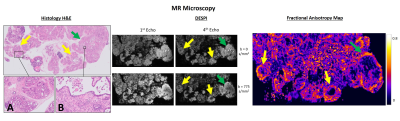 |
Ex vivo and In vivo Diffusion Weighted MRI highlights the Microarchitecture of mouse Pancreatic Intraepithelial Neoplasia
Carlos Bilreiro1,2, Francisca F Fernandes1, Rui V Simões1, Mireia Castillo-Martin1,3, Andrada Ianus1, Cristina Chavarrias1, Celso Matos1,2, and Noam Shemesh1
1Champalimaud Research, Champalimaud Centre for the Unknown, Lisbon, Portugal, 2Department of Radiology, Champalimaud Clinical Centre, Lisbon, Portugal, 3Department of Pathology, Champalimaud Clinical Centre, Lisbon, Portugal
As current therapeutic options for pancreatic cancer are mostly ineffective, developing diagnostic tools for early detection of precursor lesions (mainly pancreatic intraepithelial neoplasia – PanIN) could change the course of this disease. Here, we investigated Diffusion-MRI (dMRI) contrasts for this purpose, using transgenic mouse models. First, we performed ex vivo dMRI Microscopy at ultrahigh field with histological validation, defining the most sensitive contrasts. Then, we performed in vivo abdominal imaging, demonstrating their applicability in the non-invasive study of PanIN and pancreatic cancer. These findings provide new tools for researching PanIN and hold promise for a future translation to clinical practice.
|
|
0701.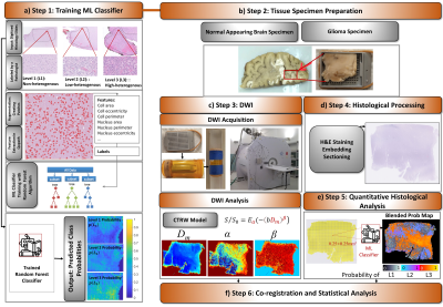 |
Looking Inside a Voxel through the Lenses of Non-Gaussian Diffusion MRI: Correlation between Imaging- and Histology-based Tissue Heterogeneity
Muge Karaman1,2, Guangyu Dan1,2, Lingdao Sha3, Tingqi Shi1, Weiguo Li4,5, Dan Schonfeld2,3,6, Tibor Valyi-Nagy7, and X. Joe Zhou1,2,8
1Center for Magnetic Resonance Research, University of Illinois at Chicago, Chicago, IL, United States, 2Department of Bioengineering, University of Illinois at Chicago, Chicago, IL, United States, 3Department of Electrical and Computer Engineering, University of Illinois at Chicago, Chicago, IL, United States, 4Research Resources Center, University of Illinois at Chicago, Chicago, IL, United States, 5Department of Radiology, Northwestern University, Chicago, IL, United States, 6Department of Computer Science, University of Illinois at Chicago, Chicago, IL, United States, 7Department of Pathology, University of Illinois at Chicago, Chicago, IL, United States, 8Departments of Radiology and Neurosurgery, University of Illinois at Chicago, Chicago, IL, United States
Studies on tissue structural heterogeneity have been the focus of a growing number of non-Gaussian diffusion models, such as the continuous-time random-walk (CTRW) model. Establishing a correlation between the voxel-level CTRW parameters and the microscopic tissue heterogeneity from gold-standard histology, however, has been challenging due to the lack of quantitative measure of histopathological heterogeneity and different spatial scales. We establish a one-to-one correspondence between imaging-based tissue heterogeneity revealed by CTRW parameters and histology-based tissue structural heterogeneity predicted by a machine-learning classifier to address an overarching question: “Can we look inside a voxel noninvasively through the lenses of the CTRW model?”.
|
||
0702.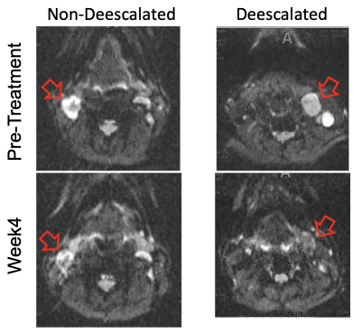 |
Diffusion MRI study of chemoradiation treatment response in patients with HPV positive oropharyngeal carcinoma
Sungheon Gene Kim1, Mehran Baboli1, Justin Fogarty2, Steven H. Baete2, Joseph Kim3, Paulina Galavis3, Moses Tam3, Kenneth Hu3, and Elcin Zan2
1Radiology, Weill Cornell Medical College, New York, NY, United States, 2Radiology, New York University School of Medicine, New York, NY, United States, 3Radiation Oncology, New York University School of Medicine, New York, NY, United States
In this study, we evaluated diffusion and kurtosis time-dependence for HPV-positive oropharyngeal squamous cell carcinoma before and during chemo-radiation treatment over a wide range for longer diffusion times (200-700 ms). The patients with less than 40% nodal volume shrinkage had significantly higher diffusivity at pretreatment and lower kurtosis at week4 than the patients with more than 40% nodal volume shrinkage. The water exchange times were 68-80 ms without a significant difference between the groups. This study demonstrates the feasibility of using diffusion MRI at relatively long diffusion times to predict and evaluate the response to chemo-radiation therapy.
|
||
0703.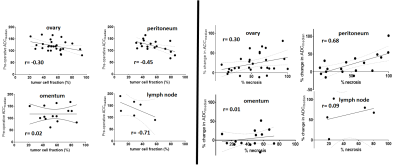 |
Relating tumor site-specific volume and ADC changes following neoadjuvant chemotherapy to histopathology in epithelial ovarian cancer
Jessica M Winfield1,2, Jennifer C Wakefield1,2, James D Brenton3,4,5, Khalid AbdulJabbar6,7, Antonella Savio8, Susan Freeman9, Erika Pace1,2, Kerryn Lutchman-Singh10, Katherine M Vroobel8, Yinyin Yuan6,7, Susana Banerjee11, Nuria Porta12, Shan E Ahmed Raza6,7,13,
and Nandita M deSouza1,2
1MRI Unit, Royal Marsden NHS Foundation Trust, London, United Kingdom, 2Division of Radiotherapy and Imaging, Institute of Cancer Research, London, United Kingdom, 3Cancer Research UK Cambridge Institute, Cambridge, United Kingdom, 4Addenbrooke’s Hospital, Cambridge University Hospitals NHS Foundation Trust, Cambridge, United Kingdom, 5Department of Oncology, University of Cambridge, Cambridge, United Kingdom, 6Centre for Evolution and Cancer, The Institute of Cancer Research, London, United Kingdom, 7Division of Molecular Pathology, The Institute of Cancer Research, London, United Kingdom, 8Department of Pathology, Royal Marsden NHS Foundation Trust, London, United Kingdom, 9Department of Radiology, Addenbrooke’s Hospital, Cambridge University Hospitals NHS Foundation Trust, Cambridge, United Kingdom, 10Swansea Gynaecological Oncology Centre, Swansea Bay University Health Board, Singleton Hospital, Swansea, United Kingdom, 11Gynaecology Unit, Royal Marsden NHS Foundation Trust, Sutton, United Kingdom, 12Clinical Trials and Statistics Unit, The Institute of Cancer Research, London, United Kingdom, 13Department of Computer Science, University of Warwick, Warwick, United Kingdom
In Epithelial Ovarian Cancer, ADCmedian demonstrates good repeatability at both primary and metastatic sites. After neoadjuvant chemotherapy, a differential increase in ADCmedian at disease sites is seen despite similar tumor shrinkage. The negative correlation between ADCmedian and tumor cell fraction after neoadjuvant chemotherapy, and positive correlation between change in ADCmedian and percentage necrosis, are driven primarily by changes in the peritoneal lesions.
|
||
0704.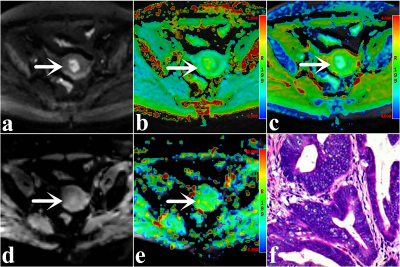 |
Endometrial Carcinoma: Assessment of Histological Features Based on Amide Proton Transfer-weighted Imaging and Diffusion Kurtosis Imaging
Nan Meng1, Zhun Huang2, Ting Fang1, Pengyang Feng2, Xuejia Wang3, Dongming Han3, Kaiyu Wang4, and Meiyun Wang*1
1Department of Radiology, Zhengzhou University People’s Hospital & Henan Provincial People’s Hospital, Zhengzhou, China, 2Department of Radiology, Henan University People’s Hospital & Henan Provincial People’s Hospital, Zhengzhou, China, 3Department of MRI, The First Affiliated Hospital of Xinxiang Medical University, Weihui, China, 4GE Healthcare, MR Research China, BeiJing, China
Our results showed that both Amide proton transfer-weighted imaging (APTWI) and Diffusion kurtosis imaging (DKI) were effective in the assessment of endometrial carcinoma in terms of clinical type, histological grade, subtype, and Ki-67 index.
|
The International Society for Magnetic Resonance in Medicine is accredited by the Accreditation Council for Continuing Medical Education to provide continuing medical education for physicians.