Oral
Diffusion Acquisition & Post-Processing
ISMRM & SMRT Annual Meeting • 15-20 May 2021

| Concurrent 6 | 14:00 - 16:00 | Moderators: Itamar Ronen & Zhe Zhang |
0103.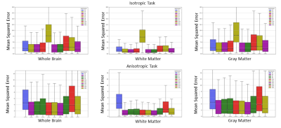 |
Resolving to super resolution multi-dimensional diffusion imaging (Super-MUDI)
Vishwesh Nath1, Marco Pizzolato2,3, Marco Palombo4, Noemi Gyori4, Kurt G Schilling5, Colin Hansen6, Qi Yang6, Praitayini Kanakaraj6, Bennett A Landman6, Soumick Chatterjee7, Alessandro Sciarra7, Max Duennwald7, Steffen Oeltze-Jafra7,
Andreas Nuernberger7, Oliver Speck7, Tomasz Pieciak 8, Marcin Baranek8, Kamil Bartocha8, Dominika Ciupek8, Fabian Bogusz8, Azam Hamidinekoo9, Maryam Afzali 10, Harry Lin4, Danny C Alexander4, Haoyu Lan11, Farshid Sepehrband11,
Zifei Liang12, Tung-Yeh Wu13, Ching-Wei Su13, Qian-Hua Wu13, Zi-You Liu13, Yi-Ping Chao13, Enes Albay14, Gozde Unal14, Dmytro Pylypenko13, Xinyu Ye13, Fan Zhang15, and Jana Hutter16
1NVIDIA Corporation, Bethesda, MD, United States, 2Department of Applied Mathematics and Computer Science, Technical University of Denmark, Kongens Lyngby, Denmark, 3École polytechnique fédérale de Lausanne (EPFL), Lausanne, Switzerland, 4CMIC, University College London, London, United Kingdom, 5Institute of Imaging Science, Vanterbilt University, Nashville, TN, United States, 6Department of Computer Science, Vanterbilt University, Nashville, TN, United States, 7Otto von Guericke University, Magdeburg, Germany, 8AGH University of Science and Technology, Krakow, Poland, 9Institute of Cancer Research, London, United Kingdom, 10CUBRIC, Cardiff University, Cardiff, United Kingdom, 11University of Southern California, Los Angeles, CA, United States, 12NYU Langone, New York, NY, United States, 13Center for Biomedical Imaging Research, Department of Biomedical Engineering, Tsinghua University, Beijing, China, 14Istanbul Technical University, Istanbul, Turkey, 15Harvard Medical School, Boston, MA, United States, 16Centre for Medical Engineering, King's College London, London, United Kingdom
Diffusion-weighted magnetic resonance imaging (DW-MRI) is a critical modality that allows characterization of microstructure of the nervous tissue in the human brain. Recent multi-parametric acquisitions expand parameter space to b-values, gradient directions, inversion and echo times. The required long scanning time could be shortened by acquiring at lower resolutions while superesolving the images during post-processing. This work embodies the evaluation of an open challenge where the objective was to upsample multi dimensional data encoding simultaneously T1, T2* and diffusion contrast to the natively acquired voxel resolution from two different down-sampled sets of the data (isotropic down-sampled and anisotropic down-sampled).
|
||
 |
0104.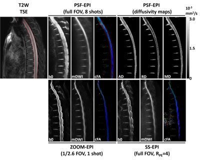 |
High-Fidelity Diffusion Tensor Imaging of the Thoracic Spinal Cord Using Point-Spread-Function Encoded EPI (PSF-EPI)
Sisi Li1, Yishi Wang2, Zhangxuan Hu3, Zhe Zhang4, Bing Wu3, and Hua Guo1
1Center for Biomedical Imaging Research, Beijing, China, 2Philips Healthcare, Beijing, China, 3GE Healthcare, MR Research China, Beijing, China, 4China National Clinical Research Center for Neurological Diseases, Beijing Tiantan Hospital, Capital Medical University, Beijing, China
Diffusion tensor imaging (DTI) holds great potential to aid the diagnosis of spinal cord pathologies. Single shot EPI (SS-EPI) is mostly implemented for DTI but is significantly limited in clinics by susceptibility inhomogeneity-induced distortions. This is especially problematic in the thoracic region for increased inhomogeneities near lungs. To solve this problem, multi-shot EPI and reduced-FOV methods have been proposed. However, these methods cannot remove distortions completely. In this study, we achieved high-fidelity DTI of the thoracic spinal cord using a distortion-free MS-EPI technique, Point-Spread-Function Encoded EPI (PSF-EPI). Both the efficacy of PSF-EPI in distortion correction and quantitative reproducibility were evaluated.
|
|
 |
0105.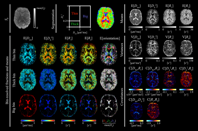 |
Nonparametric 6D D-R1-R2 distribution imaging of the human brain: Initial results on healthy volunteers
Jan Martin1, Alexis Reymbaut2, Michael Uder3, Frederik Bernd Laun3, and Daniel Topgaard1
1Lund University, Lund, Sweden, 2Random Walk Imaging AB, Lund, Sweden, 3Institute of Radiology, University Hospital Erlangen, Friedrich-Alexander-Universität Erlangen-Nürnberg (FAU), Erlangen, Germany
Diffusion-relaxation correlation NMR methods have recently received attention from the medical MRI community for their ability to characterize microstructure and local chemical composition in complex tissues containing multiple subvoxel pools of water. We here implement 6D $$$\bf{D}$$$-$$$R_1$$$-$$$R_2$$$ distribution imaging of the human brain using a 20-min acquisition protocol combining EPI signal read-out and tensor-valued diffusion encoding with varying repetition- and echo times. Monte Carlo data inversion yields nonparametric distributions, statistical descriptors, and orientation-resolved diffusion and relaxation properties of white matter fiber bundles that are in good agreement with previous results from less exhaustive 4D and 5D protocols.
|
|
0106.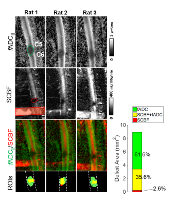 |
Diffusion-Prepared Fast Spin Echo for Artifact-free Spinal Cord Imaging
Seung-Yi Lee1, Briana Meyer1, Shekar Kurpad2, and Matthew Budde2
1Biophysics, Medical College of Wisconsin, Milwaukee, WI, United States, 2Neurosurgery, Medical College of Wisconsin, Milwaukee, WI, United States
Sagittal diffusion and perfusion magnetic resonance imaging can provide unique contrast relevant to changes in the acute spinal cord injury. However, sagittal diffusion imaging with echo planar imaging often results in severe motion and susceptibility artifacts. To improve the quality of imaging, we propose a higher-order diffusion preparation combined with a fast spin echo readout. The proposed method demonstrates high quality imaging free from artifacts. Further optimization achieved more accurate diffusivity measurements independent from direction of the cord. Lastly, we show matching diffusion and perfusion maps of the injured cord, highlighting clear spatial differences in microstructure and vasculature injuries.
|
||
|
0107.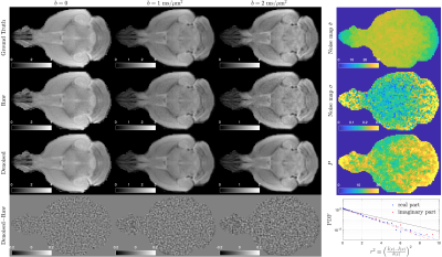 |
Universal Sampling Denoising (USD) for noise mapping and noise removal of non-Cartesian MRI
Hong-Hsi Lee1, Els Fieremans1, Jiangyang Zhang1, and Dmitry S Novikov1
1New York University School of Medicine, New York, NY, United States
Non-Cartesian MRI enables efficient coverage in k-space and is leveraged to accelerate acquisitions of images in multiple contrasts. However, denoising such data is non-trivial, since the noise statistics is neither independent nor normally distributed in reconstructed images. Here, we propose a random-matrix-theory-based denoising and noise-mapping pipeline applicable to MRI of any non-Cartesian k-space sampling. We demonstrate the denoising pipeline on diffusion MRI data, including a numerical phantom and ex vivo mouse brain data in radial trajectories. The proposed pipeline robustly estimates the noise level, removes the noise, and corrects the bias in parametric maps of diffusion and kurtosis metrics.
|
|
0108.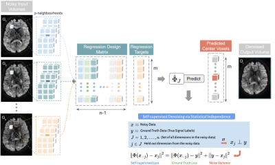 |
Patch2Self denoising reveals a new theoretical understanding of Diffusion MRI
Shreyas Fadnavis1, Joshua Batson2, and Eleftherios Garyfallidis3
1Intelligent Systems Engineering, Indiana University Bloomington, Bloomington, IN, United States, 2Chan Zuckerberg Biohub, San Francisco, CA, United States, 3Indiana University Bloomington, Bloomington, IN, United States
Diffusion MRI (dMRI) is a promising tool for evaluating the spinal cord in health and disease, however low SNR can impede accurate, repeatable, quantitative measurements. Here, we apply a recently proposed denoiser, Patch2Self, that strictly suppresses statistically independent random fluctuations in the signal originating from various sources of noise. Typical spinal cord dMRI scans have a smaller number of gradient directions (10-20) making PCA based 4D denoisers (require at least 30) inapplicable. Using self-supervised learning, Patch2Self addresses these issues which we quantitatively show with an improvement in repeatability and conspicuity of pathology in the spinal cord.
|
||
0109.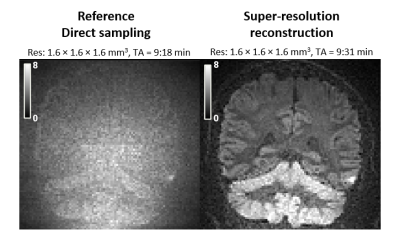 |
High-resolution visualization of isotropically restricted diffusion in brain by strong spherical dMRI and super-resolution reconstruction
Geraline Vis1, Markus Nilsson1, and Filip Szczepankiewicz1
1Diagnostic Radiology, Clinical Sciences Lund, Lund University, Lund, Sweden
Strong spherical diffusion encoding enables visualization of isotropically restricted regions in the human brain, believed to resemble densely packed cells. As this forces imaging in a low SNR regime, visualization has been limited to a low resolution to avoid deleterious signal bias caused by the noise floor. In this work, we propose a novel method based on super-resolution reconstruction to enable high-resolution visualization of isotropically restricted diffusion in human brain in vivo. We show that our method is superior over conventional methods with acquisition times that are compatible with clinical routine.
|
||
0110.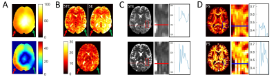 |
SNR efficiency and effectiveness of 7T high-b diffusion imaging with MESMERISED and PGSE
Alard Roebroeck1, Benedikt A Poser1, and Francisco J Fritz2
1Department of Cognitive Neuroscience, Faculty of Psychology & Neuroscience, Maastricht University, Maastricht, Netherlands, 2Department of Systems Neurosciences, University Medical Center Hamburg-Eppendorf, Hamburg, Germany, Hamburg, Germany
We compare MESMERISED and standard PGSE for their signal-to-noise efficiency in b-value = 7000 s/mm2 (b7k) dMRI at 7T and their effectiveness in supporting microstructure modeling. At current state-of-the-art gradient performance, MESMERISED’s SAR and gradient duty cycle efficiency allows it to outperform PGSE for 7T high-b dMRI and provide highly efficient and effective microstructure modeling. Its added capacity to explore diffusion times, super-accelerate quantitative T1 mapping, and also perform qT2 and B1+ mapping, makes it highly useful for quantitative multi-contrast and diffusion MRI.
|
||
0111.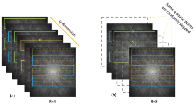 |
Incoherent k-q Under-sampled Multi-shot EPI for Accelerated Multi-shell Diffusion MRI with Model-based Deep Learning Reconstruction
Merry Mani1, Vincent Magnotta2, and Mathews Jacob2
1Radiology, University of Iowa, Iowa City, IA, United States, 2University of Iowa, Iowa City, IA, United States
We propose a new acceleration and reconstruction method for under-sampled multi-shot multi-shell dMRI. The method makes use of incoherent under-sampling in the joint k-q domain to achieve high acceleration. We develop a new model-based reconstruction that jointly recovers missing q-space points, by utilizing a q-space manifold prior that is pre-learned using deep learning. The proposed method is shown to accurately recover the DWIs from 8-fold accelerated multi-shell data. The reconstruction error is shown to be less than 3%. The proposed method enables utilization of multi-shot EPI trajectories for diffusion microstructure and connectivity studies requiring multi-shell coverage, without prolonging scan time.
|
||
|
0112.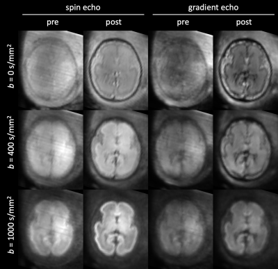 |
Combined spin echo and gradient echo slice-to-volume reconstruction in fetal diffusion MRI
Daan Christiaens1,2, Maximilian Pietsch1, Lucilio Cordero-Grande1, Anthony N Price1,3, Jana Hutter1,3, Emer Hughes1, Serena J Counsell1, Joseph V Hajnal1,3, and J-Donald Tournier1,3
1Centre for the Developing Brain, School of Biomedical Engineering and Imaging Sciences, King's College London, London, United Kingdom, 2Department of Electrical Engineering (ESAT-PSI), KU Leuven, Leuven, Belgium, 3Biomedical Engineering Department, School of Biomedical Engineering and Imaging Sciences, King's College London, London, United Kingdom
Modelling tissue microstructure during fetal and neonatal brain development relies on robustly sampling diverse, low-SNR image contrast in moving subjects. Here, we extend slice-to-volume reconstruction to multi-echo diffusion MRI, in order to correct subject motion across the combined diffusion-T2*-weighted signal. Our results suggest that the spin echo and gradient echo dMRI signal have different trends across gestational age. Tissue microstructure models could therefore leverage these multidimensional data to reveal new insights in brain development.
|
The International Society for Magnetic Resonance in Medicine is accredited by the Accreditation Council for Continuing Medical Education to provide continuing medical education for physicians.