Oral
Arterial Spin Labelling
ISMRM & SMRT Annual Meeting • 15-20 May 2021

| Concurrent 4 | 12:00 - 14:00 | Moderators: Jan Petr & Lena Vaclavu |
 |
0467.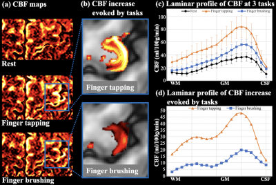 |
Layer-dependent 7T ASL reveals sensory input and motor output perfusion activity in human primary motor cortex
Xingfeng Shao1, Fanhua Guo2, Qinyang Shou1, Kai Wang1, Lirong Yan1,3, Kay Jann1,3, Peng Zhang2, and Danny JJ Wang1,3
1Laboratory of FMRI Technology (LOFT), Mark & Mary Stevens Neuroimaging and Informatics Institute, Keck School of Medicine, University of Southern California, Los Angeles, CA, United States, 2State Key Laboratory of Brain and Cognitive Science, Beijing MRI Center for Brain Research, Institute of Biophysics, Chinese Academy of Sciences, Beijing, China, 3Department of Neurology, University of Southern California, Los Angeles, CA, United States
High-resolution (iso-1mm) 7T ASL scans were performed on the primary motor cortex (M1) to characterize layer-dependent resting CBF, and perfusion activity to sensory input/motor output. Finger tapping (FT)-induced CBF increase shows a clear ‘double-peak’ pattern, consistent with the hypothesis that FT engaged neural activity of somatosensory input in the superficial layers and motor output in the deep layers. Finger brushing (FB)-induced CBF increase was overall smaller, and mainly peaked in superficial layers (somatosensory input and minimal motor output). These results demonstrate the high spatial specificity of 7T ASL, capable of resolving layer-dependent input and output activity in human M1.
|
|
 |
0468.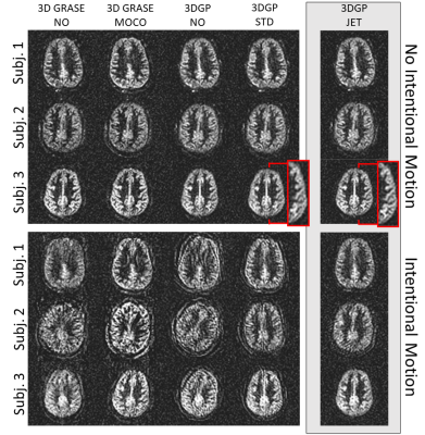 |
Joint Estimation and Correction of Motion and Geometric Distortion in Segmented 3D Arterial Spin Labeling
Jörn Huber1, Daniel Hoinkiss1, and Matthias Günther1,2
1Fraunhofer MEVIS, Bremen, Germany, 2University of Bremen, Bremen, Germany
Assessement of CBF using Arterial Spin Labeling (ASL) can yield valuable functional information regarding different neuropathological diseases like tumors and stroke without exogenous contrast agents. However, being a subtractive technique, ASL shows high sensitivity to motion artifacts. ASL in combination with a 3D GRASE PROPELLER (3DGP) readout allows self-navigated retrospective motion correction but motion estimates using the standard reconstruction are inaccurate due to geometric distortion. In this work, a novel 3DGP reconstruction algorithm is therefore demonstrated which jointly estimates the distortion field as well as motion parameters without the need for additional reference scans.
|
|
0469.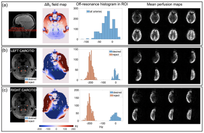 |
Regulating labeling efficiency in arterial spin labeling using a multi-coil B0 shim array: Application to territory mapping
Lincoln Craven-Brightman1, Yulin Chang2, Thomas Witzel3, Nicolas S. Arango4, Meher R. Juttukonda1,5, Luis Hernandez-Garcia6, Marta Vidorreta7, John A. Detre8,9, Lawrence L. Wald5,10, and Jason Stockmann5,10
1Massachusetts General Hospital, Charlestown, MA, United States, 2Siemens Medical Solutions USA, Inc., Malvern, PA, United States, 3Qbio Inc, San Carlos, CA, United States, 4Dept. of Electrical Engineering, Massachusetts Institute of Technology, Cambridge, MA, United States, 5Harvard Medical School, Boston, MA, United States, 6Biomedical Engineering, University of Michigan, Ann Arbor, MI, United States, 7Siemens, S.A., Madrid, Spain, 8Neurology, University of Pennsylvania, Philadelphia, PA, United States, 9Radiology, University of Pennsylvania, Philadelphia, PA, United States, 10A.A. Martinos Center for Biomedical Imaging, Department of Radiology, Massachusetts General Hospital, Charlestown, MA, United States
We apply dynamic local B0 field control with a multi-coil (MC) shim array to improve ASL labeling. Labeling efficiency can be regulated by dynamically shimming the labeling plane during a pCASL tagging pulse train. We demonstrate this capability through territory mapping with an unspecialized MC head shim array – Through shimming, we shift target arteries relative to the labeling pulse bandwidth. Implementation takes less than 10 minutes during a scan, without previous subject information. We also simulate the improvement possible with a specialized MC neck shim array, which enables regulation in more inferior labeling planes and higher field control efficiency.
|
||
|
0470.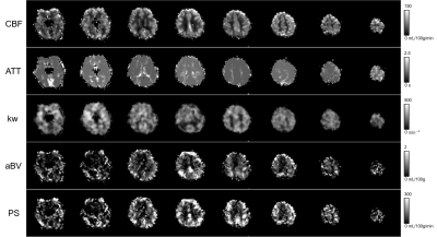 |
Quantification of blood-brain barrier water permeability and arterial blood volume with multi-slice multi-delay diffusion-weighted ASL
Hyun-Seo Ahn1, Jaeseok Park2, Chul Ho Sohn3, and Sung-Hong Park1
1Department of Bio and Brain Engineering, Korea Advnaced Institute of Science and Technology, Daejeon, Korea, Republic of, 2Department of Biomedical Engineering, Sungkyunkwan University, Suwon, Korea, Republic of, 3Department of Radiology, Seoul National University Hospital, Seoul, Korea, Republic of
Measurement of changes in blood-brain barrier permeability is important for early diagnosis of brain diseases. In this study, we propose multi-slice multi-delay diffusion-weighted arterial spin labeling for simultaneous acquisition of various quantitative perfusion estimates including the water exchange rate and the permeability surface area product, which are known to be closely related to BBB permeability, and blood perfusion and arterial blood volume. The water exchange rate in 4 Alzheimer patients were smaller than that in 6 normal subjects, opposed to common knowledges on BBB permeability. The proposed approach may work as a new biomarker for early diagnosis of Alzheimer disease.
|
|
|
0471.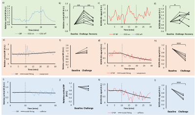 |
phMRI with Simultaneous Measurement of Cerebral Perfusion and Blood-Cerebrospinal Fluid Barrier Function using Interleaved Echo-Time ASL
Charith Perera1, Jack Wells1, Ian Harrison1, David Thomas2,3,4, and Mark Lythgoe1
1Centre for Advanced Biomedical Imaging, University College London, London, United Kingdom, 2Neuroradiological Academic Unit, Department of Brain Repair and Rehabilitation, UCL Queen Square Institute of Neurology, University College London, London, United Kingdom, 3Leonard Wolfson Experimental Neurology Centre, UCL Queen Square Institute of Neurology, University College London, London, United Kingdom, 4Wellcome Centre for Human Neuroimaging, UCL Queen Square Institute of Neurology, University College London, London, United Kingdom
We employed an interleaved short/long echo-time ASL sequence to better understand the differential response of vessels associated with the blood brain barrier (BBB), and the relatively understudied blood-cerebrospinal fluid barrier (BCSFB) to pharmacological perturbation in the healthy and aged brain. We measured changes in both cortical perfusion and the BCSFB-ASL signal in response CO2, caffeine, and vasopressin. Additionally, we demonstrated a marked decrease in BCSFB reactivity towards vasopressin in the aged vs adult brain. Together, these novel data highlight the value of this translational approach to capture simultaneous and differential pharmacological modulation of vessel tone at the BBB and BCSFB.
|
|
0472.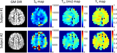 |
Venous Oxygenation Mapping using Fourier-Transform based Velocity-Selective Pulse Trains
Wenbo Li1,2, Peter van Zijl1,2, and Qin Qin1,2
1Radiology Department, Johns Hopkins University School of Medicine, Baltimore, MD, United States, 2Kirby Image Center, Kennedy Krieger Institute, Baltimore, MD, United States
A T2-oximetry method is proposed to map the venous oxygenation by using Fourier-transform based velocity-selective inversion plus non-selective inversion to null the arterial blood signal while using Fourier-transform based velocity-selective saturation to suppress the tissue signal. Compared to previous schemes, the proposed method has the benefit of high SNR and insensitivity to arterial transit delays. Using this method, the venous oxygenation values obtained from two volunteers at 3T are similar between gray matter and white matter and comparable to the values measured globally.
|
||
0473.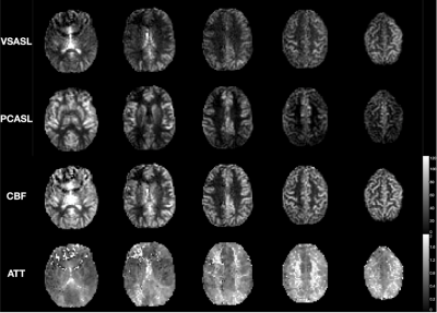 |
VESPA ASL: VElocity and SPAtially selective Arterial Spin Labeling
Joseph G Woods1, Eric C Wong1, Emma Boyd2, and Divya S Bolar1
1Radiology, UCSD, La Jolla, CA, United States, 2Neurosciences, UCSD, La Jolla, CA, United States
Velocity-selective ASL (VSASL) accurately depicts cerebral perfusion, even in regions of severely prolonged arterial transit time (ATT). In contrast, spatially-selective ASL, such as pulsed or pseudo-continuous ASL (PCASL), preserves macrovascular signal of blood flowing to these regions. Recent work highlights the importance of using both methods in a complementary way to more completely assess cerebrovascular pathology. In this study, we describe a novel ASL pulse sequence, dubbed VESPA ASL, in which VSASL and PCASL data are simultaneously acquired within a single scan. We further describe a signal model to quantify cerebral blood flow and ATT from these two data sets.
|
||
|
0474.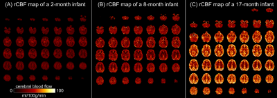 |
Faster regional cerebral blood flow increases in infant heteromodal cortex with 2.5mm3 resolution 3D multi-shot, stack-of-spirals pCASL
Minhui Ouyang1, John Detre2, Chenying Zhao1,3, Samantha Lam1, J. Christopher Edgar1,2, and Hao Huang1,2
1Department of Radiology, The Children's Hospital of Philadelphia, Philadelphia, PA, United States, 2Department of Radiology, Perelman School of Medicine, University of Pennsylvania, Philadelphia, PA, United States, 3Department of Bioengineering, School of Engineering and Applied Science, University of Pennsylvania, Philadelphia, PA, United States
During early infancy, rapid increases of regional cerebral blood flow (rCBF) supports the metabolic needs for the dramatic maturation of infant brains. In this study, we delineated the developmental pattern of infant’s rCBF from 0-18months, with an optimized 3D multi-shot, stack-of-spirals pCASL sequence for high-resolution rCBF at isotropic 2.5mm. Significant age-related rCBF increases were found during this period. The rCBF growth rates were inhomogeneous across the cortex, with faster maturation rates in the heteromodal association cortex and slower in unimodal sensorimotor cortices. Cortical regions with more rapid rCBF growth were associated with faster microstructural maturation of adjacent white matter.
|
|
|
0475.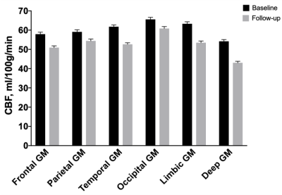 |
Spatiotemporal characteristics of longitudinal changes in cerebral blood flow across the adult lifespan
Hualu Han1,2, Zixuan Lin2,3, Melissa Rundle4, Anja Soldan5, Corinne Pettigrew5, Joshua F. Betz6, Kumiko Oishi7, Yang Li2, Binu P. Thomas8, Peiying Liu2, Marilyn Albert5, Denise Park4, and Hanzhang Lu2,3,9
1Center for Biomedical Imaging Research, Department of Biomedical Engineering, School of Medicine, Tsinghua University, Beijing, China, 2The Russell H. Morgan Department of Radiology & Radiological Science, Johns Hopkins University School of Medicine, Baltimore, MD, United States, 3Department of Biomedical Engineering, Johns Hopkins University School of Medicine, Baltimore, MD, United States, 4Center for Vital Longevity, School of Behavioral and Brain Sciences, University of Texas at Dallas, Dallas, TX, United States, 5Department of Neurology, Johns Hopkins University School of Medicine, Baltimore, MD, United States, 6Department of Biostatistics, Johns Hopkins Bloomberg School of Public Health, Baltimore, MD, United States, 7Center for Imaging Science, Johns Hopkins University, Whiting School of Engineering, Baltimore, MD, United States, 8Advanced Imaging Research Center, University of Texas Southwestern Medical Center, Dallas, TX, United States, 9F.M. Kirby Center for Functional Brain Imaging, Kennedy Krieger Institute, Baltimore, MD, United States
Characterization of age-related changes in blood supply is important in understanding brain aging. The present work reports longitudinal studies of age-related changes in cerebral blood flow (CBF) in two separate cognitively-healthy cohorts using complementary MRI techniques. We found that CBF decreased with age and the longitudinal rate of decline was faster than that from the cross-sectional data. The rate of CBF reduction was faster in younger than in older individuals, in contrast to the temporal pattern of brain volume atrophy. There were also significant spatial differences and hemispheric asymmetry in CBF decline rates.
|
|
0476.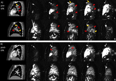 |
Quantification of arterial obstruction in pediatric patients with pulmonary embolism using arterial spin labeled perfusion MRI of the lungs
Joshua S Greer1,2,3, Mubeena Abdulkarim1, Gerald F Greil1,3, Ayesha Zia1, Ananth J Madhuranthakam2,3, and Tarique Hussain1,2
1Pediatrics, UT Southwestern Medical Center, Dallas, TX, United States, 2Radiology, UT Southwestern Medical Center, Dallas, TX, United States, 3Advanced Imaging Research Center, UT Southwestern Medical Center, Dallas, TX, United States
In this study, pulmonary perfusion imaging using arterial spin labeling (ASL) was demonstrated in pediatric patients with pulmonary embolism. A method to quantify pulmonary vascular obstruction was proposed using ASL to estimate improvements in pulmonary perfusion following treatment, which moderately agreed with obstruction measured by CTA. Perfusion defects were successfully detected in all patients. A follow-up ASL scan also showed significantly improved perfusion in a patient following treatment, and a few patients had residual perfusion defects in ASL images that were not seen by CTA, suggesting that perfusion to the microvasculature was not immediately restored following resolution of the emboli.
|
The International Society for Magnetic Resonance in Medicine is accredited by the Accreditation Council for Continuing Medical Education to provide continuing medical education for physicians.