Oral
Systems Engineering I
ISMRM & SMRT Annual Meeting • 15-20 May 2021

| Concurrent 2 | 16:00 - 18:00 | Moderators: J Hadley & Maxim Zaitsev |
0559.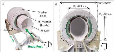 |
A portable brain MRI scanner based on a 72 mT, 35 kg "Halbach-bulb" magnet and external gradient coil
Clarissa Zimmerman Cooley1,2, Jason Stockmann1,2, and Lawrence L Wald1,2,3
1Athinoula A. Martinos Center for Biomedical Imaging, Massachusetts General Hospital, Charlestown, MA, United States, 2Harvard Medical School, Boston, MA, United States, 3Harvard–MIT Division of Health Sciences and Technology, Cambridge, MA, United States
There is clear motivation for increasing the accessibility of MRI, particularly for brain imaging. To address this, we present a portable tight-fitting MRI scanner with a geometry that is optimized for head. We use a 35kg “Halbach-bulb” permanent magnet design that combines a Halbach sphere and Halbach cylinder. The gradient coils are designed on the outer surface of the magnet, enabling a closer-fit B0 magnet. The inhomogeneous B0 magnet results image distortion, which we correct using a generalized reconstruction algorithm that employs measured field maps. We present the full scanner system and in vivo 3D images with resolution 1.5mmx2.7mmx7mm.
|
||
0560.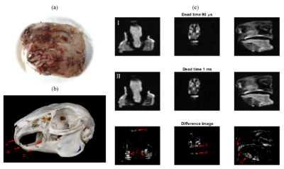 |
Simultaneous imaging of hard and soft biological tissues in a low-field MRI scanner
José Miguel Algarín1, Elena Díaz2, Pepe Borreguero1, Fernando Galve1, Daniel Grau2, Juan Pablo Rigla2, Rubén Bosch1, José Manuel González2, Eduardo Pallás1, Miguel Corberán1, Carlos Gramage1, Alfonso Ríos2, José María Benlloch1,
and Joseba Alonso1
1I3M, CSIC, Valencia, Spain, 2Tesoro Imaging, Valencia, Spain
Here we present the first demonstration of dental imaging in a low cost MRI setup at sub-Tesla fields (260 mT). We show 3D reconstructions of a rabbit head and human teeth acquired in “DentMRI – Gen I” (Fig. 1(a)), a home-made special-purpose MRI scanner designed with the goal of demonstrating dental imaging at low field strengths. We use two variations of zero echo time (ZTE) pulse sequences (Fig. 1(b)): standard PETRA , and Double Radial Non Stop Spin Echo (DRaNSSE), which we have devised to address limitations we find with PETRA. We reconstruct images by Algebraic Reconstruction Techniques (ART).
|
||
0561.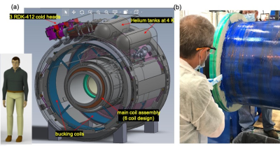 |
Design and Construction of a Low-Cryogen, Lightweight, High-Performance, Head-only Compact 7T MRI
Thomas K.F. Foo1, Mark Vermilyea2, Minfeng Xu2, Anbo Wu2, Yihe Hua2, Wolfgang Stautner2, Ye Bai2, Justin Ricci2, Doug Kelley3, John III Huston4, Yunhong Shu4, Matt A Bernstein4, Christopher Hess5, and Duan Xu5
1GE Global Research, Niskayuna, NY, United States, 2GE Research, Niskayuna, NY, United States, 3GE Healthcare, Fairfax, CA, United States, 4Mayo Clinic, Rochester, MN, United States, 5University of California - San Francisco, San Francisco, CA, United States
A design for a lightweight, low-cryogen, head-only 7T MRI system was completed with its construction underway. This new 7T system is a dedicated brain scanner with 140 mT/m and 820 T/m/s gradients and weighs about 8 tons with 18 liters of liquid helium. The fringe field is similar to the 5 Gauss limits of a clinical whole-body 3T MRI scanner.
|
||
0562.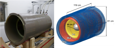 |
Design and Development of a Next-Generation 7T human brain scanner with high-performance gradient coil and dense RF arrays.
David A Feinberg1,2,3, Peter Dietz4, Chunlei Liu1,5, Kawin Setsompop6, Pratik Mukherjee7,8, Lawrence L Wald9,10,11, An T Vu7,8, Alexander JS Beckett1,2, Ignacio Gonzalez Insua4, Martin Schröder4, Stefan Stocker4, Paul H Bell12, Elmar Rummert4,
and Mathias Davids9,10,13
1Helen Wills Neuroscience Institute, University of California, Berkeley, CA, United States, 2Advanced MRI Technologies, Sebastopol, CA, United States, 3Department of Cognitive Neuroscience, Maastricht University, Maastricht, Netherlands, 4Siemens Healthcare GmbH, Erlangen, Germany, 5Department of Electrical Engineering and Computer Sciences, University of California, Berkeley, CA, United States, 6Radiological Sciences Laboratory, Stanford University, Stanford, CA, United States, 7Radiology, University of California, San Francisco, CA, United States, 8San Francisco Veteran Affairs Health Care System, San Francisco, CA, United States, 9A.A. Martinos Center for Biomedical Imaging, Dept. of Radiology, Massachusetts General Hospital, Charlestown, MA, United States, 10Harvard Medical School, Boston, MA, United States, 11Harvard-MIT Division of Health Sciences Technology, Cambridge, MA, United States, 12Siemens Medical Solutions USA, Inc, Cary, NC, United States, 13Computer Assisted Clinical Medicine, Medical Faculty Mannheim, Heidelberg University, Heidelberg, Germany
A Next-Generation 7T MRI scanner was designed to achieve higher performance in human brain imaging for the NIH BRAIN Initiative. The new 7T scanner introduces several innovative hardware designs including a PNS optimized asymmetric head gradient coil (Siemens Healthcare, Erlangen, Germany) with Gmax 200mT/m and Smax 900T/m/s. The scanner also includes 128-channel RF receiver and 16-channel transmit systems for all-in-one cumulative gains in performance. The resulting higher spatial resolution, sensitivity and image acceleration will enable in-vivo whole-brain mesoscale research on cortical layer and columnar circuitry and other brain structures.
|
||
0563.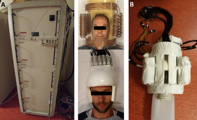 |
Towards 8ch multi transmit with high power ultrasonic spirals and 72ch receive setup for ultimate spatial encoding at 7T
Dimitri Welting1, Edwin Versteeg1, Ingmar Voogt2, Joost van Straalen3, Martijn Heintges3, Marco Rietveld3, Jeroen C.W. Siero1,4, and Dennis W.J. Klomp1
1Radiology, University Medical Center Utrecht, Utrecht, Netherlands, 2Wavetronica, Utrecht, Netherlands, 3Prodive Technologies, Son, Netherlands, 4Spinoza Centre for Neuroimaging Amsterdam, Amsterdam, Netherlands
Here we propose a setup to boost spatiotemporal encoding for fMRI in the human brain using high dense receiver arrays (72ch) and fast gradients (6000T/m/s). Moreover, we demonstrate that the operation frequency of a high-end gradient amplifier can be increased to ultrasonic frequencies so to avoid unpleasant acoustic noise. By using this amplifier with 8 transceivers, 64 receivers and a 2-axis cooled gradient coil, a light weight setup is constructed for operation in a 7 Tesla MRI system for fMRI experiments in the human brain.
|
||
 |
0564.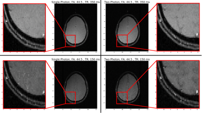 |
In vivo Two-photon Magnetic Resonance Imaging of Human Brain at 3T
Jianshu Chi1, Victor Han1, and Chunlei Liu1,2
1Electrical Engineering and Computer Sciences, University of California, Berkeley, Berkeley, CA, United States, 2Helen Wills Neuroscience Institute, University of California, Berkeley, Berkeley, CA, United States
We are exploring in vivo brain imaging with two-photon excitation on a 3T scanner. To do that, we built a customized coil that is large enough to fit a standard birdcage transceiver coil inside of it. The resulting two-photon gradient-echo brain images excited using a 25.5 kHz z-axis RF were overall similar to standard single-photon images with equivalent parameters. Interestingly, we observed some slight differences in tissue contrast whose cause are being investigated.
|
|
0565.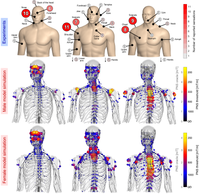 |
PNS optimization of a high-performance asymmetric gradient coil for head imaging
Mathias Davids1,2,3, Peter Dietz4, Gudrun Ruyters4, Manuela Roesler4, Valerie Klein1,3, Bastien Guerin1,2, David A Feinberg5,6, and Lawrence L Wald1,2,7
1Martinos Center for Biomedical Imaging, Boston, MA, United States, 2Harvard Medical School, Boston, MA, United States, 3Computer Assisted Clinical Medicine, Medical Faculty Mannheim, Heidelberg University, Mannheim, Germany, 4Siemens Healthineers, Erlangen, Germany, 5Advanced MRI Technologies, Sebastopol, CA, United States, 6Brain Imaging Center and Helen Wills Neuroscience Institute, University of California, Berkeley, CA, United States, 7Harvard-MIT, Division of Health Sciences and Technology, Cambridge, MA, United States
PNS modeling was utilized during the design stage of a high-strength (Gmax = 200mT/m, Smax = 900 T/m/s) head gradient for 7T fMRI research. The design-stage preview of PNS thresholds and locations allowed alteration of the winding pattern to balance head and body stimulation. This process yielded significantly improved PNS thresholds and increased usability of the coil performance space. The results were validated using PNS experiments in a constructed coil.
|
||
0566.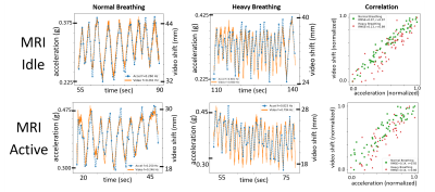 |
Wireless Body Sensor Data Acquisition Platform for Motion Tracking
Leanna Pancoast1,2, Douglas Brantner1,2, Roy Wiggins1,2, Jerzy Walczyk1,2, and Ryan Brown1,2
1Center for Biomedical Imaging, NYU Grossman School of Medicine, New York City, NY, United States, 2Center for Advanced Imaging Innovation and Research, NYU Grossman School of Medicine, New York City, NY, United States
The temporal resolution in MRI can be too slow to track respiratory or bulk subject motion. Auxiliary sensors have been developed to track motion with high temporal resolution, but can require cumbersome cabling. In this work, we describe an MRI-compatible wireless accelerometer data acquisition platform and demonstrate proof-of-concept by correlating respiratory motion data with independent, ground-truth optical measurements.
|
||
|
0567.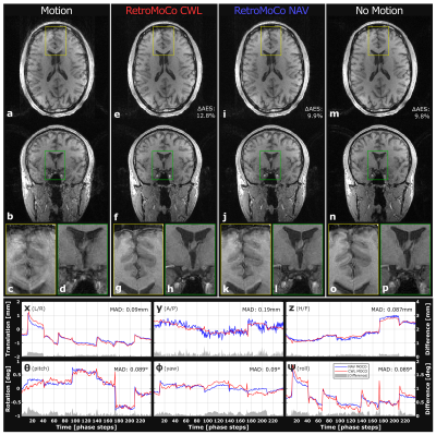 |
Head Motion Tracking using an EEG-system and Retrospective Correction of High-Resolution T1-weighted MRI
Malte Laustsen1,2,3, Mads Andersen4, Rong Xue3,5,6, Kristoffer H. Madsen2,7, and Lars G. Hanson1,2
1Section for Magnetic Resonance, DTU Health Tech, Technical University of Denmark, Kgs. Lyngby, Denmark, 2Danish Research Centre for Magnetic Resonance, Centre for Functional and Diagnostic Imaging and Research, Copenhagen University Hospital Hvidovre, Hvidovre, Denmark, 3Sino-Danish Center, University of Chinese Academy of Sciences, Beijing, China, 4Philips Healthcare, Copenhagen, Denmark, 5State Key Laboratory of Brain and Cognitive Science, Beijing MR Center for Brain Research, Institute of Biophysics, Chinese Academy of Sciences, Beijing, China, 6Beijing Institute for Brain Disorders, Beijing, China, 7DTU Compute, Technical University of Denmark, Kgs. Lyngby, Denmark
Tracking and retrospective correction of high-resolution structural 3D-GRE images is accomplished with a slightly modified EEG cap and sampling system. Carbon wire loops added to the EEG cap allow for motion tracking using gradient-induced signals from native sequence elements, without the need for sequence modification, or electrode-skin contact, while requiring only a short calibration scan, and mounting of the cap. The motion estimates closely resemble estimates from interleaved navigators (mean absolute difference: [0.13,0.33,0.12]mm, [0.28,0.15,0.22]deg). Retrospective correction using carbon wire loops yield similar improvements to Average Edge Strength (12%) for images with instructed movement, and does not degrade images without motion.
|
|
 |
0568.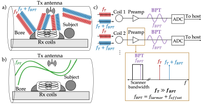 |
Beat Pilot Tone: Exploiting Preamplifier Intermodulation of UHF/SHF RF for Improved Motion Sensitivity over Pilot Tone Navigators
Suma Anand1 and Michael Lustig1
1Electrical Engineering and Computer Sciences, University of California, Berkeley, Berkeley, CA, United States
Pilot Tone (PT) navigators are tones within the MR receiver bandwidth that are used to estimate subject motion. Unfortunately, PTs are bound to the Larmor frequency with a wavelength of 1-4.7m (7T to 1.5T), limiting their sensitivity to motion. We propose a new approach, Beat Pilot Tone (BPT), which overcomes this limitation using second order intermodulation in MR coil preamplifiers (preamps). Any two tones separated by the desired PT frequency are mixed at the preamp and digitized by the receiver. We demonstrate our approach at 2.4GHz, obtaining improved sensitivity (20x) compared to PT without reduction of image SNR.
|
The International Society for Magnetic Resonance in Medicine is accredited by the Accreditation Council for Continuing Medical Education to provide continuing medical education for physicians.