Oral
Safety: Hitting a Nerve?
ISMRM & SMRT Annual Meeting • 15-20 May 2021

| Concurrent 4 | 18:00 - 20:00 | Moderators: Christoph Aigner & Natalia Gudino |
 |
0407.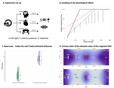 |
Going Below The Neck: Physiological limits on use of 300 mT/m gradients in the human body
Malwina Molendowska1, Fabrizio Fasano2,3, Umesh Rudrapatna1, Ralph Kimmlingen3, Derek K. Jones1,4, Slawomir Kusmia1, Chantal M. W. Tax1,5, and C. John Evans1
1Cardiff University Brain Research Imaging Centre (CUBRIC), Cardiff University, Cardiff, United Kingdom, 2Siemens Healthcare Ltd, Camberly, United Kingdom, 3Siemens Healthcare GmbH, Erlangen, Germany, 4Mary McKillop Institute for Health Research, Faculty of Health Sciences, Australian Catholic University, Melbourne, Australia, 5Image Sciences Institute, University Medical Center Utrecht, Utrecht, Netherlands
Increasing-available ultra-strong gradient systems introduce a new challenge in MRI: the unknown interaction of time-varying magnetic fields with the human body. We characterise the physiological effects of deploying 300 mT/m gradients Siemens Connectom system when imaging regions below the neck (i.e., heart and prostate). We show gradient amplitude thresholds for PNS (ramp times < 2ms) are first reached on the Y-gradient. Moreover, landmarking on the heart gives the highest probability of generating magnetophosphenes (all gradient-axes). This study establishes: (i) limitations in greatest system performance; and (ii) that the so-far head-only Connectom system can be safely used in ‘below-the-neck’ applications.
|
|
0408.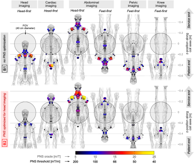 |
Fast PNS characterization of MRI gradient coils using a Huygens’ PNS model: Application to multiple patient positions and orientations
Mathias Davids1,2,3, Bastien Guerin1,2, and Lawrence L Wald1,2,4
1Martinos Center for Biomedical Imaging, Boston, MA, United States, 2Harvard Medical School, Boston, MA, United States, 3Computer Assisted Clinical Medicine, Mannheim, Germany, 4Harvard-MIT, Division of Health Sciences and Technology, Cambridge, MA, United States
We apply a fast PNS model evaluated on a Huygens’ surface to characterize PNS performance as a function of patient position and pose in previously described PNS optimized gradients. The coils were initially PNS-optimized for a female body model in a standard brain imaging position. The Huygens’ approach allows us to assess other body positions and demonstrate the PNS benefits/penalties associated with other imaging applications and in both a female and male body model by dramatically speeding up the PNS characterization (to a couple of seconds per body position/orientation). The findings support a broad benefit from the PNS optimized windings.
|
||
0409.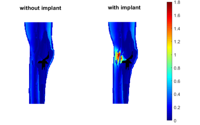 |
Enhancement of PNS risk in the presence of a metallic knee prosthesis
Luca Zilberti1, Alessandro Arduino1, Riccardo Torchio2, Umberto Zanovello1, Fabio Baruffaldi3, Paolo Bettini2, Piergiorgio Alotto2, Mario Chiampi1, and Oriano Bottauscio1
1Istituto Nazionale di Ricerca Metrologica, Torino, Italy, 2Dipartimento Ingegneria Industriale, Università degli Studi di Padova, Padova, Italy, 3Istituto Ortopedico Rizzoli, Bologna, Italy
Peripheral nerve stimulation is a typical source of concern in MRI, which, in principle, could be significantly affected by the presence of a foreign object, like an orthopaedic prosthesis, in the body. This work investigates this possibility through a computational approach, making use of a human model where a knee implant has been placed, realistically, through a “virtual surgery”. Results indicate that the presence of the prosthesis can give rise to local enhancement of the electric field induced by the operation of the gradient coils, hence increasing the risk.
|
||
0410.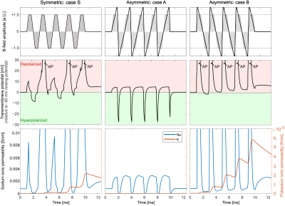 |
Exploiting Nerve Membrane Dynamics to Reduce Peripheral Nerve Stimulation using Asymmetric Readout Gradient Waveforms
Natalie G Ferris1,2, Mathias Davids3,4,5, Valerie Klein3,5, Bastien Guérin3,4, and Lawrence L Wald3,4
1Harvard Graduate Program in Biophysics, Harvard University, Cambridge, MA, United States, 2Harvard-MIT Division of Health Sciences and Technology, MIT, Cambridge, MA, United States, 3A. A. Martinos Center for Biomedical Imaging, Department of Radiology, Massachusetts General Hospital, Charlestown, MA, United States, 4Harvard Medical School, Boston, MA, United States, 5Computer Assisted Clinical Medicine, Heidelberg University, Heidelberg, Germany
Peripheral Nerve Stimulation (PNS) limits the use of high-performance gradient systems. We exploit nerve membrane hyperpolarization and depolarization in readout gradient waveform design to achieve a higher gradient moment (area) in a given time without initiating PNS. In a typical symmetric trapezoidal readout waveform, the slew ramps have identical rise times but opposite dB/dt (E-field sign). Asymmetrizing the waveform enables achieving the same gradient moment while emphasizing membrane hyperpolarization over depolarization. The disparate effects of hyperpolarizing and depolarizing pulses are sufficient to impact the nerve’s PNS threshold, potentially increasing the spatial resolution.
|
||
|
0411.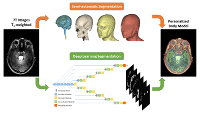 |
Numerical Body Model Inference for Personalized RF Exposure Prediction in Neuroimaging at 7T
Wyger Brink1, Sahar Yousefi1,2, Prernna Bhatnagar1, Marius Staring2, Rob Remis3, and Andrew Webb1
1C.J. Gorter Center, dept. of Radiology, Leiden University Medical Center, Leiden, Netherlands, 2Division of Image Processing, dept. of Radiology, Leiden University Medical Center, Leiden, Netherlands, 3Circuits and Systems, dept. of Microelectronics, Delft University of Technology, Delft, Netherlands
Compliance with RF exposure limits in ultra-high field MRI is typically based on “one-size-fits-all” safety margins to account for the intersubject variability of local SAR. In this work we have developed a semantic segmentation method based on deep learning, which is able to generate a subject-specific body model for personalized RF exposure prediction at 7T.
|
|
 |
0412.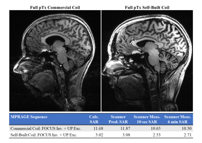 |
SAR Management in pTx Sequence Design: The Impact of Electromagnetic-Field-Derived Virtual Observation Points
Sydney Nicole Williams1, Jürgen Herrler2, Patrick Liebig3, Paul McElhinney1, Shajan Gunamony1,4, Armin M. Nagel5, and David A. Porter1
1Imaging Centre of Excellence, University of Glasgow, Glasgow, United Kingdom, 2Department of Neuroradiology, University Hospital Erlangen, Erlangen, Germany, 3Siemens Healthineers, Erlangen, Germany, 4MR CoilTech Limited, Glasgow, United Kingdom, 5Institute of Radiology, University Hospital Erlangen, Erlangen, Germany
We compare two methods for estimating local SAR with virtual observation points (VOPs) in a commercial and a self-built 8Tx/32Rx head coil, respectively. The commercial coil relies on a constant safety factor that reduces the hardware power limits in each transmit channel, where the self-built coil derives the VOPs from coil electromagnetic field (EMF) simulations. We show the use of both coils in vivo with MPRAGE using Universal and subject-specific pTx pulses. The EMF-based VOPs, used with the self-built coil, resulted in a lower local SAR estimate for a similar image quality, providing more flexibility in pulse sequence design.
|
|
0413.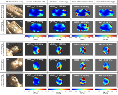 |
Uncertainty Estimation of subject-specific Local SAR assessment by Bayesian Deep Learning
E.F. Meliadò1,2,3, A.J.E. Raaijmakers1,2,4, M. Maspero2,5, M.H.F. Savenije2,5, P.R. Luijten1, and C.A.T. van den Berg2,5
1Department of Radiology, University Medical Center Utrecht, Utrecht, Netherlands, 2Computational Imaging Group for MR diagnostics & therapy, Center for Image Sciences, University Medical Center Utrecht, Utrecht, Netherlands, 3Tesla Dynamic Coils BV, Zaltbommel, Netherlands, 4Biomedical Image Analysis, Dept. Biomedical Engineering, Eindhoven University of Technology, Eindhoven, Netherlands, 5Department of Radiotherapy, Division of Imaging & Oncology, University Medical Center Utrecht, Utrecht, Netherlands
Residual error/uncertainty is always present in the estimated local SAR, therefore it is essential to investigate and understand the magnitude of the main sources of error/uncertainty. Last year we presented a Bayesian deep learning approach to map the relation between subject-specific complex B1+-maps and the corresponding local SAR distribution, and to predict the spatial distribution of uncertainty at the same time. The preliminary results showed the feasibility of the proposed approach. In this study, we show its ability to reliably capture the main sources of uncertainty and detect deviations in the MR examination scenario not included in the training samples.
|
||
|
0414.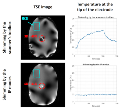 |
Implant-Friendly Excitation Strategies for Imaging DBS Electrodes at 7T
Alireza Sadeghi-Tarakameh1, Lance DelaBarre1, Nur Izzati Huda Zulkarnain1, Noam Harel1, and Yigitcan Eryaman1
1Center for Magnetic Resonance Research (CMRR), University of Minnesota, Minneapolis, MN, United States
We mitigated the radiofrequency heating at the contacts of a DBS electrode by utilizing an implant-friendly (IF) excitation scenario at 7T. IF modes of a 16-channel transmit/receive coil are calculated by minimizing electrode-shaft current close to the contacts. An IF excitation is calculated by shimming the B1+ field in an ROI using individual IF modes of the array. The proposed approach is able to mitigate the shaft current and RF heating at the contacts.
|
|
|
0415.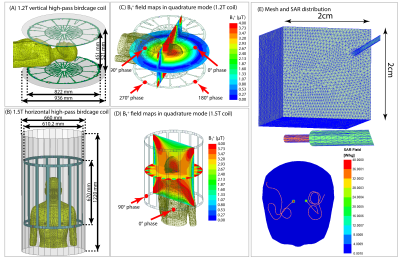 |
Open-bore vertical MRI scanners generate significantly lower RF heating around DBS implants: A Simulation study with experimental validation
Bhumi Bhusal1, Ehsan Kazemivalipour2, Jasmine Vu1, Stella LIn1, Bach Thanh Nguyen1, John Kirsch3, Elizabeth Nowac4, Julie Pilitsis5, Joshua Rosenow1, Ergin Atalar2, and Laleh Golestanirad1
1Northwestern University, Chicago, IL, United States, 2Bilkent University, Ankara, Turkey, 3Massachusetts General Hospital, Charlestown, MA, United States, 4Illinois Bone and Joint Institute, Wilmette, IL, United States, 5Albany Medical College, Albany, NY, United States
Though there are many studies reporting RF heating of implants in horizontal MRI scanners, there is almost no literature on vertical scanners that have 90° rotated transmit coils and a fundamentally different distribution of RF fields. Here we evaluate RF heating of deep brain stimulation (DBS) implants during MRI in a 1.2T open-bore vertical scanner compared to a 1.5T horizontal system with both numerical simulations and experimental measurements. We found a significant reduction in RF heating using vertical vs horizontal RF coils which are attributable to the orthogonal orientation of RF electric fields.
|
|
0416.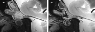 |
The Feasibility of Radiofrequency Rhizotomy Lesion Visualization in the Trigeminal Ganglion using 7.0-Tesla MRI
Kellen Mulford1, David Darrow2, Sean Moen2, Samuel Ndoro2, Bharathi D. Jagadeesan3, Andrew W. Grande2, Donald R. Nixdorf4, and Pierre-Francois Van de Moortele1
1Center for Magnetic Resonance Imaging, University of Minnesota, Minneapolis, MN, United States, 2Department of Neurosurgery, University of Minnesota, Minneapolis, MN, United States, 3Department of Radiology, University of Minnesota, Minneapolis, MN, United States, 4Department of Diagnostic and Biological Science, University of Minnesota, Minneapolis, MN, United States
Pain recurrence following invasive treatments for trigeminal neuralgia is common, yet the treatment decisions in these situations lack objective guidance. The goal of this work was to establish the feasibility of using 7.0-Tesla MRI to visualize treatment related effects from percutaneous radiofrequency rhizotomy procedures. We scanned two patients with trigeminal neuralgia and one human cadaver specimen at 7.0-Tesla after radiofrequency rhizotomy procedures. Both acute and long-term treatment related effects were successfully visualized by our protocol. Future work will recruit a large cohort of trigeminal neuralgia patients to correlate imaging findings with clinical outcomes.
|
The International Society for Magnetic Resonance in Medicine is accredited by the Accreditation Council for Continuing Medical Education to provide continuing medical education for physicians.