Oral
RF Design II
ISMRM & SMRT Annual Meeting • 15-20 May 2021

| Concurrent 2 | 18:00 - 20:00 | Moderators: Alexander Raaijmakers & Pierre-Francois Van de Moortele |
 |
0175.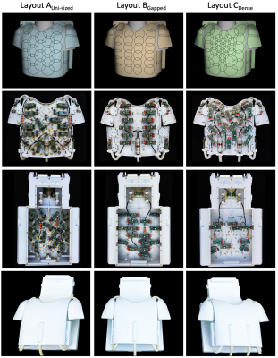 |
Coil Design Impacts Image Encoding: Optimized 64-Channel Array Configurations for Diffusion-Weighted Imaging in 3T Cardiac MRI
Robin Etzel1,2, Choukri Mekkaoui3, Ekaterina S Ivshina4, Alina Scholz1, Markus W May1, Nicolas Kutscha1, Matthäus Poniatowski1, Chaimaa Chemlali1, Anpreet Ghotra1, Sam-Luca JD Hansen1, Timothy G Reese3, Lawrence L Wald3, Andreas H Mahnken2,
and Boris Keil1
1Institute of Medical Physics and Radiation Protection, Department of Life Science Engineering, TH Mittelhessen University of Applied Sciences, Giessen, Germany, 2Philipps-University of Marburg, Department of Diagnostic and Interventional Radiology, Marburg, Germany, 3Harvard Medical School, Massachusetts General Hospital, Department of Radiology, A.A. Martinos Center for Biomedical Imaging, Boston, MA, United States, 4Princeton University, Princeton, NJ, United States
To determine the optimum configuration of a 64-channel array coil for highly accelerated in vivo cardiac diffusion-weighted imaging (DWI), the encoding capability and sensitivity of three 64-channel arrays were designed, constructed, and evaluated. Simultaneous multi-slice (SMS) acceleration enabled enhanced efficiency of the diffusion acquisitions. The array configuration with a non-uniformly distributed loop density showed the most favorable cardiac imaging performance in both SNR and SMS encoding power. Cardiac DWI of a healthy volunteer mirrors the findings from phantom evaluation and testing. Such technically advanced coils are an important component for translation of cardiac DWI to clinical settings.
|
|
|
0176.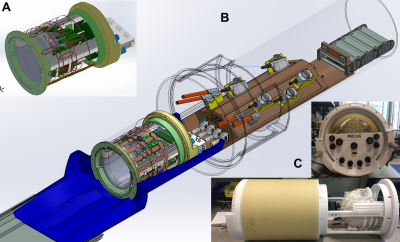 |
A 128-Channel head coil array for Cortical Imaging at 7 Tesla
Bernhard Gruber1,2, Jason P. Stockmann1, Azma Mareyam1, Boris Keil3, Anpreet Ghotra3, David A. Feinberg4, and Lawrence L. Wald1
1Department of Radiology, A.A. Martinos Center for Biomedical Imaging, Massachusetts General Hospital, Harvard Medical School, Charlestown, MA, United States, 2High Field MR Center, Center for Medical Physics and Biomedical Engineering, Medical University Vienna, Vienna, Austria, 3Department of Life Science Engineering, Institute of Medical Physics and Radiation Protection, Mittelhessen University of Applied Science, Giessen, Germany, 4Helen Wills Neuroscience Institute, University of California, Berkeley, Berkeley, CA, United States
A 128-channel receive array for cortical brain imaging at 7T was simulated and constructed. The tight-fitting brain-only coil was designed for use with a recently constructed head gradient system (Gmax = 200 mT/m and Smax = 900mT/m/s) for use with either a single channel birdcage Tx or a 24-channel pTx coil. The goal was to maximize cortical SNR and achieve the high acceleration needed for single-shot EPI based fMRI at high sub-millimeter isotropic resolution.
|
|
0177.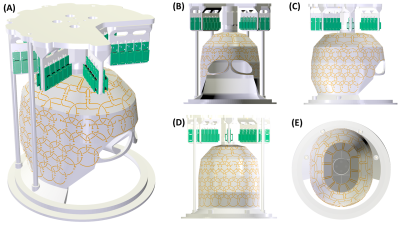 |
A 128-channel receive array for 10.5T human head imaging
Russell Luke Lagore1, Steve Jungst1, Jerahmie Radder1, Edward J Auerbach1, Steen Moeller1, Andrea Grant1, Lance DelaBarre1, Matthew Waks1, Pierre-Francois Van de Moortele1, Gregor Adriany1, and Kamil Ugurbil1
1Center for Magnetic Resonance Research, University of Minnesota, Minneapolis, MN, United States
A 128-channel receive array has been designed and prototyped for human head imaging at 10.5T. The coil employs miniaturized electronics and flexible PCB laminates for loop conductors. Bench measurements and MR experimental results for an 8-channel cluster of loops are presented which show low coil coupling and good stability during echo-planar imaging. Electromagnetic simulations will provide an estimate for the SNR gain that can be expected from this 128-channel receive array over a 64-channel receive array tuned to the same frequency. This simulation estimate will be verified with experimental results which will include intrinsic SNR and g-factor maps.
|
||
 |
0178.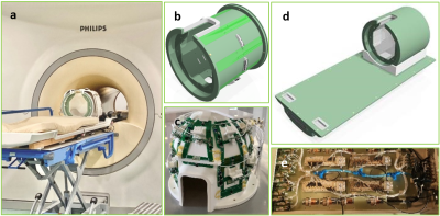 |
A Quintuple-Tuned RF Coil for Whole Brain Multi-Nuclei Magnetic Resonance Imaging and Spectroscopy at 7T
Jiying Dai1,2, Tijl A. van der Velden1, Johannes M. Hoogduin1, Fabian Bartel2, Ettore F Meliadò1,2, Mark van Uden2, Catalina S. Arteaga de Castro2, Evita C. Wiegers1, Martijn Froeling1, Mark Gosselink1, Alexander J. E. Raaijmakers1,3, and Dennis W. J. Klomp1,4
1Radiology, UMC Utrecht, Utrecht, Netherlands, 2Tesla Dynamic Coils, Zaltbommel, Netherlands, 3Biomedical Engineering, Eindhoven University of Technology, Eindhoven, Netherlands, 4Tesla Engineering Ltd, West Sussex, United Kingdom
Multi-nuclei magnetic resonance imaging and spectroscopy are interesting techniques to study metabolism, treatment efficacy tracking, and early diagnoses of many diseases. However, there are challenges to acquire high-quality MR images or spectra of nuclei other than proton, for example, much lower signal level, as well as the inconvenience of switching RF hardware in between scans, which leads to repositioning of the subject and extra preparation time. To overcome the above issues, we developed a quintuple-tuned RF coil for sensitive whole-brain scans, targeting five nuclei: 1H, 19F, 31P, 23Na and 13C. Bench tests and in-vivo scans have shown promising SNR.
|
|
0179.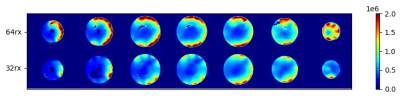 |
A Self-decoupled 64 Channel Receive Array for Human Brain MRI at 10.5T
Nader Tavaf1, Steve Jungst1, Russell L. Lagore1, Jerahmie Radder1, Steen Moeller1, Andrea Grant1, Edward Auerbach1, Kamil Ugurbil1, Gregor Adriany1, and Pierre-Francois Van de Moortele1
1Center for Magnetic Resonance Research (CMRR), University of Minnesota Twin Cities, Minneapolis, MN, United States
Receive arrays play a major role in capturing the signal-to-noise ratio (SNR) and parallel imaging performance improvements at ultrahigh-field MRI. A novel self-decoupled 64-channel receive array was built for human brain imaging and compared to a 32-channel receive array at 10.5T/447MHz. Experiments demonstrated a maximum noise correlation of 0.31 in the 64-channel receiver. Experimental SNR comparisons showed 1.95 times more SNR averaged over the sample relative to the 32-channel array at 10.5T. The 10.5T 32 channel receiver was previously shown to have 1.81 times more average SNR gain over the industry standard 32 channel array at 7T.
|
||
0180.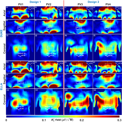 |
Development of Two Antisymmetric 16-Element Transceiver Coil Arrays for Parallel Transmit Cardiac MRI in Humans at 7T
Ibrahim A. Elabyad1, Maxim Terekhov1, and Laura M. Schreiber1
1Chair of Molecular and Cellular Imaging, Comprehensive Heart Failure Center (CHFC), University Hospital Wuerzburg, Wuerzburg, Germany
Two antisymmetric, 16-elements, transceiver RF-coil arrays were developed for improved $$${B_1^+}$$$-shimming and parallel imaging for cardiac-MRI in humans at 7T. The first array (Design1) comprised of an 8-loops for both anterior and posterior sections. The second array (Design2) was composed of 12-loops for the anterior section and 4-loops for the posterior section. Electromagnetic-field (EMF) simulations were carried out for both arrays loaded with an elliptical phantom and two human models (Duke and Ella). Static-phase $$${B_1^+}$$$-shimming has been carried out for both arrays with two different optimization cost functions to maximize the transmit-efficiency and weighted combination of $$${B_1^+}$$$-field homogeneity and transmit-efficiency.
|
||
|
0181.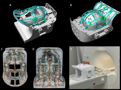 |
A Patient-Friendly 16ch Tx / 64ch Rx Array for Combined Head and Neck Imaging at 7 Tesla
Markus W. May1, Sam-Luca J.D. Hansen1, Nicolas Kutscha1, Gurinder Kaur Multani1, Mirsad Mahmutovic1, Matthäus Poniatowski1, Rene Gumbrecht2, Ralph Kimmlingen2, Markus Adriany2, Yulin Chang3, Bastien Guerin4, Christina Triantafyllou2, Lawrence L. Wald4,
and Boris Keil1
1Institute of Medical Physics and Radiation Protection (IMPS), TH Mittelhessen University of Applied Sciences, Giessen, Germany, 2Siemens Healthcare GmbH, Erlangen, Germany, 3Siemens Medical Solutions USA, Inc., Malvern, PA, United States, 4A.A. Martinos Center for Biomedical Imaging, Dept of Radiology, Massachusetts General Hospital, Harvard Medical School, Boston, MA, United States
A 16chTx / 64chRx head-neck array coil was designed, constructed, and validated at 7T ultra-high field (UHF) MRI. The clinical benefits of UHF neuroimaging were increased by extending coil coverage from the brain region to include the cervical spine. To increase patient compliance, the commonly employed separated transmit and receive coil array functionalities at UFH were combined into one anatomically shaped close-fitting housing which is fully splitable for patient access.
|
|
0182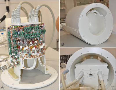 |
A 16-channel transmit 96-channel receive head coil for NexGen 7T scanner Video Permission Withheld
Shajan Gunamony1,2, Roland Müller3, Paul McElhinney1, Sydney Nicole Williams1, Nicolas Groß-Weege3,4, Nikolaus Weiskopf3,5, Harald E Möller3, and David Feinberg6
1Imaging Centre of Excellence, University of Glasgow, Glasgow, United Kingdom, 2MR CoilTech Limited, Glasgow, United Kingdom, 3Max Planck Institute for Human Cognitive and Brain Sciences, Leipzig, Germany, 4Siemens Healthcare GmbH, Erlangen, Germany, 5Faculty of Physics and Earth Sciences, Felix Bloch Institute for Solid State Physics, Leipzig, Germany, 6Helen Wills Neuroscience Institute, University of California, Berkeley, CA, United States
The NexGen 7T scanner has been designed to reach an unprecedented microscale resolution in fMRI studies of the cerebral cortex. A radiofrequency coil setup with a high density receive array is essential to capture the promised benefits. The coil design, which is already constrained by the limited space on the patient table inside the head gradient, must be robust, allow routine scanning and offer a visual field to support fMRI studies. We have developed a compact 16-channel transmit 96-channel receive head coil for the NexGen 7T scanner. In this abstract, we present the coil development and preliminary phantom results.
|
||
|
0183.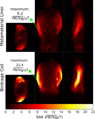 |
Whole-body Metamaterial Liner RF Coil for 1H at 4.7 T with Reduced SAR Compared to Birdcage Coil
Adam Maunder1, Ashwin Iyer2, and Nicola De Zanche1
1Oncology, Medical Physics, Cross Cancer Institute, University of Alberta, Edmonton, AB, Canada, 2Electrical and Computer Engineering, University of Alberta, Edmonton, AB, Canada
The maximum local specific absorption rate relative to B1+ field produced increases with B0 field strength. This relationship greatly constrains the parameter space in sequence optimization due to safety limits. The metamaterial liner simulated here produces lower SAR for the same transmit excitation compared to conventional birdcage coils for whole body imaging, thereby permitting more power-intensive scan parameters to be used. The transmit efficiency and homogeneity is found to be similar between the metamaterial liner and comparable birdcage coil, while the metamaterial liner produces only 41% of the 10g localized SAR for the same transmit field.
|
|
|
0184.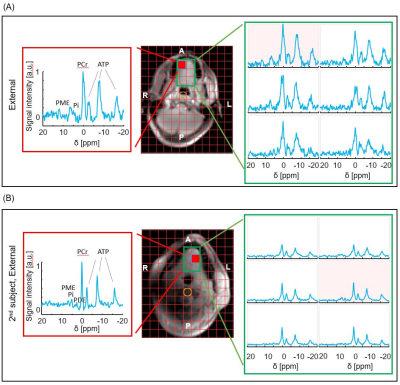 |
Novel Setup for 31P MRSI of the Human Tongue In Vivo at 7T
Ria Forner1, Kyung Min Nam1, Tijl van der Velden1, and Dennis Klomp1
1UMC Utrecht, Utrecht, Netherlands
Here we present the very first 31P MRS data in the human tongue at 7T. Three different receive coils were designed and evaluated to compare their performance: two each inside the mouth: a loop and also a saddle coil and a 3-channel loop array positioned externally. The transmit setup consists of an integrated 31P body birdcage coil for 31P transmit and an array of 8 1H dipoles for image registration. The loop coil seems to outperform the other two coils albeit that signals may originate from jaw. For voxels inside the tongue, relatively high phosphomonoesters were observed in all volunteers.
|
The International Society for Magnetic Resonance in Medicine is accredited by the Accreditation Council for Continuing Medical Education to provide continuing medical education for physicians.