Oral
RF Design I
ISMRM & SMRT Annual Meeting • 15-20 May 2021

| Concurrent 2 | 16:00 - 18:00 | Moderators: Zhipeng Cao & Irena Zivkovic |
 |
0129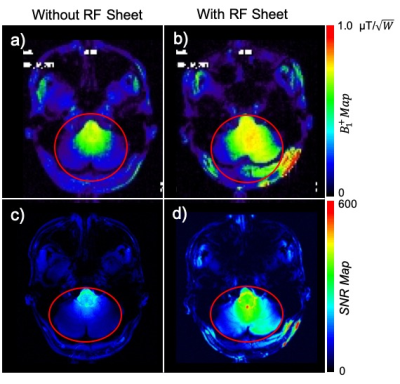 |
Enhanced Ultra-High Field Brain MRI Using a Wireless Radiofrequency Sheet Video Permission Withheld
Akbar Alipour1, Alan C Seifert1, Bradley Delman1, Raj Shrivastava2, Gregor Adriany3, Zahi Adel Fayad1, and Priti Balchandani1
1Radiology, Icahn School of Medicine at Mount Sinai, New York, NY, United States, 2Neurosurgery, Icahn School of Medicine at Mount Sinai, New York, NY, United States, 3Radiology, University of Minnesota-Medical School, Minneapolis, MN, United States
Ultra-high field (UHF) MRI, such as 7T can visualize the brain in significantly improved detail through enhanced signal-to-noise ratio and contrast mechanisms. However, when the wavelength becomes comparable with the body dimensions, excitation radiofrequency (RF) field homogeneity at UHF systems is impaired by wavelength effects. Here we report a novel RF resonator sheet design with a simple circuit structure that couples inductively to the RF coil to enhance RF transmit homogeneity, efficiency, and signal sensitivity. In-vivo human experiment results demonstrate the feasibility and effectiveness of this method in brain MRI at 7T.
|
|
0130.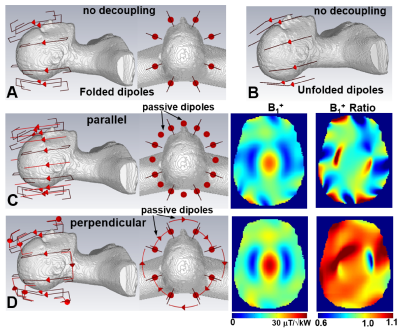 |
Unshielded Bent Folded-End Dipole 9.4 T Human Head Transceiver Array Decoupled Using Modified Passive Dipoles.
Nikolai Avdievich1, Georgiy Solomakha2, Loreen Ruhm1, Anke Henning1,3, and Klaus Scheffler1
1High-field Magnetic Resonance, Max Planck Institute for Bilogical Cybernetics, Tübingen, Germany, 2Physics and Engineering, ITMO University, St. Petersburg, Russian Federation, 3Advanced Imaging Research Center, University of Texas Southwestern Medical Center, Dallas, TX, United States
Dipole antennas have been used for human imaging at ultra-high field (UHF, >7T). However, for head imaging, dipoles must be substantially shortened, which often cause poor (~ -10dB) decoupling. Common decoupling methods are difficult to use due to distant location of dipoles. Alternatively, adjacent transmit dipoles can be decoupled using passive dipole antennas placed parallel between them. Such passive dipoles may interact destructively with the RF field of the transmit array. In this work, we developed a novel decoupling method of adjacent transmit dipoles by using modified perpendicular passive dipole antennas. The constructed array demonstrated good decoupling and whole-brain coverage.
|
||
 |
0131.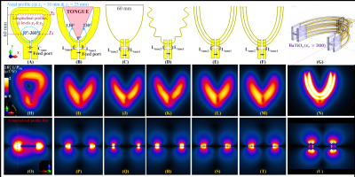 |
Novel Intraoral Dipole Antenna for Dental Applications
Ali Caglar Özen1,2, Djaudat Idiyatullin3, Gregor Adriany3, Steve Jungst3, Naoharu Kobayashi3, Beth R. Groenke4, Michael Bock1, Michael Garwood3, and Donald R. Nixdorf4,5
1Deptartment of Radiology, Medical Physics, University Medical Center Freiburg, University of Freiburg, Freiburg, Germany, 2German Consortium for Translational Cancer Research Partner Site Freiburg, German Cancer Research Center (DKFZ), Heidelberg, Germany, 3Center for Magnetic Resonance Research and Department of Radiology, University of Minnesota, Minneapolis, MN, United States, 4Division of TMD & Orofacial Pain, School of Dentistry, University of Minnesota, Minneapolis, MN, United States, 5Department of Neurology and Radiology, Medical School, University of Minnesota, Minneapolis, MN, United States
Previous studies showed that in dental MRI intraoral loop coils provide higher signal-to-noise ratio (SNR) than extraoral coils. An intraoral dipole that fits the dental arch can be used for reduced FOV and high transmit efficiency. Besides, dipoles do not restrict tongue movement. The design approach is based on comparative FDTD field simulations. The best transmit efficiency and homogeneity was achieved with a multi-wire curved dipole antenna. Additional high-permittivity cap further improved the transmit field inhomogeneity. When combined with extraoral flexible shielded loop resonators, SNR was increased and the coupling between the coils was less than -32dB.
|
|
0132.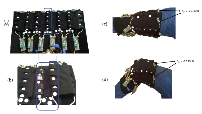 |
Wearable knee receive array coil for imaging at different flexion angles
Syed Saad Siddiq1,2, Justin Ho2,3, Billie Wang2,3, Jerzy Walczyk2,3, Karthik Lakshmanan2,3, and Ryan Brown2,3
1Department of Electrical & Computer Engineering, New York University Tandon School of Engineering, Brooklyn, NY, United States, 2Bernard and Irene Schwartz Center for Biomedical Imaging, Department of Radiology, New York University Grossman School of Medicine, New York, NY, United States, 3Center for Advanced Imaging Innovation and Research, Department of Radiology, New York University Grossman School of Medicine, New York, NY, United States
Weight-bearing and kinetic MRI are important for measuring tibialfemoral joint dynamics, but are difficult to carry out using rigid knee coils that are typically designed to restrict, rather than enable, flexion motion. We explored off-the-shelf components and constructed a six-channel flexible knee coil with an elastic shell to maintain critical geometric overlap between neighbor coils. The array enables MRI during knee flexion while providing similar SNR compared to a state-of-the-art rigid commercial coil. We anticipate that the coil will be useful for weight-bearing or kinetic knee imaging in which rigid coils provide suboptimal SNR and/or severely restrict the desired posture.
|
||
|
0133.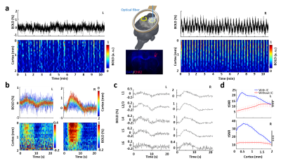 |
Inductively coupled detectors for optogenetic-driven focal and multiregional fMRI signal enhancement
Yi Chen1, Qi Wang1,2, Hang Zeng1,2, Kengo Takahashi1,2, Sangcheon Choi1,2, Chunqi Qian3, and Xin Yu1,4
1Max Planck Institute for Biological Cybernetics, Tuebingen, Germany, 2Graduate Training Centre of Neuroscience, University of Tuebingen, Tuebingen, Germany, 3Department of Radiology, Michigan State University, East Lansing, MI, United States, 4Athinoula A. Martinos Center for Biomedical Imaging, Massachusetts General Hospital and Harvard Medical School, Charlestown, MA, United States
To improve MR detection sensitivity of multi-modal fMRI platform, we present inductive coil which could relay locally detected MR signals to the external surface coil. Under sensory stimulation, evoked whole brain EPI-based fMRI and enhanced focal laminar-specific fMRI signals were acquired with high spatiotemporal resolution (100 μm and 100 ms) using a single experimental setup. Moreover, embedding inductive coil beneath the glue-secured optical fiber and the projection-mirrored cortex on the other hemisphere boosted multiregional fMRI sensitivity to investigate the interhemispheric connectivity with laminar-specificity. This is particularly helpful to study the optogenetic-driven brain connectivity with circuit specificity at multi-modal fMRI platform.
|
|
|
0134.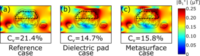 |
Metasurface for B1+ homogenization in abdominal MRI at 3T
Vsevolod Vorobyev1, Alena Shchelokova1, Aleksander Efimtcev1,2, Juan Domingo Baena3, Pavel Belov1, and Stanislav Glybovski1
1ITMO University, Saint-Petersburg, Russian Federation, 2Federal Almazov North-West Medical Research Center, Saint-Petersburg, Russian Federation, 3Universidad Nacional de Colombia, Bogota, Colombia
A novel approach for improving B1+ homogeneity in the abdominal area at 3T MRI is proposed and demonstrated numerically and experimentally. The approach is implemented via the ultralight and thin metasurface. The metasurface consists of metal wires loaded with capacitors printed on a flexible dielectric substrate of polyimide. Numerical studies and imaging of a volunteer covered with the proposed metasurface showed the same homogeneity of the transmit radiofrequency field distribution at the region-of-interest as the conventional dielectric pads.
|
|
|
0135.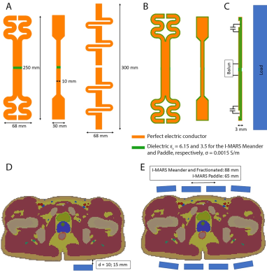 |
A novel type of radiofrequency antenna for multi-regional 7T MRI
Aurelien Destruel1, Ewald Weber1, Mingyan Li1, Jin Jin1,2, Craig Engstrom3, Feng Liu1, and Stuart Crozier1
1School of Information Technology and Electrical Engineering, The University of Queensland, Brisbane, Australia, 2Siemens Healthcare Pty Ltd, Brisbane, Australia, 3School of Human Movement and Nutrition Sciences, The University of Queensland, Brisbane, Australia
A novel radiofrequency coil element, named integrated multi-modal antenna with coupled radiating structures (I-MARS), is presented in numerical simulations and experiments for 7T MRI. Simulated comparisons of two variations of the proposed design with fractionated dipoles show I-MARS elements have advantageous robustness to load changes, inter-element isolation and are optimizable for power and SAR efficiency. Imaging of volunteers and phantom in different configurations (unilateral hip, prostate and shoulder imaging) did not require adjustments to tuning and matching, showing excellent stability and high performance for multi-anatomy 7T MRI.
|
|
|
0136 |
Self-tuning stretchable RF receive coil concept using liquid metal encapsulated within an elastic polymer
Elizaveta Motovilova1,2, Jana Vincent3, Victor Taracila3, Fraser Robb3, Ek Tsoon Tan2, James Shin1, Hollis G. Potter2, Darryl B. Sneag2, and Simone Angela Winkler1
1Radiology, Weill Cornell Medicine, New York, NY, United States, 2Radiology, Hospital for Special Surgery, New York, NY, United States, 3GE Healthcare, Aurora, OH, United States
Commercial coils, built to accommodate a wide range of anatomical dimensions, are rigid and of fixed size, thus yielding sub-optimal SNR and patient comfort. Existing flexible/stretchable solutions suffer from resonance detuning due to inductance changes under stretch/deformation. In this work, we propose an alternative coil concept using liquid metal microchannel conductors encapsulated in a stretchable polymer matrix. We developed a self-tuning coil using a stretchable, adaptively compensating, interdigital capacitor. We observed a <0.5% of frequency stability in silico and in vitro. In vivo results were demonstrated on 3T wrist imaging.
|
|
0137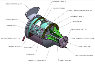 |
An RF Coil for a Head-Only MR System Video Permission Withheld
J. Thomas Vaughan1, Brandon Tramm2, Scott Schillak2, Michael Garwood3, Michael Mullen4, Lance DelaBarre4, Djaudat Idiyatullin4, and Matt Waks4
1Biomedical Engineering, Radiology, Columbia University, New York, NY, United States, 2Virtumed, LLC, Minneapolis, MN, United States, 3University of Minnesota, Minneaoplis, MN, United States, 4University of Minnesota, Minneapolis, MN, United States
A new RF coil and frontend for a head-only, 1.5T fMRI system was designed, built and demonstrated. This coil features the ability for single and multichannel transceiver operation for transmit-receive switched and simultaneous transmit and receive (STAR) operation. It includes a window and an integrated shield. First applications will be for voluntary motor control studies in humans.
|
||
|
0138.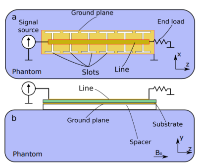 |
A non-resonant leaky-wave coil for UHF body imaging
Georgiy Solomakha1, Jan Taro Svejda2,3, Carel van Leeuwen4, Rustam Balafendiev1, Andreas Rennings2,3, Alexander Raaijmakers4,5, Stanislav Glybovski1, and Daniel Erni2,3
1The Department of Physics and Engineering, ITMO University, Saint Petersburg, Russian Federation, 2General and Theoretical Electrical Engineering (ATE), Faculty of Engineering and General and Theoretical Electrical Engineering (ATE), Faculty of Engineering, University of Duisburg-Essen, Duisburg, Germany, 3Center for Nanointegration Duisburg-Essen, Duisburg, Germany, 4Imaging Division, UMC Utrecht, Utrecht, Netherlands, 5Medical Image Analysis, Biomedical Engineering, Technical University of Eindhoven, Eindhoven, Netherlands
Ultra-high field (field strength higher than 7 Tesla) body imaging is an extensively developing field. Since at such Larmor frequencies of 298 MHz or higher volume (birdcage or TEM) coils are not efficient due to interference effects, surface or volume transmit arrays are commonly used. To obtain homogenous filed in ROI so-called RF-shimming procedure is commonly used. In this work, we present a new radiative RF-coil array for UHF MRI of the human body, based on a wideband non-resonant leaky wave antenna.
|
The International Society for Magnetic Resonance in Medicine is accredited by the Accreditation Council for Continuing Medical Education to provide continuing medical education for physicians.