Oral
High-Resolution fMRI
ISMRM & SMRT Annual Meeting • 15-20 May 2021

| Concurrent 3 | 18:00 - 20:00 | Moderators: Sriranga Kashyap & Jonathan Polimeni |
0629.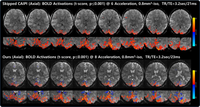 |
Highly Accelerated Sub-millimeter Resolution 3D EPI using Variable Density CAIPI Sampling with Temporal Random Walk for Functional MRI at 7 Tesla
Suhyung Park1,2, Sugil Kim3, Hankyeol Lee4, Seulgi Eun4, Seong-Gi Kim4,5, and David Feinberg6,7
1Department of Computer Engineering, Chonnam National University, Gwangju, Korea, Republic of, 2Department of ICT Convergence System Engineering, Chonnam National University, Gwangju, Korea, Republic of, 3Siemens-Healthineers, Seoul, Korea, Republic of, 4Center for Neuroscience Imaging Research (CNIR), Institute for Basic Science (IBS), Suwon, Korea, Republic of, 5Department of Biomedical Engineering, Sungkyunkwan University, Suwon, Korea, Republic of, 6University of California, Berkeley, Berkeley, CA, United States, 7Advanced MRI Technologies, Sebastopol, CA, United States
With ultra-high fields, 3D EPI has been used by improving imaging efficiency. Nevertheless, there have been some limitations: 1) the regular sampling limits the use of temporal structure in the data and 2) parallel imaging allows up to 6-fold acceleration in 3D acquisition. Here, we developed an accelerated 3D EPI using VD-CAPI sampling with temporal random walk. Experimental studies confirm advantages in acceleration, SNR, and sensitivity of the proposed method: 1) temporal random walk allows extra spatial encoding across time, 2) temporal prior provides high SNR, and 3) the temporal incoherent sampling and high SNR result in higher BOLD activations.
|
||
0630.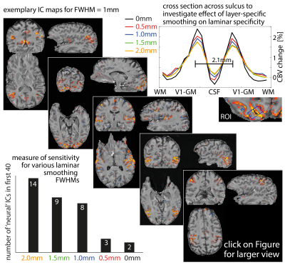 |
Whole brain layer-fMRI: An open dataset for methods benchmarking
Anna K Mueller1, Miriam Heynckes2, Christopher J Wiggins3, Omer Faruk Gulban4, Yuhui Chai5, Benedikt Poser2, and Renzo Huber2
1Goethe-Universität Frankfurt am Main, Mainz, Germany, 2Faculty of Psychology and Neuroscience, Maastricht University, Maastricht, Netherlands, 3Scannexus, Maastricht, Netherlands, 4Brain Innovation, Maastricht, Netherlands, 5NIH, Bethesda, MD, United States Laminar-specific fMRI allows neuroscientists to address research questions of directional functional connectivity within and across brain areas. While recent sequence developments allow considerable improvements in coverage, resolution, and mitigation of venous biases, it is not established how routinely useful those methods are for everyday neuroscientific application. We present an open dataset of whole-brain CBV-sensitive layer-dependent fMRI during free movie watching. Its purpose is to:
|
||
 |
0631.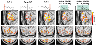 |
Simultaneous pure spin-echo and gradient-echo BOLD fMRI using Echo Planar Time-resolved Imaging (EPTI) for mapping laminar fMRI responses
Fuyixue Wang1,2, Zijing Dong1,3, Lawrence L. Wald1,2, Jonathan Polimeni1,2, and Kawin Setsompop4,5
1Athinoula A. Martinos Center for Biomedical Imaging, Massachusetts General Hospital, Charlestown, MA, United States, 2Harvard-MIT Health Sciences and Technology, MIT, Cambridge, MA, United States, 3Department of Electrical Engineering and Computer Science, MIT, Cambridge, MA, United States, 4Department of Radiology, Stanford University, Stanford, CA, United States, 5Department of Electrical Engineering, Stanford University, Stanford, CA, United States
We introduced a novel imaging approach SE-EPTI to address the T2’-contamination in SE-EPI for higher specificity of BOLD fMRI. EPTI resolves multi-contrast distortion/blurring-free images to simultaneously obtain: a pure SE image with minimal T2’-contamination, multiple GE images with various T2’-weightings, and conventional SE-EPI images with different levels of T2’-contamination. We demonstrated at 7T that the pure SE can significantly reduce the draining-vein-effect, and by using shorter ETLs, less T2’-contamination was introduced. A new echo-train-shifting method is also proposed for SE-EPTI to offer flexibility of achieving shorter TEs, allowing us to examine the TE dependence of the signal contribution.
|
|
0632.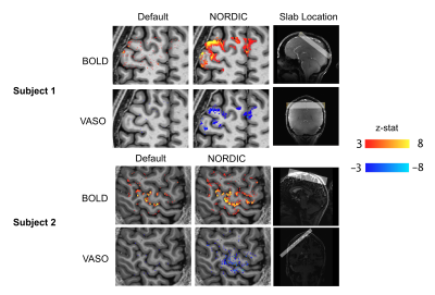 |
VASO-fMRI with Nordic-PCA for laminar sensory testing at 7 Tesla
Nils Dennis Nothnagel1, Alison Symon1, Andrew Tyler Morgan1,2, Renzo Huber3, John Riddell1, and Jozien Goense1
1Institute of Neuroscience & Psychology, University of Glasgow, Glasgow, United Kingdom, 2NIH, Bethesda, MD, United States, 3Faculty of Psychology and Neuroscience, Maastricht University, Maastricht, Netherlands
The most commonly used contrasts in laminar fMRI are blood-oxygen-level-dependent (BOLD) and vascular-space-occupancy (VASO). However, at laminar resolution, brain activity is often buried under noise, complicating the detection of small changes in activation.Here, we show the successful extraction of laminar brain activity of a complex-valued BOLD- and VASO-fMRI time series during a somatosensory task using NORDIC-PCA denoising. We expect this method to be a great asset for laminar sensory fMRI experiments as it reduces the need for anatomically informed smoothing or anisotropic filtering, which might be helpful for very small voxel sizes or when small activated areas are studied.
|
||
0633.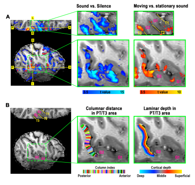 |
Topographical and Laminar Distribution of Audiovisual Processing within Human Planum Temporale
Yuhui Chai1, Tina Liu1, Sean Marrett1, Linqing Li1, Arman Khojandi1, Daniel Handwerker1, Arjen Alink2, Lars Muckli3, and Peter Bandettini1
1NIMH, Bethesda, MD, United States, 2University Medical Centre Hamburg-Eppendorf, Hamburg, Germany, 3University of Glasgow, Glasgow, United Kingdom
Multisensory interplay can occur in areas that are commonly considered unisensory, such as planum temporale (PT). The roles of different afferents to PT in multisensory processing are not well understood. Using sub-millimeter fMRI at 7T, we compared laminar activity patterns across topographical subfields of PT under unimodal and multisensory stimuli. We found (1) anterior PT was activated more by auditory inputs and received feedback in superficial layers, likely coming from higher-order multimodal areas; (2) posterior PT was preferentially activated by visual inputs and received visual feedback in both superficial and deep layers, likely projected directly from the early visual cortex.
|
||
|
0634.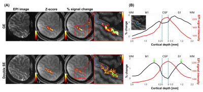 |
Double spin-echo EPI improves sensitivity and specificity for cortical depth-dependent BOLD fMRI in the human somatosensory cortex at 7 T
SoHyun Han1,2, HyungJoon Cho3, Kâmil Uludaǧ1,2, and Seong-Gi Kim1,2
1Center for Neuroscience Imaging Research, Suwon, Korea, Republic of, 2Department of Biomedical Engineering, Sungkyunkwan University, Suwon, Korea, Republic of, 3Department of Biomedical Engineering, Ulsan National Institute of Science and Technology, Ulsan, Korea, Republic of
Spatial-specificity is important for high spatial-resolution-fMRI to determine neuronal activity laminar-profiles. GE-BOLD-signals have low specificity because the highest signals originate from draining-veins at the surface of the cortex, not from capillaries nearby active neurons. However, SE-BOLD-signal has been proposed to be a better indicator of the location of neural activity. In this study, double SE-EPI-sequence was developed to achieve increased sensitivity in SE-BOLD-fMRI and demonstrated its feasibility for fMRI with 0.8-mm in-plane resolution. The results confirm that dSE-BOLD has higher specificity than GE-BOLD and better sensitivity than conventional-SE-BOLD and its potential to probe the function of cortical-circuits with high specificity.
|
|
0635. |
Mapping digit-representations in BA3b during stimulation and investigating their intrinsic connectivity at rest using VASO
Sebastian Dresbach1, Renzo Huber1, Rainer Goebel1, and Amanda Kaas1
1Cognitive Neuroscience, Faculty of Psychology and Neuroscience, Maastricht University, Maastricht, Netherlands While inter-digit interactions are crucial for manual abilities like tool use or object manipulation, individual digits seem to be distinctly represented in the primary somatosensory cortex. Here, we used cortical depth-resolved high-resolution CBV-measurements to investigate these representations in the putative S1-subregion “BA3b” and depth-dependent temporal correlation during rest, as a potential index for layer-specific integration between digit representations. We found that we can
|
||
0636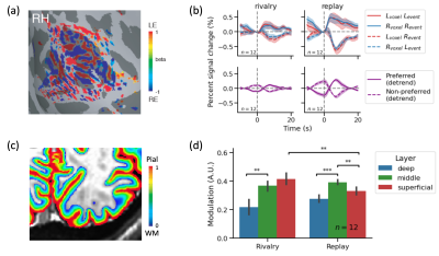 |
Layer- and column-resolved 7T fMRI reveals neural correlates of consciousness in human visual cortex and thalamus Video Permission Withheld
Chencan Qian1,2, Chengwen Liu3, Jinyou Zou4, Yan Zhuo1,2, Sheng He1,2,5, and Peng Zhang1,2
1State Key Laboratory of Brain and Cognitive Science, Institute of Biophysics, Chinese Academy of Sciences, Beijing, China, 2University of Chinese Academy of Sciences, Beijing, China, 3Department of Neurosurgery, Xiangya Hospital, Central South University, Changsha, China, 4Max-Planck-Institute for Biological Cybernetics, Tübingen, Germany, 5Department of Psychology, University of Minnesota, Minneapolis, MN, United States
Binocular rivalry is a unique window to study the neural correlates of consciousness. Where and how does binocular rivalry arise in the human brain remains an open question. Using laminar fMRI at 7T, we found that eye-specific modulation of BOLD signal peaked in the middle layer of primary visual cortex (V1) during simulated replay, but stronger in the superficial layer during rivalry. Furthermore, eye-specific modulation of lateral geniculate nucleus (LGN) activity was robust in the replay but minimal in the rivalry condition. These findings support that binocular rivalry mainly arises from interocular interaction in the superficial layer of V1.
|
||
0637. |
Submillimeter Arterial Blood Contrast fMRI at 7T
Nikos Priovoulos1, Icaro Agenor Ferreira de Oliveira1, Benedikt Poser2, David G Norris3,4, and Wietske van der Zwaag1
1Spinoza Center, Amsterdam, Netherlands, 2Maastricht University, Maastricht, Netherlands, 3Donders Institute for Brain, Cognition and Behaviour, Radboud University Nijmegen, Nijmegen, Netherlands, 4Erwin L. Hahn Institute for MRI, University of Duisburg-Essen, Essen, Germany
BOLD-fMRI has transformed human neuroscience, but is limited in its spatial specificity compared to cerebral blood-volume approaches. Arterial-Blood-Contrast was recently suggested as a cerebral-blood-volume method based on Magnetization transfer. Here, we apply Arterial-Blood-Contrast in the SAR-constrained 7T environment and examine its submillimeter usage. The results suggest good localization compared to BOLD and high-sensitivity.
|
||
0638.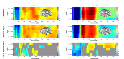 |
Correlation between inter-cortical depth fMRI signals and oscillatory neuronal responses during music listening
Hsin-Ju Lee1,2, Pu-Yeh Wu1, Hankyeol Lee3, Kamil Uludag3,4, Hsiang-Yu Yu5,6,7, Cheng-Chia Lee6,7,8, Chien-Chen Chou5,6, Chien Chen5,6, Wen-Jui Kuo7,9, and Fa-Hsuan Lin1,2,10
1Physical Sciences Platform, Sunnybrook Research Institute, Toronto, ON, Canada, 2Department of Medical Biophysics, University of Toronto, Toronto, ON, Canada, 3Center for Neuroscience Imaging Research, Institute for Basic Science, Suwon, Korea, Republic of, 4Techna Institute & Koerner Scientist in MR Imaging,, Joint Department of Medical Imaging and Krembil Brain Institute, University Health Network, Toronto, ON, Canada, 5Department of Epilepsy, Neurological Institute, Taipei Veterans General Hospital, Taipei, Taiwan, 6School of Medicine, National Yang-Ming University, Taipei, Taiwan, 7Brain Research Center, National Yang-Ming University, Taipei, Taiwan, 8Department of Neurosurgery, Neurological Institute, Taipei Veterans General Hospital, Taipei, Taiwan, 9Institute of Neuroscience, National Yang-Ming University, Taipei, Taiwan, 10Department of Neuroscience and Biomedical Engineering, Aalto University, Espoo, Finland
We explored the correlation between cortical depth-dependent fMRI signal and oscillatory neural activity during music listening using high-resolution fMRI (7T with 0.8 mm and 3T with 1.5 mm isotropic resolution, respectively) and invasive electrode recording on epilepsy patients. The hemodynamic responses in the auditory cortex were found positively and negatively correlated with neural oscillations in the gamma and alpha/beta band at right and both hemispheres, respectively. These correlations were highest at the intermediate cortical depth. Core and non-core areas of the auditory cortex had different correlations.
|
The International Society for Magnetic Resonance in Medicine is accredited by the Accreditation Council for Continuing Medical Education to provide continuing medical education for physicians.