Oral
fMRI Data Acquisition & Analysis
ISMRM & SMRT Annual Meeting • 15-20 May 2021

| Concurrent 3 | 12:00 - 14:00 | Moderators: Benedikt Poser & Olivia Viessmann |
 |
0457.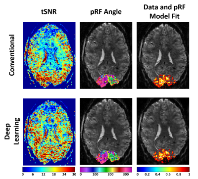 |
Improved Accelerated fMRI Reconstruction using Self-supervised Deep Learning
Omer Burak Demirel1,2, Burhaneddin Yaman1,2, Steen Moeller2, Logan Dowdle2, Luca Vizioli2, Kendrick Kay2, Essa Yacoub2, John Strupp2, Cheryl Olman2, Kâmil Uğurbil2, and Mehmet Akçakaya1,2
1Electrical and Computer Engineering, University of Minnesota, Minneapolis, MN, United States, 2Center for Magnetic Resonance Research, University of Minnesota, Minneapolis, MN, United States
There are significant benefits to HCP-style fMRI acquisitions, which acquires high spatial and temporal resolution across the whole brain in an effort to better understand the human brain. This can be achieved through simultaneous multi-slice (SMS)/Multiband (MB) imaging, which provides rapid whole-brain coverage using high acceleration rates albeit with increased noise amplification. Deep learning reconstruction techniques have recently gained substantial interest in improving accelerated MRI. Here we utilize a physics-guided self-supervised deep learning reconstruction on a 5-fold SMS and 2-fold in-plane accelerated whole brain 7T fMRI acquisition to reduce the reconstruction noise without altering the subsequent fMRI result.
|
|
 |
0458.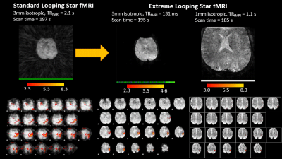 |
Extreme Looping Star: Quiet fMRI at high spatiotemporal resolution
Andrew Palmera Leynes1,2, Nikou Louise Damestani3, David John Lythgoe3, Ana Beatriz Solana4, Brice Fernandez5, Brian Burns1,6, Steven Charles Rees Williams3, Fernando Zelaya3, Peder E.Z. Larson1,2, and Florian Wiesinger3,4
1Department of Radiology and Biomedical Imaging, University of California San Francisco, San Francisco, CA, United States, 2UC Berkeley - UC San Francisco Joint Graduate Program in Bioengineering, Berkeley and San Francisco, CA, United States, 3King's College London, London, United Kingdom, 4GE Healthcare, Munich, Germany, 5GE Healthcare, Paris, France, 6GE Healthcare, Menlo Park, CA, United States
The Looping Star pulse sequence was recently introduced as an acoustically silent alternative to EPI-based fMRI pulse sequences. In this abstract, we present improvements to the spatiotemporal resolution of Looping Star using the “extreme MRI” approach, without sacrificing functional sensitivity. We demonstrate the application of silent fMRI with increased temporal resolution and increased spatial resolution using extreme Looping Star, in comparison with standard Looping Star, on a motor task and a visual task across two sites.
|
|
|
0459.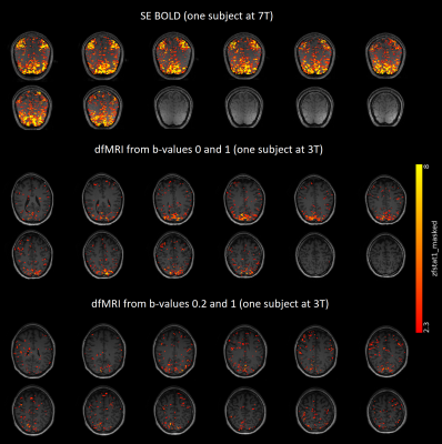 |
Beyond BOLD: in search of genuine diffusion fMRI contrast in human brain
Wiktor Olszowy1,2 and Ileana O Jelescu1,2
1CIBM Center for Biomedical Imaging, Lausanne, Switzerland, 2Animal Imaging and Technology, EPFL, Lausanne, Switzerland
Diffusion fMRI (dfMRI) is an alternative to BOLD fMRI. Here, we present the first dfMRI study in humans attempting to minimize all sources of BOLD contamination and comparing functional responses at two field strengths, both for task and resting-state fMRI. Our study benefits from unprecedented high spatiotemporal resolution. We observed task-induced water diffusivity decreases in the perfusion-free b-value regime. Furthermore, we found that positive correlations were largely preserved while anti-correlations were suppressed in dfMRI functional connectivity compared to BOLD. We conclude that dfMRI contrast is genuine and distinct from BOLD mechanisms.
|
|
0460.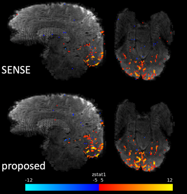 |
Respiratory fluctuations in 3D fMRI from inter-shot phase variations can be reduced by low-rank reconstruction of segmented CAIPI sampling
Xi Chen1, Wenchuan Wu1, and Mark Chiew1
1Wellcome Centre for Integrative Neuroimaging, FMRIB, Nuffield Department of Clinical Neurosciences, University of Oxford, Oxford, United Kingdom
Multi-shot 3D EPI is one of the most popular 3D imaging techniques for fMRI, which can provide higher SNR than 2D single-shot EPI. However, the vulnerability to inter-shot signal variations arising from physiological fluctuations, like respiration-related phase variations, has limited the tSNR benefits of 3D multi-shot acquisitions. To improve the temporal stability of 3D multi-shot EPI for fMRI at 7T, we investigated both the optimization of sampling trajectories which show varying vulnerabilities to the inter-shot inconsistencies, and the use of a low-rank annihilating filter constrained reconstruction which can reduce unwanted temporal variance induced by the inter-shot phase inconsistencies.
|
||
0461.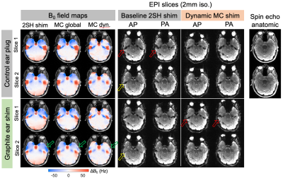 |
Combined active and passive shimming of the temporal lobes using graphite-silicone earplugs and a multi-coil B0 shim array
Andrew Lithen1,2, Albert Tamashausky3, Berkin Bilgic1,4, Kawin Setsompop5, Bryan Kennedy1, Lilianne Mujica-Parodi1,2, Lawrence Wald1,4, Shahin Nasr1,4, and Jason Stockmann1,4
1A. A. Martinos Center for Biomedical Imaging, Massachusetts General Hospital, Charlestown, MA, United States, 2Dept. of Biomedical Engineering, Stony Brook University, Stony Brook, NY, United States, 3Asbury Carbons, Asbury, NJ, United States, 4Harvard Medical School, Boston, MA, United States, 5Dept. of Electrical Engineering, Stanford University, Stanford, CA, United States
Neuroimaging of the human brain temporal lobes has long been impeded by severe B0 inhomogeneity arising from air-tissue susceptibility interfaces around the ear canals and temporal bone. Here, we propose localized passive shimming using graphite-embedded silicone ear plugs, which replace standard ear plugs used during MRI. We further synergistically combine the passive shims with active multi-coil B0 shimming to boost shim performance. Our results show improvements in B0 homogeneity as assessed using field maps and signal loss and distortion in gradient-echo EPI slices.
|
||
0462.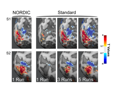 |
A Paradigm Change in Functional Brain Mapping: Suppressing the Thermal Noise in fMRI
Luca Vizioli1,2, Steen Moeller1, logan T Dowdle1, Mehmet Akcakaya1, Federico De Martino3, Essa Yacoub1, and Kamil Ugurbil1
1CMRR, University of Minnesota, minneapolis, MN, United States, 2Department of Neurosurgery, University Of Minnesota, minneapolis, MN, United States, 3University Of Maastricht, Maastricht, Netherlands
Functional imaging with the BOLD contrast is an essential tool to investigate human brain functions, however the contribution of thermal noise compromises the signal, particularly at higher resolutions. A recently developed technique, NORDIC, aims to suppress this noise source. Here we show that NORDIC produces data with substantially higher signal-to-noise and functional contrast to noise ratios, resulting in improved detection of functional activation with no change in spatial precision. One run of denoised data is equivalent to combining 3 to 5 runs of conventional data. These effects create new opportunities for functional neuroimaging, while reducing participant or patient burden.
|
||
0463.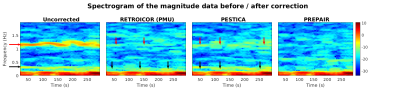 |
Unsupervised Correction of Sub-TR Physiological Noise using Phase and Magnitude fMRI data
David Bancelin1, Beata Bachrata1,2, Pedro Lima Cardoso1, Siegfried Trattnig1,2, and Simon Daniel Robinson1,3,4
1High-Field MR Centre, Medical University of Vienna, Vienna, Austria, 2Christian Doppler Laboratory for Clinical Molecular MR Imaging, Vienna, Austria, 3Department of Neurology, Medical University of Graz, Graz, Austria, 4Centre for Advanced Imaging, University of Queensland, Queensland, Australia
External physiological recordings can be used to filter out cardiac and respiration fluctuations in fMRI data but these can be unreliable. We propose an unsupervised method which derives physiological noise regressors, including cardiac fluctuations with a period much less than the volume TR, from phase and magnitude fMRI data. We compare its efficacy with a correction method which uses external recordings (RETROICOR), and a rival physiological data-free method (PESTICA).
|
||
|
0464.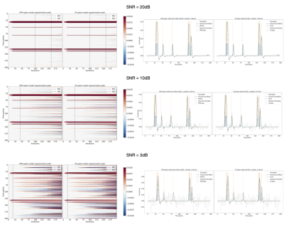 |
fMRI deconvolution with synthesis-based Paradigm Free Mapping and analysis-based Total Activation operate identically
Eneko Uruñuela1, Stefano Moia1, and César Caballero-Gaudes1
1Basque Center on Cognition, Brain and Language, Donostia - San Sebastián, Spain
Functional MRI deconvolution algorithms are gaining popularity to study the dynamicss of functional brain activity and connectivity at short timescales. This work sheds light on our understanding of two state-of-the-art approaches based on L1-norm regularized estimators: Paradigm Free Mapping (synthesis model) and Total Activation (analysis model). Through simulations with varying signal-to-noise ratios, and an experimental motor task dataset, we demonstrate that both formulations produce identical estimates of the innovation and activity-inducing signals underlying BOLD events when identical hemodynamic response and regularization parameters are used. These observations open up the possibility for future developments without questioning their core formulation and performance.
|
|
0465. |
Brain function induces alteration in the autocorrelation of the fMRI signal
Ali Golestani1, Nichole R Bouffard1, Morgan D Barense1,2, and Morris Moscovitch1,2
1Department of Psychology, University of Toronto, Toronto, ON, Canada, 2Rotman Research Institute at Baycrest, Toronto, ON, Canada
The autocorrelation (AC) of the fMRI signal is assumed irrelevant to the brain function and is eliminated by fMRI preprocessing. Recent findings have suggested that the brain function may alter the AC value of the fMRI signal. We used fMRI data acquired during cognitive processes of working memory (WM), mathematical computations, video watching, and resting-state, and showed that cognitively demanding tasks decrease the AC values in functionally related brain regions. Decrease in AC is related to performance on the target tasks. The AC of the fMRI signal is affected by cognitive processes and can provide complementary information about brain function.
|
||
0466.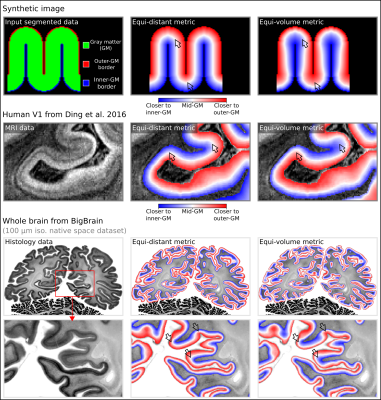 |
LayNii: A software suite for layer-fMRI
Renzo Huber1, Benedikt Poser1, Peter A Bandettini2, Kabir Arora1, Konrad Wagstyl3, Shinho Cho4, Jozien Goense5, Andrew T Morgan2,5, Nils Nothnagel5, Anna K Mueller6, Job van den Hurk7, Richard C Reynolds2, Daniel R Glen2,
Rainer Goebel1,8, and Omer Faruk Gulban8
1Faculty of Psychology and Neuroscience, Maastricht University, Maastricht, Netherlands, 2NIH, Bethesda, MD, United States, 3UCL, London, United Kingdom, 4CMRR, Minneapolis, MN, United States, 5University of Glasgow, Glasgow, United Kingdom, 6Uni Mainz, Mainz, Germany, 7Scannexus, Maastricht, Netherlands, 8Brain Innovation, Maastricht, Netherlands
|
The International Society for Magnetic Resonance in Medicine is accredited by the Accreditation Council for Continuing Medical Education to provide continuing medical education for physicians.