Oral
Pediatric MRI
ISMRM & SMRT Annual Meeting • 15-20 May 2021

| Concurrent 3 | 16:00 - 18:00 | Moderators: Cristina Cudalbu |
 |
0347.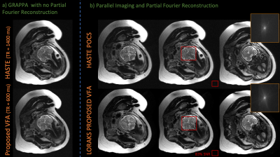 |
Rapid fetal HASTE imaging using variable flip angles and simultaneous multislice wave-LORAKS
Yamin Arefeen1, Tae Hyung Kim2, Justin Haldar3, Ellen Grant4,5, Borjan Gagoski6,7, Berkin Bilgic2,7, and Elfar Adalsteinsson1,8,9
1Massachusetts Institute of Technology, Cambridge, MA, United States, 2Athinoula A. Martinos Center for Biomedical Imaging, Charlestown, MA, United States, 3Department of Electrical Engineering, University of Southern California, Los Angeles, CA, United States, 4Boston Children’s Hospital, Boston, MA, United States, 5Harvard Medical School, Boston, MA, United States, 6Fetal-Neonatal Neuroimaging and Developmental Science Center, Boston Children’s Hospital, Boston, MA, United States, 7Department of Radiology, Harvard Medical School, Cambridge, MA, United States, 8Harvard-MIT Health Sciences and Technology, Cambridge, MA, United States, 9Institute for Medical Engineering and Science, Cambridge, MA, United States
Fetal MRI utilizes Half-Fourier-acquisition-single-shot-turbo-spin-echo (HASTE) for rapid T2-weighted imaging to mitigate motion. However, specific-absorption-rate (SAR) constraints from the refocusing pulse train reduce acquisition efficiency. Variable refocusing flip angle (VFA) acquisitions can improve efficiency, but may suffer from low signal-to-noise ratios (SNR). Here, we propose a VFA scheme and incorporate a rapid, low-SAR calibration scan. We simulate and prospectively evaluate the SNR and SAR properties of the VFA scheme and utilize the calibration scan for LORAKS parallel imaging and retrospective evaluation of wave-encoded simultaneous-multislice (SMS). VFA prospectively reduces acquisition time by ~2.3-2.5x and incorporating SMS could further improve efficiency.
|
|
 |
0348.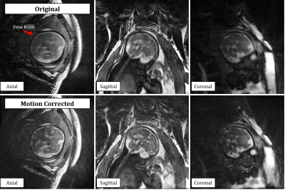 |
Motion Compensated Free-Running 3D Fetal Magnetic Resonance Imaging: Initial Feasibility
Christopher W Roy1, Leonor Alamo1, Estelle Tenisch1, John Heerfordt1,2, Milan Prsa3, Meritxell Bach Cuadra1,4,5, Davide Piccini1,2, Jérôme Yerly1,4, and Matthias Stuber1,4
1Radiology, Lausanne University Hospital (CHUV) and University of Lausanne (UNIL), Lausanne, Switzerland, 2Advanced Clinical Imaging Technology, Siemens Healthcare, Lausanne, Switzerland, 3Division of Pediatric Cardiology, Department Woman-Mother-Child, Lausanne University Hospital (CHUV) and University of Lausanne (UNIL), Lausanne, Switzerland, 4Center for Biomedical Imaging (CIBM), Lausanne, Switzerland, 5Signal Processing Laboratory 5 (LTS5), Ecole Polytechnique Fédérale de Lausanne (EPFL), Lausanne, Switzerland
A novel framework for 3D MRI of the fetus with retrospective motion compensation is developed and its initial feasibility demonstrated. This approach enables imaging with high isotropic resolution and allows for retrospective evaluation of the complex fetal anatomy in arbitrary scan planes, setting the stage for new possibilities in the assessment of fetal diseases in utero.
|
|
0349.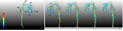 |
Clinical fetal cardiovascular MRI based on Doppler ultrasound gating at 3T and 1.5T: On a technical aspect of imaging pulse sequence optimization
Shuo Zhang1, Janine Knapp2, Roland Cronenberg3, Björn Schönnagel2, Manuela Tavares de Sousa4, Barbara Ulm5, Daniela Prayer3, Vanessa Berger-Kulemann3, and Fabian Kording2,6
1Philips, Hamburg, Germany, 2Department of Diagnostic and Interventional Radiology and Nuclear Medicine, University Hospital Hamburg-Eppendorf, Hamburg, Germany, 3Department of Biomedical Imaging and Image-Guided Therapy, Division of Neuroradiology and Musculoskeletal Radiology, Medical University of Vienna, Vienna, Austria, 4Department of Obstetrics and Fetal Medicine, University Hospital Hamburg-Eppendorf, Hamburg, Germany, 5Department of Gynecology and Obstetrics, Division of Feto-Maternal Medicine, Medical University of Vienna, Vienna, Austria, 6northh medical GmbH, Hamburg, Germany
Cardiovascular MRI is considered a valuable diagnostic tool for studying congenital abnormalities in children and adults. However, simple, high-quality imaging of the fetal heart is challenging due to lack of direct in-utero cardiac gating. We aimed to employ a recently introduced Doppler ultrasound (DUS) device and combine with optimized routine imaging techniques for structural and functional studies of the fetal heart and to establish a standard acquisition approach for high-quality fetal cardiovascular MRI in the clinical practice.
|
||
0350.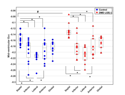 |
Early Non-Contrast Biomarkers of Left Ventricular Cardiomyopathy in Children with Duchenne Muscular Dystrophy
ZHAN-QIU LIU1, Nyasha Maforo2, Seraina Dual3, Ashley Prosper4, Pierangelo Renella4, Nancy Halnon5, Holden Wu4, and Daniel Ennis3
1Cardiovascular Institute, Stanford University, Stanford, CA, United States, 2Physics and Biology in Medicine Interdepartmental Program, University of California, Los Angeles, Los Angeles, CA, United States, 3Department of Radiology, Stanford University, Stanford, CA, United States, 4Department of Radiological Sciences, University of California, Los Angeles, Los Angeles, CA, United States, 5Department of Pediatrics, University of California, Los Angeles, Los Angeles, CA, United States
Left ventricular(LV) peak mid-wall circumferential strain (Ecc) is a sensitive early biomarker for evaluating the subtle and highly variable onset and progression of cardiomyopathy in pediatric subjects with Duchenne muscular dystrophy (DMD). Cine Displacement Encoding with Stimulated Echoes (DENSE) has proven sensitive to changes in Ecc. Using cine DENSE we show a significantly decreased septal Ecc in LGE negative(-) boys with DMD absent identifiable focal fibrosis compared with controls. A binomial logistic regression model that combines septal Ecc, LV pre-contrast T1, and LV ejection fraction can sensitively distinguish LGE(-) boys with DMD from healthy boys without the need for contrast.
|
||
0351.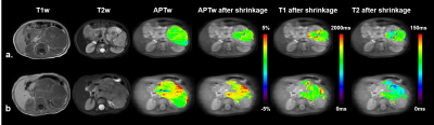 |
Evaluating the Risk of Pediatric Neuroblastoma in the Abdomen with Amide Proton Transfer Imaging
Wenqi Wang1, Xuan Jia2, Jiawei Liang2, Xiaohui Ma2, Weibo Chen3, Dan Wu1, Can Lai2, and Yi Zhang1
1Key Laboratory for Biomedical Engineering of Ministry of Education, Department of Biomedical Engineering, College of Biomedical Engineering & Instrument Science, Zhejiang University, Hangzhou, Zhejiang, China, 2Department of Radiology, Children’s Hospital, Zhejiang University School of Medicine, Hangzhou, Zhejiang, China, 3Philips Healthcare, Shanghai, China
Neuroblastoma (NB) most often occurs in young children, and accurate diagnosis of NB with anatomical MRI remains challenging. Here, we explored the potential of amide proton transfer (APT) imaging in evaluating the risk of pediatric abdominal NB. A total of 25 patients were enrolled, including 12 with low-risk NB and 13 with high-risk NB. An automatic shrinkage algorithm was applied to the initial region of interest delineated by an experienced radiologist to focus on the most aggressive part of tumors. We obtained an AUC of 0.917 for stratifying the risk of NB with APT, demonstrating the potential of clinical application.
|
||
0352.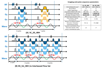 |
Liver Stiffness in a Single Breath-hold Using Wave Polarity-Inversion Motion Encoding and Compressed SENSE: Coverage Equivalent to 5 Slices
Amol Pednekar1, Deep B. Gandhi2, Hui Wang3, Jean A. Tkach1, Andrew T. Trout1, and Jonathan R. Dillman1
1Department of Radiology, Cincinnati Children's Hospital Medical Center, Cincinnati, OH, United States, 2Imaging Research Center, Department of Radiology, Cincinnati Children's Hospital Medical Center, Cincinnati, OH, United States, 3MR Clinical Science, Philips, Cincinnati, OH, United States
2D GRE MRE liver images acquired at 4 transverse levels in breath-hold times >13s per slice is currently standard of care (SC_2D_4BH). The combination of wave polarity-inversion motion encoding and compressed-SENSE enables volumetric three-dimensional (3D) MRE data acquisition in a single breath-hold of <16 s, with identical spatial resolution and 5 slice equivalent coverage (3D_5Sl_SBH). In 19 participants, mean liver shear stiffness values estimated with SC_2D_4BH and 3D_5sl_SBH correlated very strongly (ICC=0.96) with a mean bias of 0.13 kPa (<5 %). 3D_5sl_SBH MRE provides similar stiffness estimates as SC_2D_4BH with increased coverage in a single breath-hold compared to 4 breath-holds.
|
||
0353.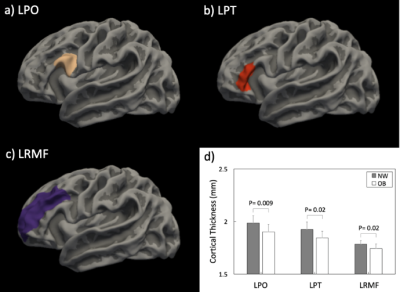 |
Maternal Obesity during Pregnancy is Associated with Lower Cortical Thickness in the Newborn Brain
Xiaoxu Na1, Natalie E. Phelan1, Marinna R. Tadros1, Aline Andres2,3, Thomas M. Badger2,3, Charles M. Glasier1, Raghu H. Ramakrishnaiah1, Amy C. Rowell1, Li Wang4, Gang Li4, Zhengwang Wu4, David K. Williams5, and Xiawei Ou1,3,6
1Radiology, University of Arkansas for Medical Sciences, Little Rock, AR, United States, 2Pediatrics, University of Arkansas for Medical Sciences, Little Rock, AR, United States, 3Arkansas Children's Nutrition Center, Little Rock, AR, United States, 4Radiology, University of North Carolina at Chapel Hill, Chapel Hill, NC, United States, 5Biostatistics, University of Arkansas for Medical Sciences, Little Rock, AR, United States, 6Arkansas Children's Research Institute, Little Rock, AR, United States
This study examined the relationships between maternal obesity during pregnancy and newborn’s brain cortical development. Healthy normal weight or obese pregnant women were recruited at early pregnancy and their newborns underwent a brain MRI examination at 2 weeks of age. Structural MR images of the brain were post-processed to reconstruct cortical surfaces, and mean cortical thickness in different brain regions was measured. Significant differences in cortical thickness between infants born to normal weight vs. obese mothers were found in multiple brain regions, and negative correlations between maternal body fat mass percentage and infant cortical thickness were also observed.
|
||
|
0354.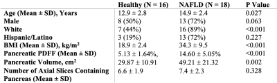 |
Using Free-Breathing MRI to Characterize Heterogeneity of Pancreatic Fat in Children with Nonalcoholic Fatty Liver Disease
Jacob Story1, Sevgi Gokce Kafali2,3, Shu-Fu Shih2,3, Kara L. Calkins4, Shahnaz Ghahremani3, and Holden H. Wu2,3
1David Geffen School of Medicine at UCLA, Los Angeles, CA, United States, 2Department of Bioengineering, University of California Los Angeles, Los Angeles, CA, United States, 3Department of Radiological Sciences, University of California Los Angeles, Los Angeles, CA, United States, 4Department of Pediatrics, University of California Los Angeles, Los Angeles, CA, United States
Metabolic dysfunction in children is a major health issue. Pancreatic fat is a potential biomarker of metabolic dysfunction, but methodological difficulties have limited research on its role in at-risk children. This work used free-breathing abdominal MRI to quantify pancreatic fat and to characterize its heterogeneity in children with and without nonalcoholic fatty liver disease (NAFLD). Children with NAFLD had increased pancreatic fat that was heterogeneously distributed and predominantly located in the superior region. A simple region-of-interest-based measurement method failed to account for this heterogeneity, and thus underestimated pancreatic fat. Full segmentation pancreatic fat measurements correlated with markers of metabolic dysfunction.
|
|
|
0355.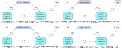 |
Regional changes in brain development and cognitive outcome in infants with Congenital Heart Disease
Alexandra F Bonthrone1, Ralica Dimitrova1,2, Andrew Chew1, Christopher J Kelly1, Lucilio Cordero-Grande1,3, Olivia Carney1, Alexia Egloff1, Emer Hughes1, Katy Vecchiato1,2, John Simpson4, Joseph V Hajnal1,5, Kuberan Pushparajah4, Suresh Victor1,
Chiara Nosarti1,6, Mary A Rutherford1, A. David Edwards1, Jonathan O’Muircheartaigh1,2, and Serena J Counsell1
1Centre for the Developing Brain, School of Biomedical Engineering and Imaging Sciences, King's College London, London, United Kingdom, 2Department for Forensic and Neurodevelopmental Sciences, Institute of Psychiatry, Psychology and Neuroscience, King's College London, London, United Kingdom, 3Biomedical Image Technologies, ETSI Telecomunicación, Universidad Politécnica de Madrid and CIBER-BBN, Madrid, Spain, 4Paediatric Cardiology Department, Evelina London Children's Healthcare, London, United Kingdom, 5Biomedical Engineering Department, School of Biomedical Engineering and Imaging Sciences, King's College London, London, United Kingdom, 6Department of Child and Adolescent Psychiatry, Institute of Psychiatry, Psychology and Neuroscience, King's College London, London, United Kingdom
Infants with Congenital Heart Disease (CHD) are at high risk of neurodevelopmental disorders. We acquired presurgical neonatal T2-weighted MRI (N=66), cerebral oxygen delivery (CDO2; N=53), and 22-month cognitive and motor scores (N=44). Atypicality indices, representing the degree of deviation of a regional brain volume from the normative neonatal mean for a given gestational age, sex and postnatal age, were calculated. Reduced CDO2 was indirectly associated with lower cognitive scores through the mediating effect of negative bilateral caudate and thalami atypicality indices. The aetiology of cognitive impairments in CHD may encompass poor CDO2 leading to impaired caudate and thalamus growth.
|
|
|
0356.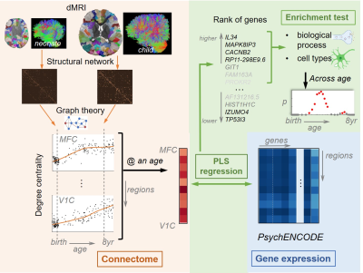 |
Transcriptomic decoding of the human brain structural connectome through the 3rd trimester and early childhood
Chenying Zhao1,2, Gabriel Santpere3, Minhui Ouyang1, David Andrijevic4, Nenad Sestan4, and Hao Huang1,5
1Department of Radiology, Children's Hospital of Philadelphia, Philadelphia, PA, United States, 2Department of Bioengineering, School of Engineering and Applied Science, University of Pennsylvania, Philadelphia, PA, United States, 3Neurogenomics group, Research Programme on Biomedical Informatics (GRIB), Hospital del Mar Medical Research Institute (IMIM), DCEXS, Universitat Pompeu Fabra, Barcelona, Spain, 4Department of Neuroscience and Kavli Institute for Neuroscience, Yale School of Medicine, New Haven, CT, United States, 5Department of Radiology, Perelman School of Medicine, University of Pennsylvania, Philadelphia, PA, United States
Transcriptome, the set of gene expression, is spatiotemporally heterogeneous across brain development. Under its regulation, dramatic changes in brain connectivity estimated through diffusion MRI is observed in early childhood. However, the association between the macroscopic structural connectome and microscopic transcriptome across early postnatal years is not clear. Here, we revealed this dynamic association between structural connectome and gene expression from a large cohort of 200 neonates and children through the 3rd trimester and early childhood. The changes of associated genes’ enrichment in cell types and biological processes across different ages shed light into the dynamic transcriptomic roles in connectome development.
|
The International Society for Magnetic Resonance in Medicine is accredited by the Accreditation Council for Continuing Medical Education to provide continuing medical education for physicians.