Oral
Making MRI More Accessible: Speed, Cost & New Developments
ISMRM & SMRT Annual Meeting • 15-20 May 2021

Oral
Making MRI More Accessible: Speed, Cost & New Developments
| Concurrent 3 | 14:00 - 16:00 | Moderators: Samantha By & Clarissa Cooley |
0741.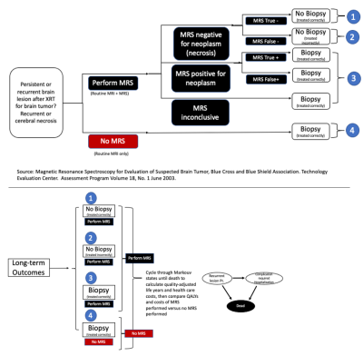 |
Cost-Effectiveness Analysis of Magnetic Resonance Spectroscopy for the Differentiation of Recurrent Glioma from Radiation Necrosis
Huijun Liao1, So Wing Lum1, and Alexander P. Lin1
1Center for Clinical Spectroscopy, Brigham and Women's Hospital, Boston, MA, United States
To date there have been no formal studies of the cost-effectiveness of magnetic resonance spectroscopy. Cost-effectiveness analysis was conducted by utilizing a decision-analytic model that compared MRS and standard of care to standard of care alone for recurrent glioma and necrosis differentiation. Our results showed that performing MRS was highly cost-effective with incremental cost-effectiveness ratios of -$98,978, -$50,666 and -$41,349 per quality-adjusted life-years over 5-year, 10-year and lifetime horizons. At the willingness-to-pay threshold of $50,000, performing MRS was more cost-effective with certainty of 96% to 97% over those time-horizons. MRS should be considered reimbursable by policy makers.
|
||
 |
0742.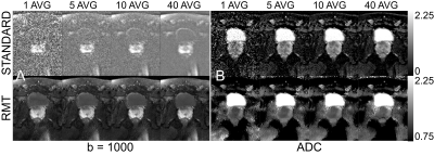 |
Feasibility of accelerated diffusion weighted imaging for prostate cancer screening on prototype 0.55T system enabled with random matrix theory
Gregory Lemberskiy1, Dmitry S Novikov1, Mary Bruno1, Mahesh Bharath Keerthivasan2, Els Fieremans1, and Hersh Chandarana1
1Radiology, NYU School of Medicine, New York, NY, United States, 2Siemens Medical Solutions, New York, NY, United States
We compared standard and RMT reconstructions for a prostate DWI exam on a prototype 0.55T for varying number of averages (1 to 40) to evaluate the image quality and overall loss. For standard reconstructions, regardless of number of averages, the Rician bias is present even at 40 averages for high-b value data, whereas 1 average on RMT is sufficient to produce diagnostic grade DWI at 0.55T.
|
|
0743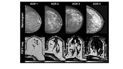 |
Assessing breast density using the standardized proton density fat fraction based on chemical shift encoding-based water-fat separation Video Permission Withheld
Tabea Borde1, Mingming Wu1, Stefan Ruschke1, Christof Böhm1, Kilian Weiss2, Stephan Metz1, Marcus R Makowski1, and Dimitrios Karampinos1
1Department of Diagnostic and Interventional Radiology, Technical University of Munich, Munich, Germany, 2Philips Healthcare, Hamburg, Germany
Breast density is confirmed as a strong, independent risk factor of breast cancer which is why there is a clinical need for a robust, reader-independent, non-ionizing, quantitative assessment of breast density. This retrospective study proposes the proton density fat fraction (PDFF) derived from chemical shift encoding-based water-fat separation as a novel quantitative MRI biomarker of breast density. As a clinically highly practicable biomarker that is automatedly obtainable and insensitive to acquisition parameters and the partial volume effect, the PDFF significantly negatively correlated with the most commonly used conventional radiographic mammogram breast density estimations.
|
||
0744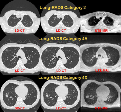 |
Can pulmonary thin-section MRI with UTE<200μs be applied for lung cancer screening similar to CT? Video Permission Withheld
Yoshiharu Ohno1,2,3, Masao Yui4, Takeshi Yoshikawa3,5, Daisuke Takenaka5, Kaori Yamamoto4, Yoshimori Kassai4, Kazuhiro Murayama2, and Hiroshi Toyama1
1Radiology, Fujita Health University School of Medicine, Toyoake, Japan, 2Joint Research Laboratory of Advanced Medical Imaging, Fujita Health University School of Medicine, Toyoake, Japan, 3Division of Functional and Diagnostic Imaging Research, Department of Radiology, Kobe University Graduate School of Medicine, Kobe, Japan, 4Canon Medical Systems Corporation, Otawara, Japan, 5Diagnostic Radiology, Hyogo Cancer Center, Akashi, Japan
No report has been found to compare the capability for lung cancer screening among pulmonary MR imaging with ultra-short TE (UTE), low-dose CT (LDCT) and standard-dose CT (SDCT). We hypothesized that pulmonary MR imaging with UTE has a similar potential to detect pulmonary nodules and evaluate Lung-RADS classification and can apply lung cancer screening as well as CT. The purpose of this study was to compare the capability for lung cancer screening among pulmonary MR imaging with UTE and both dose CTs.
|
||
0745.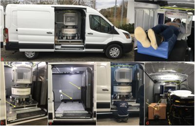 |
Residential MRI: Fully Mobile Neuroimaging for Community and Population-Based Studies
Sean Deoni1, John Rogers2, Jennifer Beauchemin2, Viren D'Sa3, Eddy Boskamp4, Samantha By4, Chris McNulty4, William Mileski4, Brian Welch4, and Paul Medeiros5
1Bill & Melinda Gates Foundation, Seattle, WA, United States, 2Advanced Baby Imaging Lab, Rhode Island Hospital, Providence, RI, United States, 3Pediatrics, Rhode Island Hospital, Providence, RI, United States, 4Hyperfine, Guildford, CT, United States, 5New England Collision, Seekonk, MA, United States
The continued increase in magnetic field strength and gradient capabilities of MRI systems has allowed ever more sophisticated interrogation of brain structure and function. However, these systems are often limited to high resource imaging research centers. To extend to more general population-studies, there is need for low-cost mobile imaging systems that allow for anywhere/everywhere imaging. In this work, we built a mobile MRI lab using the 64mT Hyperfine SwoopTM system and a customized Ford Transit cargo van, and demonstrated the ability to perform routine and rapid at-home MRI for the first time.
|
||
0746.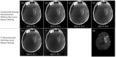 |
The Impact of Acceleration on Radiologists' Confidence in Point-Of-Care 0.5T MRI For Triage of Acute Stroke
Michelle Pryde1,2, Sarah Reeve2,3, Taylor Bouchie2,4, Elena Adela Cora5,6, David Volders5,6, Chris Bowen2,3,5, James Rioux2,3,5, and Steven Beyea1,2,3,5
1School of Biomedical Engineering, Dalhousie University, Halifax, NS, Canada, 2Biomedical Translational Imaging Centre, QEII Health Sciences Centre, Halifax, NS, Canada, 3Physics and Atmospheric Science, Dalhousie University, Halifax, NS, Canada, 4Medicine, Dalhousie University, Halifax, NS, Canada, 5Diagnostic Radiology, Dalhousie University, Halifax, NS, Canada, 6Diagnostic Imaging, Nova Scotia Health, Halifax, NS, Canada
Accelerated MR image acquisition is key for emergency medicine situations, such as acute stroke, but yields degraded image quality. Therefore, our aim in this study was to calibrate a relationship between IQMs and radiologists' confidence in answering pointed clinical questions relevant to triaging of stroke patients so as to develop a protocol at low-field that is “as fast as clinically useful”. We observed that upon increasing R and decreasing NEX, radiologists’ confidence scores in their ability to identify diagnostically relevant features of both acute and chronic stroke decreased; however, radiologists’ confidence remained high despite retrospective acceleration of up to R=6.
|
||
0747.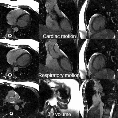 |
Running Free on a Low-Field: a Proof of Principle
Davide Piccini1,2, Jerome Yerly2,3, Tobias Kober1,2,4, Lorenzo Di Sopra2, Aurélien Bustin2,5,6, Daniel Giese7, Mario Bacher7, Michaela Schmidt7, Peter Speier7, Christian Geppert7, Rainer Schneider7, David Grodzki7, and Matthias Stuber2,3
1Advanced Clinical Imaging Technology, Siemens Healthcare AG, Lausanne, Switzerland, 2Department of Radiology, Lausanne University Hospital and University of Lausanne, Lausanne, Switzerland, 3CIBM Center for Biomedical Imaging, Lausanne, Switzerland, 4LTS5, École Polytechnique Fédérale de Lausanne (EPFL), Lausanne, Switzerland, 5IHU LIRYC, Electrophysiology and Heart Modeling Institute, Fondation Bordeaux Université, Pessac-Bordeaux, France, 6Department of Cardiovascular Imaging, Hôpital Cardiologique du Haut-Lévêque, CHU de Bordeaux, Pessac, France, 7Magnetic Resonance, Siemens Healthcare, Erlangen, Germany
Low-field MR has recently attracted considerable attention because of reduced overall cost, lower field inhomogeneity, a lower specific absorption rate, and the potential for a more widespread global use. Owing to the simplicity and scalability of our recently developed free-running framework (FRF) for cardiovascular imaging, we here present the first results obtained with FRF at 0.55T. We demonstrate that FRF is scalable to this field strength by successfully reconstructing 5D cardiac- and respiratory motion-resolved whole heart images without the need of any gating or triggering devices, while reducing scan planning to a single mouse-click.
|
||
0748.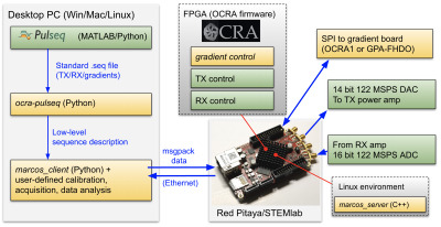 |
Research and educational applications of an open source, low cost MRI console with an accessible pulse sequence programming environment
Lincoln Craven-Brightman1, Thomas O'Reilly2, Benjamin Menkuec3, Marcus Prier4, Rubén Pellicer-Guridi5,6, Joseba Alonso5,6, Lawrence L. Wald1,7, Maxim Zaitsev8, Jason Stockmann1,7, Thomas Witzel9, Andrew Webb2, and Vlad Negnevitsky10
1A.A. Martinos Center for Biomedical Imaging, Department of Radiology, Massachusetts General Hospital, Charlestown, MA, United States, 2Department of Radiology, Leiden University Medical Center, Leiden, Netherlands, 3University of Applied Sciences and Arts Dortmund, Dortmund, Germany, 4Otto von Guericke Universität Magdeburg, Magdeburg, Germany, 5Universitat Politècnica de València, Valencia, Spain, 6Spanish National Research Council, Valencia, Spain, 7Harvard Medical School, Boston, MA, United States, 8Medizinische Universität Wien, Vienna, Austria, 9Qbio Inc, San Carlos, CA, United States, 10Independent researcher, Zürich, Switzerland
The hardware and software for a low cost programmable MR console has been developed, characterized and tested in various setups at multiple sites for both educational and research applications. A new Python-based wrapper allows easy pulse programming of different sequences and k-space trajectories using PulSeq, with output data also being processed via Python. The first two- and three-dimensional in vivo images have also been acquired using this hardware on a large bore Halbach array system.
|
||
0749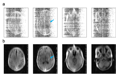 |
Freeing MRI from its Faraday cage with Interference Rejection Video Permission Withheld
Hadrien Dyvorne1, Todd Rearick1, Michael Poole1, Carole Lazarus1, Pierre Weiss2, Laura Sacolick1, Jeremy Jordan1, Cedric Hugon1, William Mileski1, Gang Chen1, Rafael O'Halloran1, Chris McNulty1, Jonathan Lowthert1, Anne
Nelson1, Aristito Lorenzo1, Nicholas Zwart1, Prantik Kundu1, Scott Martin1, Andrei Loutchouk1, E. Brian Welch1, Samantha By1, Bradley Cahn3, Matthew Yuen3, Mercy Mazurek3, Anjali Pranhat3, Matthew Rosen4, Kevin Sheth3,
and Jonathan Rothberg1
1Hyperfine, Guilford, CT, United States, 2ITAV, CNRS, Toulouse, France, 3Neurology, Yale University School of Medicine, New Haven, CT, United States, 4Athinoula A. Martinos Center for Biomedical Imaging, Massachusetts General Hospital, Charlestown, MA, United States
We introduce methods and hardware for rejection of high levels of interference from MR data, allowing the acquisition of MR imaging scans outside the shielded room and with no disruption to patient care. Interference cancellation relies on deriving a transfer function between “primary” sensors that detect MR signal and interference, and “reference” sensors that detect interference only. Interference rejection up to 67 dB is demonstrated, along with imaging results from scans acquired at the patient bedside in a Neuro ICU.
|
||
0750.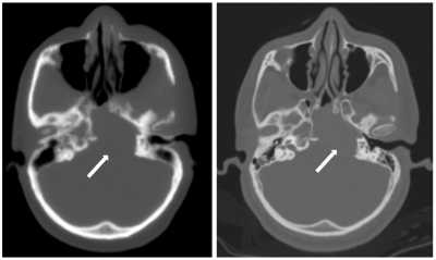 |
Comparing Zero Time of Echo (ZTE) MRI sequence and Computed Tomography for assessing bony lesions of skull base and calvarium : A Pilot study
Vikas Chauhan1, Sandeep Kaushik2, Florian Wiesinger2, Cristina Cozzini2, Michael Carl2, Maggie Fung2, Bhairav Mehta2, Bejoy Thomas1, and Kesavadas Chandrasekharan1
1Imaging Sciences and Interventional Radiology, Sree Chitra Tirunal Institute for Medical Sciences and Technology, Thiruvananthapuram, India, 2GE Healthcare, GE Healthcare, Bangalore, India
ZTE is a novel MRI sequence which can be used for the imaging of the bone. In this pilot study we present our initial experience with this sequence. The pseudoCT images generated after post-processing of raw ZTE data were similar to the real CT images in bone windows and were able to give the relevant clinical information about the lesion size, margins, erosions and bony expansion. As this technique is free of ionising radiation, it has a huge potential for bone imaging. Thus the MRI becomes one stop shop for the imaging of patients with bone lesions.
|
The International Society for Magnetic Resonance in Medicine is accredited by the Accreditation Council for Continuing Medical Education to provide continuing medical education for physicians.