SMRT Edu Session
Data & Post-Processing I
ISMRM & SMRT Annual Meeting • 15-20 May 2021

| SMRT Session | 18:00 - 20:00 | Moderators: George Bouzalis & Joseph Joslin |
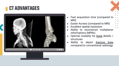 |
Post-Processing MR into CT-Like Images
Jeff Chen
MRI may be able to replace the need for CT for osseous injury, significantly reducing exposure to ionizing radiation and enabling MRI to be a one-stop imaging modality for pediatric trauma studies and further prevent any delays of patient’s treatment or having more scans that may prolong hospital stay. 3D spoiled gradient echo sequences such as the T1 eTHRIVE / LAVA / VIBE and the T2* mFFE / MERGE / MEDIC can produce CT-like images has demonstrated its ability to show fractures and surrounding soft tissue structures.
|
|
 |
Automatic Post-Processing in the Cloud Space
Kevin King
The rise of cloud-based computing has heralded a fundamental shift in the way radiology evaluates images. In large part, this is not due to development of new imaging analysis tools, but rather automation of these tools using basic imaging analysis tools which have been well developed in the research setting. The standardization brought about by lack of user interaction has impacted how these imaging tools are validated and how findings are disseminated. Further benefits from automation may help to reduce monotony while supporting the high efficiency expected of radiology.
|
|
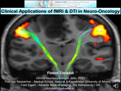 |
Clinical Applications of fMRI & DTI in Neuro-Oncology
Foteini Christidi
Functional MRI and diffusion tensor imaging are increasingly incorporated in clinical practice in order to identify eloquent brain regions. The latter is important in case of brain tumors where pre-operative identification of these regions may affect the surgical procedure and predict post-operative clinical outcome. In this 30min educational session, we will explore the fundamental clinical applications of task and resting-state fMRI and DTI/Tractography in neuro-oncology by processing pre-operative fMRI and DTI data of real brain tumor cases and discussing pre-operative findings with regards to findings from electrical stimulation and patients’ post-operative clinical outcome.
|
The International Society for Magnetic Resonance in Medicine is accredited by the Accreditation Council for Continuing Medical Education to provide continuing medical education for physicians.
 Watch the Video