Sebastian Theilenberg1, Chathura Kumaragamage2, Scott McIntyre2, Terry W. Nixon2, Christoph Juchem1,3, and Robin A. de Graaf2
1Biomedical Engineering, Columbia University, New York, NY, United States, 2Department of Radiology and Biomedical Imaging, Magnetic Resonance Research Center, Yale University School of Medicine, New Haven, CT, United States, 3Radiology, Columbia University Medical Center, New York, NY, United States
1Biomedical Engineering, Columbia University, New York, NY, United States, 2Department of Radiology and Biomedical Imaging, Magnetic Resonance Research Center, Yale University School of Medicine, New Haven, CT, United States, 3Radiology, Columbia University Medical Center, New York, NY, United States
A proposed low-angle combined-echo sequence is capable of producing high quality images in the presence of strong B0 magnetic field inhomogeneity with good contrast for human gray and white brain matter.
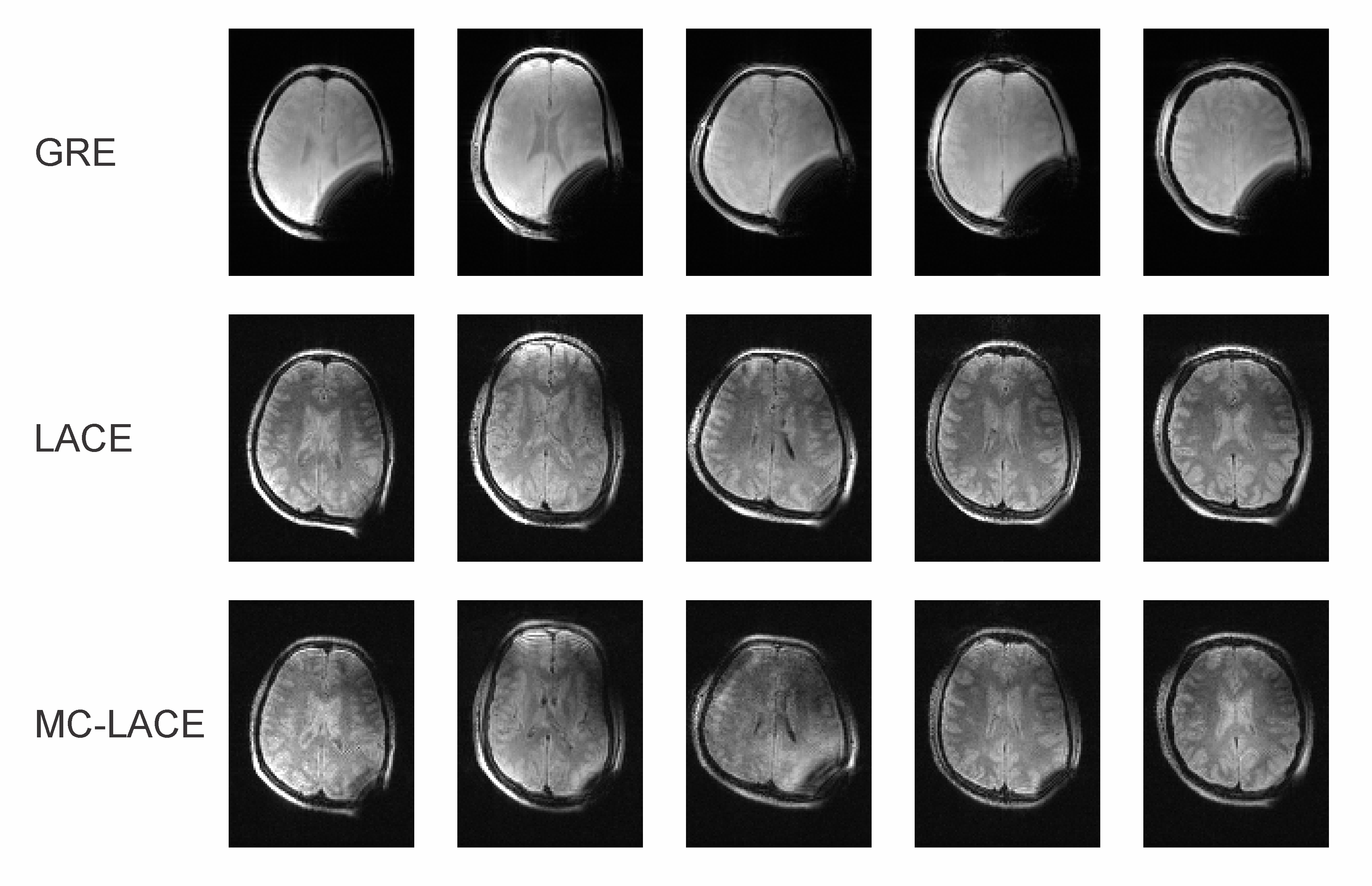
Figure 5: LACE brain images of 5 human subjects in the presence of B0 inhomogeneity induced by a paper clip (FoV 256x192x128 mm3 at 2x2x4 mm resolution). 3D gradient echo images show large areas of signal dropout near the paper clip (top row). LACE images recover signal in this area, but exhibit image deformations due to the extreme inhomogeneity (middle row). Multi-Coil generated LACE images show largely the same image quality than the ones acquired with standard gradient coils (lower row). No image distortion correction was applied.
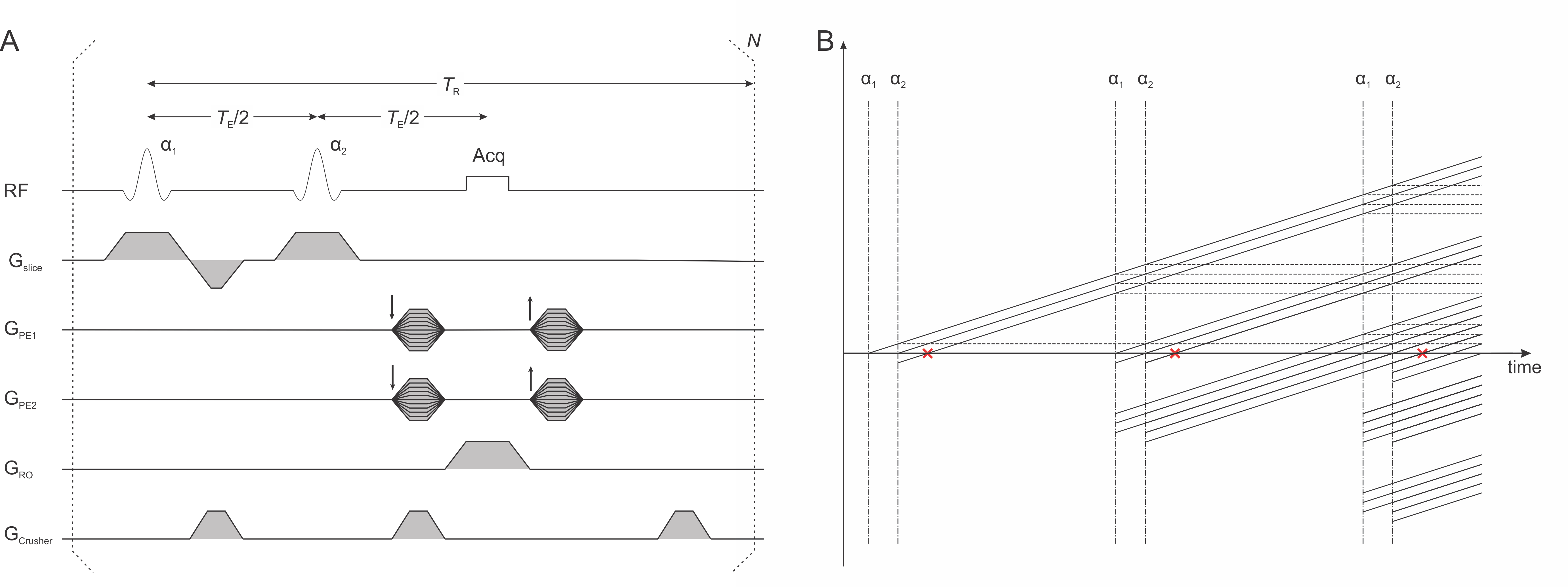
Figure 1: LACE sequence. A) Sequence diagram. B) Phase graph.
The sequence consists of two broadband excitation pulses separated by TE/2.
Signal is acquired at the time of the spin echo of the two RF pulses (red crosses
in B), which in the steady-state comprises a combination of signal from spin and stimulated echoes.
Crusher gradients symmetrically around the 2nd RF pulse and at the
end of TR ensure a consistent signal independent of the background B0
field.