Yanning Liu1, Henk M. De Feyter1, Scott McIntyre1, Terence W. Nixon1, and Robin A. de Graaf1
1MRRC Yale University, New Haven, CT, United States
1MRRC Yale University, New Haven, CT, United States
We propose an interleaved MRI+DMI routine, including the hardware and sequence modifications to integrate DMI with clinical MRI. Using interleaved FLAIR+DMI, we demonstrate that MR image quality, DMI sensitivity, information content are preserved in phantoms and in the human brain in vivo.
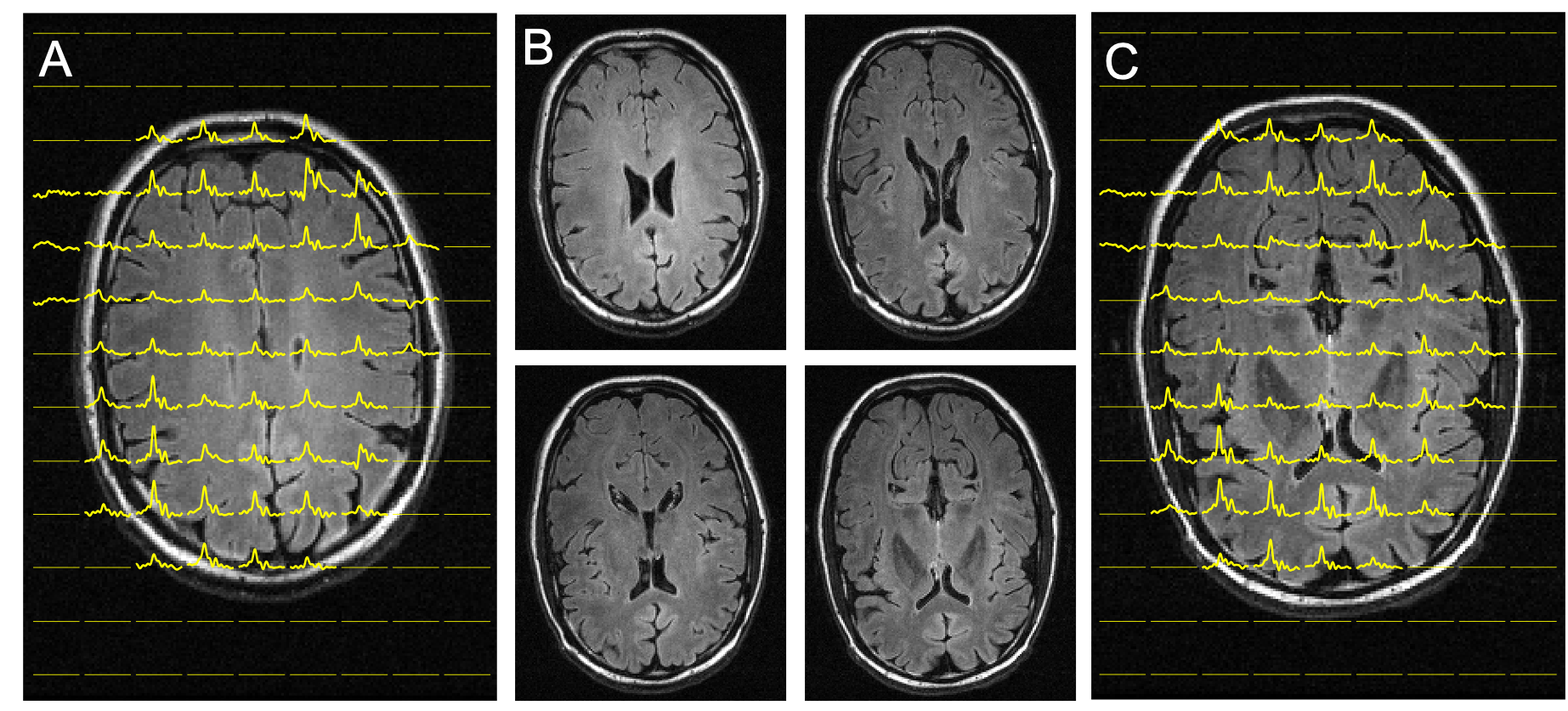
Interleaved FLAIR and DMI of human brain 75 min following the oral administration of [6,6’-2H2]-glucose. DMI was acquired as a 13 x 9 x 11 matrix (YXZ) over a 260 x 180 x 220 mm3 FOV, whereas the FLAIR images were acquired as 14 slices and a 256 x 192 matrix over a 256 x 192 mm2 FOV (TR/TI/TE = 8800/2200/90 ms). During the ~ 7 min FLAIR acquisition, the DMI data could be acquired twice (i.e. NA = 2). (A-C) FLAIR images spanning circa 40 mm, covering two axial planes from the 3D DMI dataset as shown in (A) and (C).
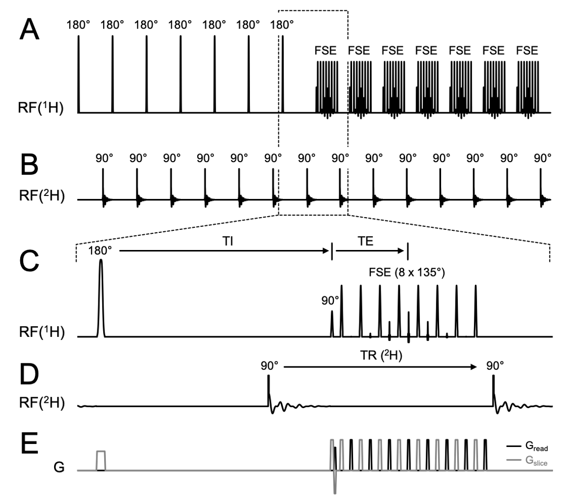
Interleaved FLAIR MRI+DMI sequence. (A) 1H pulse acquisition scheme. 7 slices are inverted consecutively followed by a fast spin-echo (FSE) acquisition (TR/TI/TE = 8800/2200/90 ms). The (B) 2H pulse acquisitions are placed in the dead time during TI and right after FSE with equal TR (= 314 ms). (E) Shows the gradients used during acquisition. Note the plot only shows the first half of FLAIR TR, where half of the slices (odd number slices) were excited. The pulse acquisition scheme is repeated on the even number slices in the second half of TR. 28 DMI acquisitions are achieved in one FLAIR TR.